Delayed Repair of Aortic Dissection in Sickle Cell Anaemia as a Combined Cardiac and Vascular Surgical Approach
←
→
Page content transcription
If your browser does not render page correctly, please read the page content below
CASE REPORT

              Delayed Repair of Aortic Dissection in Sickle Cell
              Anaemia as a Combined Cardiac and Vascular
              Surgical Approach

              Massimo Capoccia a,*, Maziar Mireskandari b,
              Nicholas J. Cheshire b, Ulrich P. Rosendahl a

              a
                  Aortic and Cardiac Surgery, Royal Brompton Hospital, London, United Kingdom
              b
                  Vascular Surgery, Royal Brompton Hospital, London, United Kingdom

              Abstract

                 We discuss a patient who presented with a type B aortic dissection with a retrograde progression in the context of
              sickle cell anaemia. Given the involvement of the superior mesenteric artery and concern for bowel ischaemia, a delayed
              approach was considered. Subsequently, a frozen elephant trunk was performed in the hybrid theatre with the back-up
              of the vascular surgeon for mesenteric protection. A technically demanding procedure followed by a prolonged and
              challenging postoperative course finally led to a successful outcome. We argue that the case presented is an example of
              how a close cooperation between professionals can offer additional options to treatment based on a mixture of skills and
              background to achieve the desired outcome.

              Keywords: Aortic aneurysm, Aortic dissection, Frozen elephant trunk, Cardiac surgeons, Vascular surgeons

              1. Introduction                                                                    2. Case presentation
                                                                                                   A 60-year-old patient was referred to our hospital
              T    raditionally, aortic disease has been
                   approached by cardiac and vascular sur-
                                                                                                 with features suggestive of type A aortic dissection
                                                                                                 on initial CT-scan assessment made by the radi-
              geons individually where each professional has                                     ology team of the referring Hospital (Fig. 1 and
              focused on a specific section of the vessel.                                        Fig. 2). Co-morbidities included arterial hyperten-
              Nevertheless, aortic disease frequently involves                                   sion, sickle cell anaemia and chronic renal impair-
              the whole vessel with particular reference to                                      ment requiring dialysis for which a left brachio-
                                                                                                 cefalic fistula had been created. Following transfer
              dissection and aneurysm. A close cooperation
                                                                                                 and further review of the available images, it was
              between cardiac and vascular surgeons for the                                      clear that the dissection extended to the superior
              treatment of aortic disease is currently being                                     mesenteric artery (SMA) giving matter of concern
              acknowledged as an effective approach although                                     for potential bowel ischemia postoperatively (Fig. 3).
              not completely accepted by many. Here we pre-                                      Therefore, a decision was made to maintain blood
              sent a case of aortic dissection whose treatment                                   pressure control with b-blocker intravenously and
              has benefited from our established and well                                         intervene 24 h later. The aim would be to perform a
              amalgamated aortic team.                                                           digital subtraction angiography (DSA) in our hybrid

              Received 30 November 2019; revised 12 January 2020; accepted 12 January 2020.
              Available online 20 May 2020

              * Corresponding author. Aortic and Cardiac Surgery, Royal Brompton Hospital, Sydney Street, Chelsea, London, SW3 6NP, United
              Kingdom.
              E-mail address: capoccia@doctors.org.uk (M. Capoccia).

              https://doi.org/10.37616/2212-5043.1043
              2212-5043/© 2020 Saudi Heart Association. This is an open access article under the CC-BY-NC-ND license (http://creativecommons.org/licenses/by-nc-nd/4.0/).
JOURNAL OF THE SAUDI HEART ASSOCIATION 2020;32:208e212                                            209

                                                                                                                                               CASE REPORT
theatre to further assess the visceral vessels and
insert a wire in the SMA for stenting after the
planned surgical procedure. Following contrast in-
jection, it became evident that the features of the
disease were more consistent with a type B dissec-
tion that had progressed in a retrograde manner
with lower entry points in the descending thoracic
aorta (Fig. 4). These findings allowed us to plan a
definitive surgical strategy following a period of
stabilisation. Aortic arch replacement with a frozen
elephant trunk under hypothermic circulatory arrest
with potential TEVAR extension and mesenteric
protection should the need arise to address the SMA
was considered an appropriate delayed course of
action on this occasion1,2. In the meanwhile, the
patient would be nursed in the high dependency
unit with strict blood pressure control using b-
blocker and ACE-inhibitor. Initial medical man-
agement was relatively successful although serial
CT-scan imaging showed further dilatation of the
ascending aorta, which triggered the final timing for                   Fig. 2. Preoperative contrast CT-scan showing false and true lumen
                                                                       relationship in ascending and descending thoracic aorta.
intervention.
   Plasmapheresis was required to address the sickle
cell anaemia preoperatively.                                           myocardial protection. Body temperature at 24  C
   The patient was transferred once again to our                       was considered as a compromise in view of the
hybrid theatre and prepped in the supine position.                     sickle cell anaemia. Antegrade cerebral perfusion
Invasive arterial and venous monitoring was ob-                        was achieved with cannulation of each epiaortic
tained through the left and right radial artery, left                  vessel following longitudinal incision of the
femoral artery and right internal jugular vein. Car-                   ascending aorta and arch. Continuous, non invasive
diopulmonary bypass was established through right                      monitoring of cerebral oxygen saturation was
femoral and right atrial cannulation. Antegrade and                    maintained with near infrared spectroscopy (NIRS)
retrograde cold blood cardioplegia was delivered for

                                                                       Fig. 3. Preoperative contrast CT-scan highlighting involvement of the
Fig. 1. Preoperative contrast CT-scan highlighting arch involvement.   superior mesenteric artery.
210 JOURNAL OF THE SAUDI HEART ASSOCIATION 2020;32:208e212
CASE REPORT
physiotherapy and CPAP delivery. Regular filtration
was used to address the renal impairment. Strict
blood pressure control was addressed. Close liaison
with haematology and intensivist colleagues was
maintained as far as postoperative management of
the sickle cell anaemia was concerned. Finally, the
patient was discharged to the local hospital 19 days
postoperatively for continuity of care. The histo-
logical findings were consistent with features of
cystic medial necrosis and atherosclerosis in the
absence of granuloma and vasculitis.
A postoperative echocardiographic and CT-scan
assessment was considered extremely satisfactory
(Fig. 5). The innominate and left common carotid
arteries remained well perfused. The left subclavian
artery still showed some residual dissection without
obstruction. Residual degree of perfusion of the
false lumen was observed with celiac axis and SMA
appropriately perfused from both true and false
lumen as preoperatively. Follow-up appointment
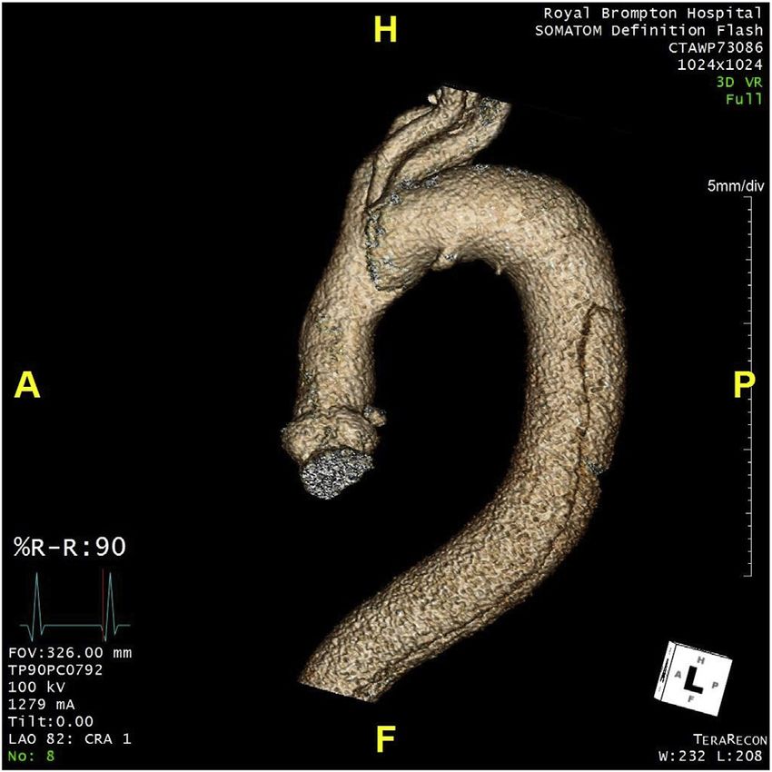
Fig. 4. Preoperative 3D reconstruction of the key areas of the dissection. was arranged at 3 months with further CT-aortic
angiogram. Next step would be another CT-aortic
angiogram at 6 months with the aim to keep yearly
imaging surveillance according to our established
using INVOS™ 5100C Cerebral/Somatic Oximeter routine.
monitoring system. A frozen elephant trunk with a
28 mm Thoraflex™ Hybrid Plexus 4 device (Vascu- 3. Discussion
tek, TERUMO Aortic, Inchinnan, UK) was per-
Traditionally, the treatment of acute type A aortic
formed with distal anastomosis within zone 2
dissection has been immediate surgery whereas
sparing the left subclavian artery under circulatory
type B would be addressed more conservatively.
arrest. Subsequently, distal reperfusion was
Over the years the attitude has shifted towards a
commenced through the side arm of the Thoraflex.
The true lumen placement of the stent of the
Thoraflex was guaranteed by the presence of a stiff
wire previously inserted through the left femoral
artery under image intensifier. Then, the proximal
anastomosis was completed maintaining further
myocardial protection. Finally, the innominate and
left common carotid arteries were de-branched and
anastomosed to the Thoraflex device. The patient
was rewarmed and gradually weaned off cardio-
pulmonary bypass. Blood products, surgical seal-
ants and additional suturing were required to ach-
ieve satisfactory haemostasis. Atrial and ventricular
pacing wires were used for temporary pacing as
required. Three chest drains inserted and the chest
closed in layers. An on table angiogram was per-
formed, which confirmed appropriate placement of
the Thoraflex stented component in the descending
thoracic aorta with satisfactory occlusion of the
distal entry point. Therefore, further stenting as
previously planned was not required at this stage.
Also the SMA did not require further attention. The
postoperative course was complicated by temporary Fig. 5. Postoperative 3D reconstruction following deployment of the
impairment of gas exchanges requiring aggressive stented portion of the Thoraflex device.JOURNAL OF THE SAUDI HEART ASSOCIATION 2020;32:208e212 211
CASE REPORT
more delayed and planned repair3,4, which is every possible option is being offered. Although
currently our approach as reported in this case. The communication between teams takes time and may
challenge related to the presence of sickle cell lead to treatment delay, it remains an important
anaemia even more required a careful delayed and element when dealing with patients with complex
planned intervention. The role of the aortic team is background requiring the input of multiple spe-
being acknowledged5 but still not completely and cialists. The benefit of operating in the same room
widely accepted. A close cooperation between the means that the original plan may evolve according
two disciplines allows sharing of different skills to the difficulty of the procedure and patient's need.
settings with a real time joint operating by senior We can do things together that could not be done
surgeons to shorten procedure time. individually. Needless to say, this unique type of
The Thoraflex™ is a multi-branched hybrid de- work requires the right support from anaesthetist,
vice with a high degree of versatility, which makes it operating room technicians and intensive care unit
suitable for the management of complex aortic dis- staff intra-operatively and postoperatively. There
ease involving the arch and the proximal descend- are not just surgeon-based factors: the system has to
ing thoracic aorta like the case here discussed. It is a be built around what can be done to achieve the
short device with a malleable shaft which allows desired outcome. The case here discussed is only an
shaping of the stented component to adapt to the example of a different way of working towards
isthmus and the descending thoracic aorta reducing successful outcome where a true team effort re-
potential trauma to the aortic wall. The sewing collar mains the key element.
between the Dacron tube and the stented compo-
nent facilitates the distal anastomosis. The separate 4. Conclusion
branches allow re-implantation of the epiaortic
Communication and willingness to cooperate
vessels with better haemostatic control. Such a
remain an essential element towards progress and
hybrid device allows single-stage completion of
complex aortic procedures involving the proximal development. The case discussed in this context is
an example of a different approach to treatment
descending thoracic aorta6. We aimed at zone 2 to
based on an established cooperation between pro-
avoid unnecessary complications with the distal
fessionals with a common aim.
anastomosis. On this occasion, we managed to spare
the left subclavian artery because it did not require
attention. Ligation is appropriate if the vessel is Informed consent
severely affected with the option of an extra- The patient had been consented at the time of
anatomical by-pass between the left subclavian ar- surgery for his anonymised information to be pub-
tery and the left common carotid artery if necessary. lished in this article.
Re-implantation of the left subclavian artery re-
mains the ideal procedure when feasible. Funding
The back-up of the vascular colleagues was
particularly important on this occasion should The authors did not receive any financial support.
additional stenting be required to extend the frozen
elephant trunk graft if it had not covered the large Author contribution
defect in the descending thoracic aorta. In addition, M. Capoccia: Conception, Design, Supervision,
a real time intervention to detect and treat visceral Materials, Data collection and/or processing, Anal-
ischaemia would be available if needed given the ysis and/or interpretation, Literature review, Writer,
involvement of the SMA. Although treatment in the Critical review. U Rosendahl: Conception, Analysis
presence of visceral malperfusion remains contro- and/or interpretation, Critical review. N. Cheshire:
versial without a definitive agreement, our delayed Conception, Analysis and/or interpretation, Critical
approach has achieved the desired outcome and is review. M. Mireskandari: Conception, Analysis
supported by others7e9. When working as individ- and/or interpretation, Critical review. All authors
ual specialists, we tend to have a limited approach have approved the final version of the manuscript.
without considering the aorta as a whole entity,
which can be diffusely diseased. For this reason, an
Declaration of Competing Interest
aortic aneurysm or a dissection may well benefit
from a multidisciplinary approach where there is The authors declare no conflict of interest.
not competition for patients but on the contrary212 JOURNAL OF THE SAUDI HEART ASSOCIATION 2020;32:208e212
CASE REPORT
References Association for Cardio-Thoracic Surgery (EACTS) and the Eu-
ropean Society for Vascular Surgery (ESVS). Eur J Cardio
Thorac Surg 2019;55:133e62.
1 Girdauskas E, Kuntze T, Borger MA, Falk V, Mohr F-W. Sur- 6 Ruggieri VG, Vola M, Anselmi A, Verhoye JP. Multibranched
gical risk of preoperative malperfusion in acute type A aortic hybrid device for frozen elephant trunk: what does it change?
dissection. J Thorac Cardiovasc Surg 2009;138:1363e9. J Thorac Cardiovasc Surg 2015;150:253e5.
2 Tsagakis K, J anosi RA, Frey UH, Schlosser T, Chiesa R, 7 Di Eusanio M, Trimarchi S, Patel HJ, Hutchison S, Suzuki T,
Rassaf T, et al. True lumen stabilization to overcome malper- Peterson MD, et al. Clinical presentation, management, and
fusion in acute type I aortic dissection. Semin Thorac Car- short-term outcome of patients with type A acute dissection
diovasc Surg 2019;31:740e8. complicated by mesenteric malperfusion: observations from
3 Estrera AL, Huynh TTT, Porat EE, Miller III CC, Smith JJ, the International Registry of Acute Aortic dissection. J Thorac
Safi HJ. Is acute type A aortic dissection a true surgical emer- Cardiovasc Surg 2013;145:385e90.
gency? Semin Vasc Surg 2002;15(2):75e82. 8 Yang B, Patel HJ, Williams DM, Dasika NL, Deeb GM. Man-
4 Fleck T, Hutschala D, Czerny M, Ehrlich MP, Kasimir M-T, agement of type A dissection with malperfusion. Ann Car-
Cejna M, et al. Combined surgical and endovascular treatment diothorac Surg 2016;5(4):265e74.
of acute aortic dissection type A: preliminary results. Ann 9 Yang B, Norton EL, Rosati CM, Wu X, Kim KM, Khaja MS, et al.
Thorac Surg 2002;74:761e6. Managing patients with acute type A aortic dissection and
5 Czerny M, Schmidli J, Adler S, van den Berg JC, Bertoglio L, mesenteric malperfusion syndrome: a 20-year experience.
Carrel T, et al. Current options and recommendations for the J Thorac Cardiovasc Surg 2019;158(3):675e87. e4.
treatment of thoracic aortic pathologies involving the aortic
arch: an expert consensus document of the EuropeanYou can also read