Heatmap-based 2D Landmark Detection with a Varying Number of Landmarks
←
→
Page content transcription
If your browser does not render page correctly, please read the page content below

A3093
          Heatmap-based 2D Landmark Detection with a
                Varying Number of Landmarks

          Antonia Stern1 , Lalith Sharan1 , Gabriele Romano2 , Sven Koehler1 ,
         Matthias Karck2 , Raffaele De Simone2 , Ivo Wolf3 , Sandy Engelhardt1
    1
        Group Artificial Intelligence in Cardiovasular Medicine (AICM), Department of
                Internal Medicine III, Heidelberg University Hospital, Heidelberg
         2
           Department of Cardiac Surgery, Heidelberg University Hospital, Heidelberg
           3
             Faculty of Computer Science, University of Applied Sciences, Mannheim
                          sandy.engelhardt@med.uni-heidelberg.de

           Abstract. Mitral valve repair is a surgery to restore the function of the
           mitral valve. To achieve this, a prosthetic ring is sewed onto the mitral
           annulus. Analyzing the sutures, which are punctured through the annu-
           lus for ring implantation, can be useful in surgical skill assessment, for
           quantitative surgery and for positioning a virtual prosthetic ring model
           in the scene via augmented reality. This work presents a neural network
           approach which detects the sutures in endoscopic images of mitral valve
           repair and therefore solves a landmark detection problem with varying
           amount of landmarks, as opposed to most other existing deep learning-
           based landmark detection approaches. The neural network is trained sep-
           arately on two data collections from different domains with the same
           architecture and hyperparameter settings. The datasets consist of more
           than 1, 300 stereo frame pairs each, with a total over 60, 000 annotated
           landmarks. The proposed heatmap-based neural network achieves a mean
           positive predictive value (PPV) of 66.68 ± 4.67% and a mean true posi-
           tive rate (TPR) of 24.45 ± 5.06% on the intraoperative test dataset and
           a mean PPV of 81.50 ± 5.77% and a mean TPR of 61.60 ± 6.11% on a
           dataset recorded during surgical simulation. The best detection results
           are achieved when the camera is positioned above the mitral valve with
           good illumination. A detection from a sideward view is also possible if
           the mitral valve is well perceptible.

1       Introduction
Mitral valve reconstruction is a complicated surgery, where the surgeon implants
a prosthetic ring to the tissue by placing approx. 12 to 15 sutures on the mitral
annulus [1]. Analysing how sutures are placed (e.g., their pattern and distances)
can help in improving the quality and consistency of the procedure. This could
be useful in surgery itself and in preoperative simulation for trainees [2]. Besides
that, the position of the sutures may be utilized to reconstruct the 3D posi-
tion of the mitral annulus in a stereo-setting and a virtual ring model can be
superimposed onto the endoscopic video stream prior to ring implantation [3,4].
2                                Stern et al.

     Several deep learning-based methods exist for landmark detection in computer-
vision and medical applications. In general, they can be classified into heatmap
approaches [5,6], coordinate regression and patch-based [7] approaches. Gilbert et
al. [5] used a U-Net-like architecture to detect anatomical landmarks by predict-
ing one heatmap per landmark. The input label heatmaps contain 2D Gaussians
centred at the annotated point location. In contrast to that, coordinate regres-
sion approaches directly output the coordinates instead of heatmaps, which is
less computationally expensive as no upsampling is necessary. Usually, these ap-
proaches consist of a sequence of convolutional layers along with a fully connected
layer or a 1 × 1-convolution. Another common approach is to detect landmarks
in global and local patches with the help of displacement vectors [7].
     State-of-the-art literature on deep learning-based landmark detection focuses
mainly on detecting a predefined number of keypoints. In medical applications,
landmarks are often used to define a fixed number of anatomical points. These
approaches output one heatmap for each landmark or use a vector representa-
tion of fixed length with the length corresponding to the number of keypoints.
However, these methods cannot be applied to a setting with an arbitrary num-
ber of keypoints. Furthermore, in our scenario, unlike in most other works, the
landmarks can lie in close proximity to each other, which makes the application
of patch-based approaches difficult.
     The work presented in this paper uses a network architecture that is able to
deal with a varying number of landmarks, since, in principle, a varying number
of sutures can be placed. This work uses a U-Net based architecture with a single
foreground heatmap as output. Thresholding this heatmap allows us to detect
a non-predefined number of points in endoscopic images, which represent the
positions where the sutures enter the tissue.

2     Materials and methods
2.1   Network Architecture

This work uses a U-Net-based [8] architecture with a depth of 4. After each
3×3-convolution, batch normalisation is applied. The padded 3×3-convolutional
layers use the ELU activation function. The final layer is a 1 × 1-convolutional
layer with a sigmoid activation function. The first convolutional layer has 16
filter maps, while the bottleneck layer has 256 filter maps. The number of
filters is doubled/halved after every maxpooling/upsampling in the downsam-
pling/upsampling path. The dropout rate is set to values between 0.3 and 0.5
with the lowest dropout rate in the first downsampling/last upsampling block.
Unlike the original U-Net [8], our architecture applies zero-padding in the con-
volutional layers. Therefore, the input size has to be divisible by 4depth , which
equals 16 in this work. The loss function is of the form M SE − SDC, where
M SE is the mean squared error function and SDC, the Sørensen dice coefficient.
     To preserve the aspect ratio of the images, a width of 512 pixels and a height
of 288 pixels was chosen as the input size. The input images are RGB-images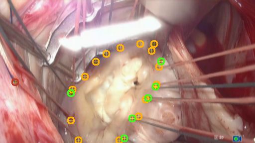
Detection of a varying number of landmarks        3

with 3 channels. The two-channel output masks contain one channel for the
suture landmarks and one for the background. This design is more efficient than
heatmaps approaches, which use one channel for each landmark. The predicted
output heatmap is converted into a binary mask by thresholding. Then, the
centre of mass for each region is determined as point of interest.

2.2   Dataset
Two instances of the same neural network approach proposed in this work were
trained separately on two different data sub-collections (Tab. 1, A.1 and B)
consisting of endoscopic images of mitral valve repair. One data collection con-
tains very heterogeneous intraoperative images extracted from videos which were
recorded during surgery (different view angles, light intensities, number of su-
tures, surgical instruments, prosthesis etc.). Different amounts of frames were
annotated from these datasets, therefore an additional hold-out test set was cre-
ated from surgeries with significantly less frames (A.2). The other simulation
data collection was extracted from videos of operations performed on an artifi-
cial mitral valve replica made of silicone, which was first introduced in [2] and
used in surgical training applications.
    The endoscopic videos were recorded from an Image S1 stereo camera (Karl
Storz SE & Co. KG, Tuttlingen, Germany) in full HD resolution or larger at
25 fps. Frames were saved in top-down format (left image top, right image bot-
tom). Relevant scenes were identified before extracting the frames from the
videos and afterwards every 120th frame was extracted. In scenes with rapid
changes, every 10th frame was extracted and in scenes with only few changes,
every 240th frame was extracted. These characteristics were identified manually.
    This work uses the two subimages of the stereo recording as separate input
images to the network, thus the final number of frames for training and testing
is twice as large as given in Tab. 1. The intraoperative dataset used for training
consists of 2654 frames, which were extracted from 5 surgeries (mean 530,8 ±
213,6 frames per surgery). An additional balanced intraoperative test dataset
was annotated, containing 200 frames extracted from 4 surgeries (mean 100 ±
0 frames per surgery). The simulator datasets consists of 2708 frames extracted
from 10 simulated surgeries (mean 270,7 ± 77,7 frames per surgery).
    The frames were manually annotated with the tool labelme [9]. During anno-
tation, corresponding suture points in the left and right image of the stereo pair
are joined with a line. If a suture point is only visible in one of the subimages it
is marked as a point. The ground truth heatmaps were then created by placing
2D Gaussians at the position of the landmarks. For a down-scaled input size of
512 × 288, a variance σ of 1 was used.
    During training the images are randomly augmented with a probability of
80% using tensorflow functions: rotation of ±60◦ , pixel shifting in a range of
±10%, mask pixel shifting in a range of ±1%, shearing in a range of ±0.1,
brightness in a range of ±0.2, contrast in a range from 0.3 to 0.5, random sat-
uration in a range from 0.5 to 2.0 and hue in a range of ±0.1. Additionally the
images were flipped horizontally and vertically with a probability of 50%.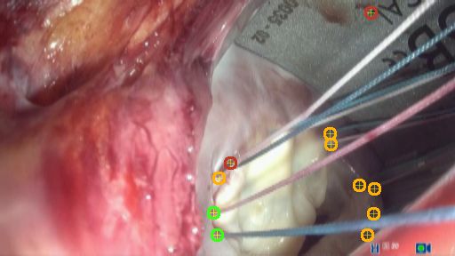
4                                 Stern et al.

Table 1. Three sub-collections from two different domains were used. Note that we
treat left and right stereo frame independently in our work, therefore training frames
are doubled. The additional intraop test set has significantly lower number of labeled
frames, hence we decided to not include it directly in the cross validation (CV).

domain       usage                    # stereo frame # suture endpoints # surgeries
A.1 intraop 5-fold CV (train, test)       1, 327           26, 937             5
A.2 intraop test                           200              4, 305             4
B simulator 5-fold CV (train, test)       1, 354           33, 893            10

2.3   Evaluation
During method development, which involved hyperparameter tuning, training
and validation was performed on dataset A.1. The best neural network had a
total of 2.1M trainable parameters. The initial learning rate was set to 10-3
with a decay factor of 0.1. After fixing these parameters, final evaluation was
conducted on A.1, A.2 and B, involving two 5-fold cross validations (CV) on
different splits of dataset A.1 and B. Furthermore, an additional disjoint test set
was used (A.2) to further assess method generalizability. To prevent data leakage,
dataset splitting was always carried out on the level of the surgeries. The results
are given for the epoch ≤ 200 with the lowest value of the validation loss. In
total, 10 different models were trained, one for each fold of each application
(intraop and simulation).
    A suture point detection is considered successful if the centres of mass of
ground truth and prediction are less than 6 pixels apart. On an image of size
512 × 288, this radius roughly corresponds to the thickness of a suture when it
enters the tissue. If a point in the produced mask or the ground truth mask
is assigned multiple times, only the matched pair with the least distance is
kept. Every matched point from the produced mask is considered a true pos-
itive (TP). Predicted points that could not be matched to any ground truth
point are defined as false positives (FP) and all ground truth points without a
corresponding point in the produced mask are false negatives (FN). To evaluate
the precision (positive predictive value, PPV) and sensitivity (true positive rate,
TPR) are used. Precision and sensitivity are defined as P P V = T P/(T P + F P )
and T P R = T P/(T P + F N ). The values of PPV and TPR are displayed over
a threshold from 0.05 to 1.0 to show the influence of thresholding the heatmap
on the detection rate.

3     Results
Some visual examples are provided in Fig. 1(a) and Fig.1(b). In intraoperative
images, green sutures are better recognized than white ones because they can be
better distinguished from the background. During surgery (not during simula-
tion), white sutures often appear red because they are soaked with blood which
makes it even harder to distinguish them from the background.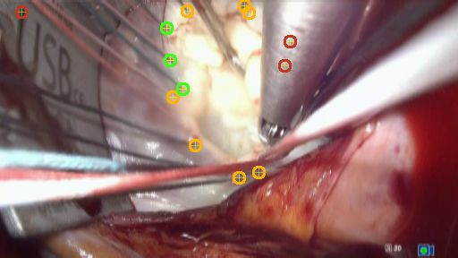
Detection of a varying number of landmarks 5
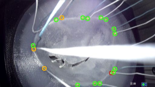
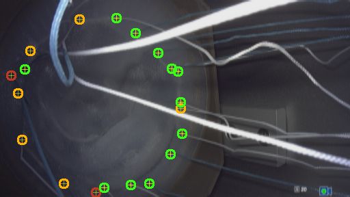
(a) simulator dataset (B)
(b) intraoperative test dataset (A.2)
Fig. 1. Example predictions on the two domains. Green circles represent true positives
(TP), red circles show false positives (FP) and orange circles show false negatives (FN).
The model achieved a mean PPV of 67.99 ± 7.69% and a mean TPR of
29.03±7.74% on the intraoperative dataset (A.1) for a threshold of t = 0.8 during
testing on the respective hold-out folds (Fig. 2(a)). All models from the CV were
also applied to the separate test set (A.2) and comparable results were obtained
with a mean PPV of 66.68 ± 4.67% and a mean TPR of 24.45 ± 5.06% (Fig.
2(b)), meaning that it generalizes beyond the surgeries where hyperparameter
tuning was performed on. When training the model on the simulator dataset
(B) with the same settings, a mean PPV of 81.50 ± 5.77% and a mean TPR of
61.60 ± 6.11% is achieved during CV (Fig. 2(c)). As can be seen in the plots, the
estimated TPR and PPV are not sensitive with regard to the chosen threshold.
Performance differences between the folds are expressed by the bars in Fig. 2.
4 Discussion
The developed approach is able to detect a varying number of landmarks, which
represent the position of sutures stitched through the mitral annulus. Evaluation
on a large dataset of two domains revealed that the neural networks detects
sutures in most of the scenes, but had difficulties in cases where the mitral
annulus is partly occluded by the prosthetic ring, the surgical tools or tissue (Fig.
1(a) 4th image). Reflections or embossings on tools has also led to an increase
in FPs (Fig. 1(b) 3rd image). The best detection results were achieved when the
(a) CV on A.1 (intraop) (b) Test on A.2 (intraop) (c) CV on B (sim)
Fig. 2. The mean PPV and TPR of all folds with variable mask threshold. The bars
show the value of the fold with the lowest and highest value respectively.
6                                   Stern et al.

        camera was positioned above the mitral valve with good illumination. Comparing
        both domains, the network performance was better on the simulation domain,
        where the camera angle is often more favorable and the silicone appearance
        is more homogeneous in comparison to intraoperative scenes with various tissue
        textures and blood. However, the performance differences could be also explained
        by the higher numbers of simulated surgery instances used during training in the
        simulator domain.
            In this work, two neural networks were trained separately on two datasets
        from different domains with the same architecture and hyperparameter settings.
        Future work includes incorporating image-to-image translation by generative
        adversarial networks [10] to adapt between the domains and to allow for joint
        landmark detection in both domains.

        Acknowledgement. The research was supported by the German Research Foun-
        dation DFG Project 398787259, DE 2131/2-1 and EN 1197/2-1 and by Infor-
        matics for Life funded by the Klaus Tschira Foundation.

        References
         1. Carpentier A, Adams D, Filsoufi F. Carpentier’s Reconstructive Valve Surgery.
            Saunders; 2010.
         2. Engelhardt S, Sauerzapf S, Preim B, et al. Flexible and comprehensive patient-
            specific mitral valve silicone models with chordae tendinae made from 3D-printable
            molds. Int J Comput Assist Radiol Surg. 2019;14(7):1177–1186.
         3. Engelhardt S, De Simone R, Zimmermann N, et al. Augmented reality-enhanced
            endoscopic images for annuloplasty ring sizing. In: Augmented Environments for
            Computer-Assisted Interventions. Springer International Publishing; 2014. p. 128–
            137.
         4. Engelhardt S, Kolb S, De Simone R, et al. Endoscopic feature tracking for
            augmented-reality assisted prosthesis selection in mitral valve repair. In: Proc
            SPIE, Medical Imaging: Image-Guided Procedures, Robotic Interventions, and
            Modeling. vol. 9786; 2016. p. 402–408.
         5. Gilbert A, Holden M, Eikvil L, et al. Automated left ventricle dimension measure-
            ment in 2D cardiac ultrasound via an anatomically meaningful CNN approach. In:
            Smart Ultrasound Imaging and Perinatal, Preterm and Paediatric Image Analysis.
            Springer International Publishing; 2019. p. 29–37.
         6. Jin H, Liao S, Shao L. Pixel-in-pixel net: towards efficient facial landmark detection
            in the wild. arXiv:200303771v1 [csCV]. 2020;.
         7. Noothout JMH, De Vos BD, Wolterink JM, et al. Deep learning-based regression
            and classification for automatic landmark localization in medical images. IEEE
            Trans on Med Imag. 2020; p. 1–1.
         8. Ronneberger O, Fischer P, Brox T. U-Net: convolutional networks for biomedical
            image segmentation. In: MICCAI. vol. 9351 of LNCS. Springer; 2015. p. 234–241.
         9. Wada K. labelme: image polygonal annotation with python; 2016.
            https://github.com/wkentaro/labelme.
        10. Engelhardt S, Simone RD, Full PM, et al. Improving surgical training phantoms
            by hyperrealism: deep unpaired image-to-image translation from real surgeries. In:
            MICCAI. Springer International Publishing; 2018. p. 747–755.
E3093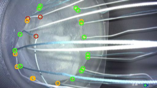

You can also read