Complement dysregulation is associated with severe COVID-19 illness
←
→
Page content transcription
If your browser does not render page correctly, please read the page content below
Complement dysregulation is associated with severe
COVID-19 illness

by Jia Yu, Gloria F. Gerber, Hang Chen, Xuan Yuan, Shruti Chaturvedi, Evan M. Braunstein ,
and Robert A. Brodsky

Haematologica. 2021; Jul 22.
doi: 10.3324/haematol.2021.279155
[Epub ahead of print]

Received: May 17, 2021.
Accepted: July 14, 2021.

Citation: Jia Yu, Gloria F. Gerber, Hang Chen, Xuan Yuan, Shruti Chaturvedi, Evan M. Braunstein,
and Robert A. Brodsky. Complement dysregulation is associated with severe COVID-19 illness.

Publisher's Disclaimer.
E-publishing ahead of print is increasingly important for the rapid dissemination of science.
Haematologica is, therefore, E-publishing PDF files of an early version of manuscripts that
have completed a regular peer review and have been accepted for publication. E-publishing
of this PDF file has been approved by the authors. After having E-published Ahead of Print,
manuscripts will then undergo technical and English editing, typesetting, proof correction and
be presented for the authors' final approval; the final version of the manuscript will then
appear in a regular issue of the journal. All legal disclaimers that apply to the
journal also pertain to this production process.
Complement dysregulation is associated with severe
                COVID-19 illness
                Short Title: Complement dysregulation & COVID-19

     Jia Yu, Gloria F. Gerber, Hang Chen, Xuan Yuan, Shruti Chaturvedi, Evan M.
                           Braunstein and Robert A. Brodsky

Division of Hematology, Department of Medicine, Johns Hopkins School of Medicine,
Baltimore, MD, USA

Corresponding author: Robert A. Brodsky
Email: brodsro@jhmi.edu
Mailing address: 720 Rutland Ave., Ross Rm 1025, Baltimore MD, 21205
Phone: 410-502-2546, Fax: 410-955-0185

Acknowledgment
This work was supported by grants from National Institutes of Health (NIH), National
Heart, Lung, and Blood Institute (NHLBI) grant R01 HL 133113 (R.A.B.); NIH Grant K08
HL138142 (E.M.B.); NHLBI T32 HL 007525 (G.F.G).

Disclosure
R.A.B has served on advisory board for Alexion Pharmaceutical Inc. S.C. has served on
boards for Alexion and Sanofi-Genzyme, and her institution has received research
funding on her behalf from Takeda.

Contributions
J.Y. designed and performed experiments, analyzed the data, enrolled patients and
wrote the first draft of the manuscript; G.F.G. enrolled patients, analyzed the data, and
edited the manuscript; H.C., X.Y., S.C. and E.M.B interpreted the data and edited the
manuscript; R.A.B designed the study, supervised the experiments, interpreted the data
and wrote portions of the manuscript.

Abstract word count: 226 words
Text word count: 3482 words
Method word count: 499 words
Reference: 46
Tables: 1
Figures: 7
Supplemental appendix: 1

                                                                                        1
Abstract
Severe acute respiratory syndrome coronavirus-2 (SARS-CoV-2) may manifest
as thrombosis, stroke, renal failure, myocardial infarction, and thrombocytopenia,
reminiscent of other complement-mediated diseases. Multiple clinical and preclinical
studies have implicated complement in the pathogenesis of COVID-19 illness. We
previously found that the SARS-CoV-2 spike protein activates the alternative pathway of
complement (APC) in vitro through interfering with the function of complement factor H,
a key negative regulator of APC. Here, we demonstrated that serum from 58 COVID-19
patients (32 patients with minimal oxygen requirement, 7 on high flow oxygen, 17
requiring mechanical ventilation and 2 deaths) can induce complement-mediated cell
death in a functional assay (the modified Ham test) and increase membrane attack
complex (C5b-9) deposition on the cell surface. A positive mHam assay (>20% cell-
killing) was present in 41.2% COVID-19 patients requiring intubation (n=7/17) and only
6.3% in COVID-19 patients requiring minimal oxygen support (n=2/32). C5 and factor D
inhibition effectively mitigated the complement amplification induced by COVID-19
patient serum. Increased serum factor Bb level was associated with disease severity in
COVID-19 patients, suggesting that APC dysregulation plays an important role.
Moreover, SARS-CoV-2 spike proteins directly block complement factor H from binding
to heparin, which may lead to complement dysregulation on the cell surface. Taken
together, our data suggest that complement dysregulation contributes to the
pathogenesis of COVID-19 and may be a marker of disease severity.
2Introduction
Complement has emerged as a potential driver of the pathogenesis of severe
coronavirus disease 2019 (COVID-19).1–3 Clinically, endothelial damage, systemic
microvascular thrombosis and recalcitrant hypercoagulability observed in COVID-19
mirror other disorders of complement regulation, such as atypical hemolytic uremic
syndrome (aHUS) and catastrophic antiphospholipid antibody syndrome (CAPS).1–6
Autopsy studies showed microvascular thrombi and complement deposition in the lungs
and kidneys of patients with severe COVID-19.7, 8 Several studies have demonstrated
elevated levels of soluble C5a and C5b-9 in COVID-19 patients.9,10 Further, plasma C3a
levels are higher in COVID-19 patients admitted to the ICU compared to non-ICU
patients.11 A small number of patients treated with eculizumab, a monoclonal anti-C5
antibody, experienced rapid improvement in hypoxia and inflammatory markers.12,13
Three patients treated with an upstream complement C3 inhibitor demonstrated
reduction in inflammatory markers and improved disease severity within 48 hours of
treatment.14 Clinical trials of complement inhibitors in COVID-19 are ongoing
(NCT04355494, NCT04390464).
The complement system, a key component of the human innate immune
response, is a collection of proteases, receptors and inhibitors that functions to
eliminate cellular debris, promote inflammation and defend against pathogens.15 The
system consists of three major pathways – the classical, lectin and alternative pathways
(APC). The three pathways converge on the cleavage of C3 and subsequently C5,
which produces anaphylatoxins (C3a and C5a, respectively) that are associated with
3the acute inflammatory response and thrombosis.16 The membrane attack complex
(C5b-9) is the terminal product of complement activation.15
Evidence for the role of complement in COVID-19 continues to emerge, but
understanding of the underlying mechanisms remains incomplete. Recently, we
demonstrated that the SARS-CoV-2 spike protein (subunits 1 and 2) converts
inactivator surfaces into activator surfaces through interference with the function of
complement factor H (CFH), a critical negative regulator of APC on host cells.17 These
studies were performed by adding recombinant spike protein to normal human serum
(NHS) and measuring complement-mediated killing on the surface of nucleated cells.
Complement inhibition with C5 and factor D inhibitors effectively prevented C5b-9
accumulation induced by the SARS-CoV-2 spike proteins.17 To confirm that complement
is a rational target for treating COVID-19, it is important to show that serum from
patients infected with SARS-CoV-2 also displays complement dysregulation. In this
study, we evaluated cell surface C5b-9 deposition and complement-mediated cell killing
induced by COVID-19 patient sera in the modified Ham (mHam) test 5,18,19 and showed
that impaired complement regulation correlates with COVID-19 disease severity and
can be mitigated with complement inhibitors.
Methods
Patients and samples collection
Serum samples and a limited clinical data set were obtained for 58 COVID-19
patients from the Clinical Characterization Protocol for Severe Infectious Diseases
repository (NCT04496466), approved by the Johns Hopkins Institutional Review Board.
4Samples were collected between April and May 2020. One sample collected outside of
the patient’s hospitalization for COVID-19 was excluded. We also recruited one COVID-
19 patient requiring mechanical ventilation and five healthy individuals who received the
Pfizer-BioNTech (BTN162b2) COVID-19 vaccine between November and December
2020. Informed written consent for sample use from the Complement Associated
Disorders Registry study was obtained. Blood was collected by venipuncture in serum
separator tubes.
Date of COVID-19 diagnosis was based on the earliest known positive SARS-
CoV-2 nucleic acid amplification test from a nasopharyngeal swab or time an infection
flag was placed on the patient’s chart. Severity of COVID-19 was graded based on
World Health Organization (WHO) eight-point ordinal outcome scale.20
The modified Ham test
The modified Ham test was used to assess complement-mediated cell killing of
TF1PIGAnull cells induced by patient serum as previously described.5,17–19,21 The assay
is detailed in Supplemental Appendix.
Detection of complement activity by flow cytometry
C5b-9 and C3c deposition after incubation of TF1PIGAnull cells with COVID-19
patient serum was measured by flow cytometry as previously described.5,17,21 The assay
is detailed in Supplemental Appendix.
Complement inhibition in patient serum
5Patient serum was incubated with 1 µM factor D inhibitor (ACH145951, Achillion
Pharmaceuticals) or 50 µg anti-C5 monoclonal antibody (anti-C5Ab, Alexion
pharmaceuticals) then added to cells. C5b-9 and C3c deposition was measured by flow
cytometry.
Quantification of serum factor Bb by ELISA
Factor Bb level in patient serum was measured using MicroVue Bb Plus EIA kit
(Quidel). NHS preincubated with 20 µg/mL cobra venom factor (Complement
Technology) served as a positive control.
Competitive heparin binding assay
Immunoprecipitation of His-tagged SARS-CoV-2 spike protein subunit 1 (S1,
RayBiotech) and subunit 2 (S2, RayBiotech) was performed using Heparin-Sepharose
beads (BioVision). We incubated 0.2 M S1 or S2 overnight at 4°C with 80 µL beads in
the presence and absence of 0.2 M CFH protein (Complement Technology) in
phosphate buffered saline. After washing, the beads were denatured with LDS buffer
(Invitrogen) and reducing agent (Invitrogen) at 70°C for 15 minutes and then centrifuged.
The supernatants were collected for western blot. We used 10 µL of input protein
solution for western blot.
We loaded 10 µL reduced proteins to mini-PROTEIN TGX Gels (Bio-Rad
Laboratories) and then transferred to PVDF membranes. The membrane was probed
with anti-CFH antibody (1:1500, Cat. Sc-53073, Santa Cruz Biotechnology) or anti-His
antibody (1:2000, Cat. Sc-53073, Santa Cruz Biotechnology). The blot was incubated
6with HRP-linked anti-mouse IgG (1:10000, Cat. 7076S, Cell Signaling Technology) and
imaged with SuperSignal West Femto Maximum Sensitivity Substrate (Thermo Fisher).
Statistical Analysis
Data was summarized as mean ± standard error (SE). Chi-squared test was
used to compare the rates of mHam positivity across WHO groups. Student’s t-test was
used to evaluate differences between unpaired groups. P < 0.05 was considered
statistically significant.
Results
Patient characteristics
All 58 patients studied required hospitalization for their COVID-19 illness (WHO
score 4-8). Thirty-two patients required minimal oxygen support (WHO score 4), 7
required high flow nasal cannula oxygen therapy (WHO score 5), 17 received
mechanical ventilation and additional organ support (WHO score 7), and 2 died (WHO
score 8) (Table 1). Five healthy individuals were recruited at the same hospital system
and their serum samples post COVID-19 vaccination were tested for complement
activation.
COVID-19 patient serum induced complement-mediated cell killing
The modified Ham test (mHam) measures the ability of a nucleated cell to protect
itself from complement-mediated cell killing in vitro in the absence of downstream cell-
surface complement regulators, CD55 and CD59.18,21 Thus, mHam exposes defects in
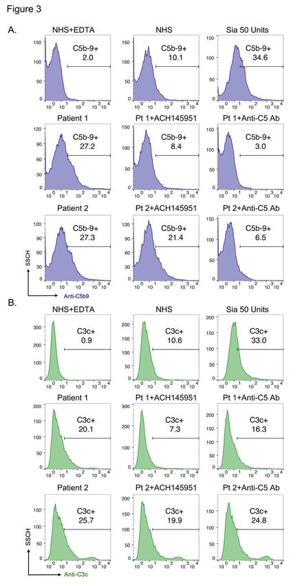
7complement regulation in patient serum. This assay has been validated in complement- mediated disorders such as aHUS, HELLP and CAPS.5,18,21,22 The mHam test was positive (> 20% cell killing) in 41.2% (7 of 17) of patients who required intubation (WHO Score 7), compared to 6.3% (2 of 32) of those who only needed minimal oxygen support (WHO Score 4) (p=0.002) (Figure 1). Serum from COVID-19 patients who required mechanical ventilation induced significantly higher cell killing compared to those on minimal oxygen support (mean 17.3% versus 7.7%, p
was completely inhibited by blocking the terminal complement pathway with an anti-C5

antibody. The factor D inhibitor (ACH145951), an APC-specific inhibitor, partially

reduced C5b-9 deposition induced by serum from patient 2 and achieved complete

inhibition in patient 1 (Figure 3A). In addition, the factor D inhibitor was more effective

than anti-C5 antibody in inhibiting C3c deposition triggered by COVID-19 patient sera

(Figure 3B), since it targets upstream of the anti-C5 monoclonal antibody.

Increased APC activation is associated with COVID-19 disease severity

       We utilized standard ELISA to measure the factor Bb level in COVID-19 patient

serum. Factor Bb, which results from cleavage of factor B by factor D, is a biomarker of

APC activation. Regardless of disease severity, the serum level of Bb was significantly

higher in COVID-19 patients compared to the healthy controls. We also found that

COVID-19 patients requiring intubation (WHO score 7) had significantly higher Bb levels

than those requiring minimal oxygen support (WHO score 4) (Figure 4). These

observations suggested that increased APC activation is associated with disease

severity in COVID-19.

SARS-CoV-2 spike proteins compete with CFH for cell surface heparan sulfate

binding

       We previously demonstrated that SARS-CoV-2 spike proteins (both subunit 1

and 2) bind heparan sulfate on the cell surface.17 Heparan sulfate also serves as a

necessary co-factor for binding of SARS-CoV-2 spike proteins to the angiotensin

receptor 2 (ACE2).23 CFH, a negative regulator of APC, also utilizes

                                                                                              9
glycosaminoglycans, such as heparan sulfate, and α2,3 N-linked sialic acid residues for
binding to nucleated cells; thus, we hypothesized that SARS-CoV-2 spike protein
competes with CFH for binding to heparan sulfate and its tissue specific, more highly-
sulfated variant, heparin. To evaluate whether SARS-CoV-2 spike proteins block CFH
from binding to heparan sulfate, we compared the heparin-binding activity of CFH in the
presence and absence of the SARS-CoV-2 spike proteins using heparin-linked beads.
CFH alone bound to the heparin-beads with high affinity (Figure 5A, lane 1). In the
presence of SARS-CoV-2 S1 and S2, binding of CFH to the heparin-beads was
markedly reduced (Figure 5A, lane 2 and 3).
We also compared the heparin-binding ability of SARS-CoV-2 spike proteins in
the presence and absence of CFH. S1 alone showed strong binding to heparin-beads
and retained high heparin-binding activity in the presence of CFH (Figure 5B). S2 bound
to heparin-beads with similarly high efficiency under both conditions (Figure 5C). These
results indicated the SARS-CoV-2 spike proteins have higher binding affinity for heparin
than CFH and interfere with the binding of CFH to heparin.
SARS-CoV-2 mRNA vaccine does not markedly increase complement activity in
healthy individuals
The mRNA COVID-19 vaccines employ the SARS-CoV-2 spike protein as an
immunogenic target. Given that SARS-CoV-2 spike proteins activate complement in
vitro, concern arises whether the COVID-19 vaccine could also trigger transient
complement dysregulation in vivo through generation of the spike protein.
10To test this, we obtained serum samples from five healthy individuals who
received both doses of the Pfizer-BioNTech (BTN162b2) COVID-19 vaccine at three
time points: before the vaccine (baseline), 24 to 48 hours after receiving the first vaccine
dose, and 24 to 48 hours after the second dose. We measured the serum level of factor
Bb in these individuals pre- and post-COVID-19 vaccination. Two out of five individuals
showed significantly higher serum Bb levels from their baseline after receiving the
COVID-19 vaccine (Figure 6). Notably, these two individuals experienced side effects
post vaccination including fever, headache and fatigue. We next performed functional
assays to assess for cell surface complement amplification post-vaccination. On
average, serum collected after the first vaccine dose did not lead to increased C5b-9
deposition on the surface of TF1PIGAnull cells compared to the individual’s serum pre-
vaccination. The second dose of the vaccine led to an 11% increase in the C5b-9
deposition. All individuals had negative mHam results at baseline, which remained
negative after receiving the COVID-19 vaccine (data not shown). These results from the
functional assays demonstrate that the Pfizer-BioNTech SARS-CoV-2 mRNA vaccine
does not sufficiently alter complement regulation in healthy individuals.
Discussion
Previously, we showed that the SARS-CoV-2 spike protein dysregulates the
alternative complement pathway in vitro by interfering with CFH function on the cell.17
Here, we extend these findings and demonstrate that the SARS-CoV-2 spike protein
directly competes with CFH for binding to heparan sulfate. In addition, we demonstrate,
for the first time, that COVID-19 patient serum induces complement dysregulation on
11the cell surface using a functional assay (the mHam). Moreover, a positive mHam is
highly associated with disease severity in COVID-19 patients, and C5b-9 cell deposition
induced by patient sera is blocked by factor D and C5 inhibitors.
Our finding that complement dysregulation is inherent to the pathogenesis of
COVID-19 is in agreement with existing pre-clinical and clinical data. Autopsy studies
revealed depositions of complement proteins in lung and other tissues co-localize with
the SARS-CoV-2 spike proteins.8,24 Markers of complement activation in sera from
COVID-19 hospitalized patients are associated with respiratory failure. Specifically,
markers of classical/lectin (C4d), alternative (C3bBbP) and common pathway (C3bc,
sC5b-9) amplification were increased in COVID-19 patient sera throughout
hospitalization, indicative of sustained activation of all complement pathways.9–11,25
Sinkovits et al26 showed that complement overactivation and consumption is predictive
of in-hospital mortality in SARS-CoV-2 infection. Specifically, these authors reported
consumption of C3 in the serum of patients with severe COVID-19 disease. This is
consistent with our finding of increased C3c deposition on TF1PIGAnull cells. In
transcriptome analysis, expression of multiple complement genes such as C2, C3, CFB
and CFH were upregulated in COVID-19 patients.27,28 Gavriilaki et al29 analyzed genetic
and clinical data from 97 patients hospitalized with COVID-19 and found an increase in
rare variants associated with thrombotic microangiopathies (several involving the
alternative pathway of complement) in patients with severe COVID-19 disease.
Complement amplification in hospitalized patients with COVID-19 is multifactorial.
Our previous data adding recombinant spike protein to normal human serum showed
that complement-mediated cell killing was almost entirely through the alternative
12complement pathway.17 Here, using serum from patients with COVID-19, we find
additional contributions from the non-APC pathways. This may be because serum
samples in this study were obtained a median of seven days after testing positive for
COVID-19, at a time when the SARS-CoV-2 viral load is decreasing,30,31 as opposed to
measuring cell surface C5b-9 deposition within minutes after adding spike protein to
normal human serum. We also demonstrated that mHam positivity (measuring the
activation of all complement pathways) from SARS-CoV-2 infected patients is
associated with the need for mechanical ventilation; however, we did not find as strong
a correlation between cell surface C5b-9 and the mHam as we did after supplementing
normal human serum with the spike protein.
Virtually all patients had elevated levels of Bb in their serum, even two of the
healthy subjects after COVID-19 mRNA vaccination, suggesting that APC activation is
an early event in the pathogenesis of SARS-CoV-2 infection. This is in agreement with
the prominent role for APC found in proteomics studies, which showed increased factor
B (CFB) levels in serum from severe COVID-19 patients.32 CFB deposition was also
observed in the lung tissue of COVID-19 patients, and a CFB inhibitor blocked the C3a
generated by infection of respiratory epithelial cells with SARS-CoV-2.33,34 Pekayvaz et
al35 further showed upregulation of complement factor D (CFD), produced mainly in
adipocytes, in monocytes of severe COVID-19 patients. Ma et al36 demonstrated that
enhanced activation of the APC is associated with markers of endothelial injury and
hypercoagulability in severe COVID-19 patients as compared to other non-COVID-19
patients admitted to the intensive care unit with acute respiratory failure.
13C5b-9 deposition induced by COVID-19 patient serum is blocked by both a
terminal complement inhibitor (anti-C5 antibody) and an alternative pathway specific
inhibitor (factor D inhibitor, ACH145951). The factor D inhibitor was more effective in
blocking C3c deposition induced by COVID-19 patient serum as compared to the anti-
C5 antibody. These results are supported by observations from case series of
eculizumab, a monoclonal anti-C5 antibody, in which treated COVID-19 patients
showed significant improvements in clinical parameters.12,13 However, the phase III trial
of Eculizumab (NCT04355494) in COVID-19 patients on mechanical ventilation was
paused due to interim analysis of 122 patients showing that the drug did not meet its
prespecified efficacy outcome of survival on day 29. Final results from this trial are
eagerly anticipated as are those for COVID-19 patients who are hospitalized but not on
mechanical ventilation. COVID-19 patients may derive benefit from complement
inhibition early in their disease course or from more proximal complement inhibition.
A comparative study of Eculizumab versus AMY-101, an upstream C3 inhibitor,
in a small number of patients showed that both decreased inflammatory markers and
led to improvements in lung functions. The three patients who received AMY-101
demonstrated greater reduction in plasma levels of C3a, sC5b-9 and CFB as compared
to patients who received eculizumab.37 This limited clinical data in addition to our in vitro
results suggests that proximal complement inhibitors may be more effective than
terminal inhibitors in reducing COVID-19 disease severity. Notably, treatment of six
severely ill COVID-19 patients with Narsoplimab, a monoclonal antibody against MASP-
2 inhibiting lectin-pathway activation, showed rapid reduction in serum inflammatory
markers and survival in all patients.38 In addition to the COVID-19 infection itself, there
14are likely multifactorial contributions from tissue damage, secondary infections, and
thrombosis, leading to complement activation from all pathways. Further studies
comparing different complement inhibitors would be valuable to identify the most
appropriate therapeutic targets.
The SARS-CoV-2 spike protein interferes with the function of CFH and this is
likely an early event in the pathogenesis of COVID-19. In our prior work, we found that
addition of purified CFH protein to serum treated with the SARS-CoV-2 spike protein
decreased C3c and C5b-9 deposition on the cell surface.17 Here, we used competitive
immunoprecipitation experiments to show that the SARS-CoV-2 spike protein directly
blocks CFH from binding to heparin, which may explain the APC dysregulation
observed in COVID-19 infection (Figure 5). When binding to glycosaminoglycans on cell
surface, including heparan sulfate, and α2-3 N-linked sialic acid residues, CFH achieves
a more active conformation that allows for C3b binding.39 Interestingly, genetic variants
in CFH, that occur in the same region where factor H binds heparan sulfate, have been
identified as an important risk factor for morbidity and mortality from COVID-19.27
COVID-19 vaccines lead to transient expression of the SARS-CoV-2 spike
protein and are effective in preventing severe infection.40,41 Our vaccine studies (Figure
6) are reassuring that mRNA vaccines should not induce clinically significant
complement amplification in healthy individuals, as suggested by the negative results
from our functional assays; however, more data is necessary in patients with disorders
of complement regulation, such as paroxysmal nocturnal hemoglobinuria (PNH), aHUS,
CAPS, HELLP and cold agglutinin disease.42 Relapse of aHUS has been reported in
patients with COVID-19 infection43, and PNH patients have experienced adverse
15reactions to COVID-19 vaccines including severe hemolysis and need for blood
transfusions even while on a C5 inhibitor.44 This evidence suggests that although
complement activation induced by COVID-19 vaccines is well-controlled in healthy
individuals, patients with disorders of complement regulation could be at higher risk for
adverse reactions to vaccine.
Limitations of our study are that we received a limited amounts of patient serum
for experiments and were unable to test other complement markers such as C4d
deposition on the cell surface. We also had limited access to clinical information from
which to draw robust conclusions regarding the association of complement activation
with clinical parameters. Further, serum sample collection was not standardized and
occurred at different time points from the initial diagnosis of COVID-19 and hospital
admission. For example, in one of the two patients who died due to multiorgan failure
from COVID-19, serum was collected near the end of his clinical course, at which point
peak amplification of complement may have passed. Finally, we do not have serial
samples from patients to estimate the persistence of complement activation over time.
In future studies, it will be important to do serial mHam, Bb, and surface C5b-9
deposition studies starting soon after infection and correlating with SARS-CoV-2 viral
load.
In summary, we showed that COVID-19 patient serum can induce complement
dysregulation on cell surfaces that tracks with disease severity. Our previous data
showed that the SARS-CoV-2 spike proteins convert inactivator surfaces to activator
surfaces. Taken together, we postulate that dysregulation of the APC is likely an early
event after SARS-CoV-2 infection. Complement amplification from classical and lectin
16pathways following tissue damage, secondary infections, and thrombosis likely
exacerbate end-organ damage similar to severe forms of aHUS and CAPS (Figure 7).
Preconditions that lead to inflammation (e.g. obesity, diabetes and vascular disease) or
contribute to complement activation (e.g. third-trimester pregnancy) or dysregulation
(age-related macular degeneration and other germline complement mutations) may
contribute to a severe phenotype. Indeed, components of the APC (CFB, CFD, and C3)
are elevated in patients with obesity and insulin resistance.45 Prospective studies
correlating SARS-CoV-2 viral load to complement-mediated cell damage over the
course of infection and additional genetic studies probing for rare variants in
complement regulatory genes are needed. Our data also suggest that for complement
inhibitors to be most effective, they should be initiated early in the disease process, but
this too requires prospective study, as is the subject of the ongoing TACTIC-R study
(NCT04390464).46
17Reference:
1. Java A, Apicelli AJ, Liszewski MK, et al. The complement system in COVID-19:
Friend and foe? JCI Insight. 2020;5(15):e140711.
2. Gavriilaki E, Brodsky RA. Severe COVID-19 infection and thrombotic
microangiopathy: success does not come easily. Br J Haematol.
2020;189(6):e227-e230.
3. Risitano AM, Mastellos DC, Huber-Lang M, et al. Complement as a target in
COVID-19? Nat Rev Immunol. 2020;20(6):343-344.
4. Campbell CM, Kahwash R. Will Complement Inhibition Be the New Target in
Treating COVID-19-Related Systemic Thrombosis? Circulation.
2020;141(22):1739-1741.
5. Chaturvedi S, Braunstein EM, Yuan X, et al. Complement activity and
complement regulatory gene mutations are associated with thrombosis in APS
and CAPS. Blood. 2020;135(4):239-251.
6. Baines AC, Brodsky RA. Complementopathies. Blood Rev. 2017;31(4):213-223.
7. Wichmann D, Sperhake JP, Lütgehetmann M, et al. Autopsy Findings and
Venous Thromboembolism in Patients With COVID-19: A Prospective Cohort
Study. Ann Intern Med. 2020;173(4):268-277.
8. Diao B, Wang C, Wang R, et al. Human kidney is a target for novel severe acute
respiratory syndrome coronavirus 2 infection. Nat Commun. 2021;12(1):1-9.
9. Carvelli J, Demaria O, Vély F, et al. Association of COVID-19 inflammation with
activation of the C5a–C5aR1 axis. Nature. 2020;588(7836):146-150.
10. Cugno M, Meroni PL, Gualtierotti R, et al. Complement activation in patients with
COVID-19: A novel therapeutic target. J Allergy Clin Immunol. 2020;146(1):215-
217.
11. de Nooijer AH, Grondman I, Janssen NAF, et al. Complement Activation in the
Disease Course of Coronavirus Disease 2019 and Its Effects on Clinical
Outcomes. J Infect Dis. 2021;223(2):214-224.
12. Diurno F, Numis FG, Porta G, et al. Eculizumab treatment in patients with COVID-
19: Preliminary results from real life ASL Napoli 2 Nord experience. Eur Rev Med
Pharmacol Sci. 2020;24(7):4040-4047.
13. Annane D, Heming N, Grimaldi-Bensouda L, et al. Eculizumab as an emergency
treatment for adult patients with severe COVID-19 in the intensive care unit: A
proof-of-concept study. EClinicalMedicine. 2020;28:100590.
14. Mastaglio S, Ruggeri A, Risitano AM, et al. The first case of COVID-19 treated
with the complement C3 inhibitor AMY-101. Clin Immunol. 2020;215:108450.
15. Ling M, Murali M. Analysis of the Complement System in the Clinical Immunology
Laboratory. Clin Lab Med. 2019;39(4):579-590.
16. Kourtzelis I, Markiewski MM, Doumas M, et al. Complement anaphylatoxin C5a
contributes to hemodialysis-associated thrombosis. Blood. 2010;116(4):631-639.
17. Yu J, Yuan X, Chen H, Chaturvedi S, Braunstein EM, Brodsky RA. Direct
activation of the alternative complement pathway by SARS-CoV-2 spike proteins
is blocked by factor D Inhibition. Blood. 2020;136(18):2080-2089.
18. Gavriilaki E, Yuan X, Ye Z, et al. Modified Ham test for atypical hemolytic uremic
syndrome. Blood. 2015;125(23):3637-3646.
1819. Vaught AJ, Braunstein EM, Jasem J, et al. Germline mutations in the alternative
pathway of complement predispose to HELLP syndrome. JCI Insight. 2018;3(6):5-
7.
20. World Health Organization. COVID-19 Therapeutic Trial Synopsis. World Health
Organization. https://cdn.who.int/media/docs/default-source/blue-print/covid-19-
therapeutic-trial-synopsis.pdf?sfvrsn=44b83344_1&download=true 2020,
Accessed May 12, 2021.
21. Yuan X, Yu J, Gerber G, et al. Ex vivo assays to detect complement activation in
complementopathies. Clin Immunol. 2020;221:108616.
22. Vaught AJ, Braunstein EM, Jasem J, et al. Germline mutations in the alternative
pathway of complement predispose to HELLP syndrome. JCI Insight.
2018;3(6):e99128.
23. Clausen TM, Sandoval DR, Spliid CB, et al. SARS-CoV-2 Infection Depends on
Cellular Heparan Sulfate and ACE2. Cell. 2020;183(4):1043-1057.e15.
24. Magro C, Mulvey JJ, Berlin D, et al. Complement associated microvascular injury
and thrombosis in the pathogenesis of severe COVID-19 infection: A report of five
cases. Transl Res. 2020;220:1-13.
25. Holter JC, Pischke SE, de Boer E, et al. Systemic complement activation is
associated with respiratory failure in COVID-19 hospitalized patients. Proc Natl
Acad Sci U S A. 2020;117(40):25018-25025.
26. Sinkovits G, Mező B, Réti M, et al. Complement Overactivation and Consumption
Predicts In-Hospital Mortality in SARS-CoV-2 Infection. Front Immunol.
2021;12:663187.
27. Ramlall V, Thangaraj PM, Meydan C, et al. Immune complement and coagulation
dysfunction in adverse outcomes of SARS-CoV-2 infection. Nat Med.
2020;26(10):1609-1615.
28. Valenti L, Griffini S, Lamorte G, et al. Chromosome 3 cluster rs11385942 variant
links complement activation with severe COVID-19. J Autoimmun.
2021;117:102595.
29. Gavriilaki E, Asteris PG, Touloumenidou T, et al. Genetic justification of severe
COVID-19 using a rigorous algorithm. Clin Immunol. 2021;226:108726.
30. Jang S, Rhee JY, Wi YM, Jung BK. Viral kinetics of SARS-CoV-2 over the
preclinical, clinical, and postclinical period. Int J Infect Dis. 2021;102:561-565.
31. Sun J, Tang X, Bai R, et al. The kinetics of viral load and antibodies to SARS-
CoV-2. Clin Microbiol Infect. 2020;26(12):1690.e1-1690.e4.
32. Messner CB, Demichev V, Wendisch D, et al. Ultra-High-Throughput Clinical
Proteomics Reveals Classifiers of COVID-19 Infection. Cell Syst. 2020;11(1):11-
24.e4.
33. Yan B, Freiwald T, Chauss D, et al. SARS-CoV-2 drives JAK1/2-dependent local
complement hyperactivation. Sci Immunol. 2021;6(58):1-20.
34. Macor P, Durigutto P, Mangogna A, et al. Multi-organ complement deposition in
COVID-19 patients. medRxiv. 2020;(165):1-13.
https://pubmed.ncbi.nlm.nih.gov/33442701/ 2020, Accessed May 12, 2021.
35. Pekayvaz K, Leunig A, Kaiser R, et al. Protective Immune Trajectories in Early
Viral Containment of Non-Pneumonic SARS-CoV-2 Infection. bioRxiv.
https://www.biorxiv.org/content/10.1101/2021.02.03.429351v1 2021, Accessed
19May 12, 2021.
36. Ma L, Sahu SK, Cano M, et al. Increased complement activation is a distinctive
feature of severe SARS-CoV-2 infection. Sci Immunol. 2021;6(59):1-18.
37. Mastellos DC, Pires da Silva BGP, Fonseca BAL, et al. Complement C3 vs C5
inhibition in severe COVID-19: Early clinical findings reveal differential biological
efficacy. Clin Immunol. 2020;220:108598.
38. Rambaldi A, Gritti G, Micò MC, et al. Endothelial injury and thrombotic
microangiopathy in COVID-19: Treatment with the lectin-pathway inhibitor
narsoplimab. Immunobiology. 2020;225(6):152001.
39. Perkins SJ, Fung KW, Khan S. Molecular interactions between complement factor
H and its heparin and heparan sulfate ligands. Front Immunol. 2014;5:1-14.
40. Polack FP, Thomas SJ, Kitchin N, et al. Safety and Efficacy of the BNT162b2
mRNA Covid-19 Vaccine. N Engl J Med. 2020;383(27):2603-2615.
41. Baden LR, El Sahly HM, Essink B, et al. Efficacy and Safety of the mRNA-1273
SARS-CoV-2 Vaccine. N Engl J Med. 2021;384(5):403-416.
42. Gavriilaki E, Brodsky RA. Complementopathies and precision medicine. J Clin
Invest. 2020;130(5):2152-2163.
43. Ville S, Le Bot S, Chapelet-Debout A, et al. Atypical HUS relapse triggered by
COVID-19. Kidney Int. 2021;99(1):267-268.
44. Gerber GF, Yuan X, Yu J, et al. COVID-19 Vaccines Induce Severe Hemolysis in
Paroxysmal Nocturnal Hemoglobinuria. Blood. 2021;137(26):3670-3673.
45. Shim K, Begum R, Yang C, Wang H. Complement activation in obesity, insulin
resistance, and type 2 diabetes mellitus. World J Diabetes. 2020;11(1):1-12.
46. Kulkarni S, Fisk M, Kostapanos M, et al. Repurposed immunomodulatory drugs
for Covid-19 in pre-ICU patients - multi-arm Therapeutic study in pre-ICU patients
admitted with Covid-19 - Repurposed Drugs (TACTIC-R): A structured summary
of a study protocol for a randomised controlled trial. Trials. 2020;21(1):20-22.
20Table 1. COVID-19 Patient Characteristics
Hi-Flow
Oxygen Oxygen Ventilation Death
(N=32) (N=7) (N=17) (N=2)
Age (years), median 55 (26-78) 58 (49-69) 59 (27-80) 57 (56-57)
Gender
Female 18 3 7 1
Male 14 4 10 1
Race
Black 11 4 9 1
White 11 2 4 0
Other 10 1 4 1
Ethnicity
Hispanic 23 6 14 1
Non-Hispanic 9 1 3 1
33.6 (20.5- 34.2 (27.1- 30.4 (16.2-
BMI, median 58.2) 51.9) 46.3) 65.3
Days from COVID-19 Diagnosis to
4 (1-13) 8 (2-16) 13 (3-34) 18
Serum Sample Collection, median
Days from Hospital Admission to
2 (1-13) 6 (1-10) 13 (2-32) 13
Serum Sample Collection, median
Length of Hospitalization (days),
9 (2-45) 11 (7-15) 28 (3-70) 17
median
21Figure legends
Figure 1. Complement-mediated cell killing induced by COVID-19 patient serum is
associated with disease severity.
TF1PIGAnull cells were treated with 20% COVID-19 patient serum, and cell killing was
measured using the modified Ham test. Complement-mediated cell killing (%) was
significantly elevated in COVID-19 patients requiring mechanical ventilation (vent), as
compared to pooled normal human serum (NHS) and COVID-19 patients who needed
minimal oxygen support (p < 0.01). The dotted line at 20% non-viable cells represented
the threshold for a positive mHam based on a previously established receiver operator
curve. All experiments were run in triplicate. NHS indicates normal human serum; Sia
indicates sialidase and is used as a positive control; stx1 indicates Shiga toxin subunit 1
and is used as a positive control.
Figure 2. COVID-19 patient serum induces C5b-9 and C3c deposition on the cell
surface.
COVID-19 patient serum led to increased cell surface C5b-9 deposition in all
complement pathway buffer (A) and alternative pathway specific buffer (B). C3c
deposition was increased compared to control normal human serum in all complement
pathway buffer (C) and alternative pathway specific buffer (D). Statistical significance
was calculated between each disease group and the NHS control group. EDTA
indicates ethylenediaminetetraacetic and is used as a negative control; NHS indicates
normal human serum; Sia indicates sialidase and is used as a positive control for APC
22activation; stx1 indicates Shiga toxin subunit 1 and is used as a positive control for all complement pathways activation. Figure 3. C5 and factor D inhibition block complement activation induced by COVID-19 patient serum. Flow cytometry demonstrated increased C5b-9 (A) and C3c (B) deposition in two representative COVID-19 patients. C5b-9 deposition was completely blocked in the presence of 50 µg anti-C5 antibody. 1 µM factor D inhibitor (ACH145951) partially reduced the C5b-9 deposition in patient 2 and achieved complete inhibition in patient 1 (A). Factor D inhibitor also effectively decreased C3c accumulation induced by both patients’ sera, whereas anti-C5 antibody did not appreciably prevent C3c deposition (B). SSCH indicates side scatter; ACH145951, indicates factor D inhibitor; Anti-C5 Ab indicates anti-C5 monoclonal antibody. Figure 4. The Bb level is increased in COVID-19 patient serum and associated with disease severity. Serum Bb level was significantly elevated in COVID-19 patients who required minimal oxygen support, high flow nasal cannula oxygen therapy and mechanical ventilation (vent) as compared to pooled normal human serum and healthy controls (healthy ctrl). Patients requiring mechanical ventilation also had significantly higher serum Bb level than patients with minimal oxygen support (p
Figure 5. SARS-CoV-2 spike proteins compete with complement factor H for the

same binding sites on heparin.

(A) The binding of CFH to heparin-linked beads was markedly reduced in the presence

of SARS-CoV-2 spike protein subunit 1 (S1) and subunit 2 (S2). The western blot was

performed using anti-CFH antibody. (B) S1 and S2 retained similarly high binding

affinity to heparin-linked beads in the presence and absence of CFH. The western blot

was performed using anti-His antibody. The input is the protein solution that is used to

incubate with the beads. CFH indicates complement factor H; S indicates SARS-CoV-2

spike protein; S1 indicates SARS-CoV-2 spike protein subunit 1; S2 indicates SARS-

CoV-2 spike protein subunit 2.

Figure 6. SARS-CoV-2 mRNA vaccine minimally induces cell surface C5b-9

deposition but increases serum Bb level in healthy individuals.

Sera from five healthy individuals were collected before they received the COVID-19

mRNA vaccine, 24-48 hours after their first vaccine dose and 24-48 hours after their

second dose. (A) On average, the first vaccine dose did not lead to elevation in C5b-9

deposition from the individual’s baseline, whereas the second dose increased cell

surface C5b-9 deposition by 11%. (B) Two out of five healthy individuals had markedly

increased serum Bb level post vaccination as compared to baseline, which correlated

with their vaccine side effects including headache, fatigue and fever.

Figure 7. Proposed model for complement dysregulation in SARS-CoV-2 infection.

                                                                                           24
Early in infection, the SARS-CoV-2 spike protein binds heparan sulfate on the
endothelial cell surface and interferes with the inhibitory function of CFH, leading to
APC dysregulation. Suppression of CFH binding results in increased cleavage of factor
B by factor D and generation of Bb. Factor Bb binds to C3b to form the alternative
pathway C3 convertase (C3bBb), leading to the cleavage of C3 and generation of the
C5 convertase (C4b2a3b or C3bBb3b). The C5 convertase cleaves C5 to generate C5a
and C5b, which complexes with C6-9 to form the membrane attack complex (C5b-9).
C3a and C5a are anaphylatoxins that recruit inflammatory cells and upregulate the
expression of acute phase proteins, such as C-reactive protein. Complement
amplification from the classical and lectin pathways follows subsequent tissue damage,
secondary infections and thromboses. Formation of antibody-antigen complexes can
activate the classical complement pathway by binding to the C1 complex and cleaving
C4 and C2 to form the classical C3 convertase (C4b2a). In the lectin pathway, MBL-
MASP binds to carbohydrates on the surface of microbes and mediates the cleavage of
C2 and C4, to generate the C3 convertase (C4b2a). Preconditions that enhance
inflammation (e.g. obesity, diabetes, and vascular disease) or contribute to complement
activation (e.g. third-trimester pregnancy) or dysregulation (age-related macular
degeneration and other germline complement mutations) may contribute to a severe
phenotype through upregulation of these pathways. HS indicates heparan sulfate; MBL-
MASP indicates mannose-binding protein-mannose-binding lectin serine protease; CRP
indicates C-reactive protein.
25Complement dysregulation is associated with severe COVID-
19 illness
Supplemental Methods
The modified Ham test
The modified Ham test was used to assess complement activation in patient
serum as described previously.1–4 Briefly, TF1PIGAnull cells were maintained at a
density of 500,000 cells/mL daily. The cells were washed with phosphate-buffered
saline (PBS) and seeded in a round-bottom 96-well plate at a density of 6700 cells/well
in 80 µL GVB++ buffer (Cat. B102, Complement Technology, Inc) in triplicate. 20 µL of
serum was added to the cells and incubated at 37°C for 45 minutes with constant
shaking. Normal human serum (Cat. NHS, Complement Technology) was heated at
56°C for 30 minutes and used as a negative control (NHS(H)). As a positive control for
the mHam, NHS was first incubated with Shiga toxin 1 (stx1, Cat. SML0562, Sigma-
Aldrich) for 15 minutes on ice before exposure to the cells. As a positive control for APC
activation, the cells were treated with 50 units of Sialidase per sample (Cat. P0720L,
New England Bio Labs) at 37°C for 30 minutes, followed by addition of NHS.
After incubation of the cells and serum, the cells were washed with PBS and
resuspended in 100 µL 10% WST-1 proliferation solution (WST-1: RPMI 1640 without
phenol red at a ratio of 1:9, WST-1 Cat. 11644807001, Roche, Switzerland) and
incubated for 2 hours at 37°C. The absorbance of the chromogenic metabolized product
was measured with a plate reader (ELX808, BioTeK, Winooski, VT) at 450 nm with a
reference wavelength at 630 nm. The sample absorbance was normalized by
subtracting the absorbance of a blank control, which contained WST-1 solution only.
1The percentage of live cells was calculated using [(Asample-Ablank)/Asample(H) -Ablank) x 100].
The percentage of non-viable cells (100- percentage of live cells) was used as a
measure of complement-mediated cell killing. All experiments were performed in
triplicate.
Detection of complement activity by flow cytometry
Cell surface deposition of C5b-9 and C3c on TF1PIGAnull cells was measured
by flow cytometry as previously described.3–5 TF1PIGAnull cells were washed with PBS
and seeded in a V-bottom 96-well plate (1.2 x 10⁵ cells/well) in 80 µL of either GVB++
buffer or GVB0·MgEGTA buffer (pH 6.4) (GVB0 Cat. B103, Complement Technology,
Inc). GVB++ allows for activation of all complement pathways while GVB 0·MgEGTA only
allows for alternative pathway activation. 20 µL patient serum was added to the cells
and incubated at 37°C for 15 minutes with constant shaking. For alternative pathway
activation, NHS was acidified to pH 6.4 with 0.2 M HCl. 5 mM of
ethylenediaminetetraacetic acid (EDTA) was added to NHS to inhibit complement
activation and used as a negative control.
After incubation, the reaction was stopped by addition of PBS supplemented with
1% BSA and 15 mM EDTA. The cells were centrifuged at 600 g for 3 minutes and the
cell pellet was collected. The cells were stained with anti-C5b-9 monoclonal antibody
(Cat. Sc-58935, Santa Cruz Biotechnology, Inc, dilution at 1:100) followed by Alexa 647
conjugated secondary antibody (Cat. Ab 172325, Abcam, dilution at 1:500). The cells
were also stained with Alexa 488 conjugated anti-C3c antibody (Cat.4212, Abcam,
dilution at 1:150). Ten thousand events per sample were recorded using BD
2FACSCalibur, and data were analyzed using FlowJo Software version 10.5.3 (FlowJo
Inc).
Supplemental References
1. Vaught AJ, Braunstein EM, Jasem J, et al. Germline mutations in the alternative
pathway of complement predispose to HELLP syndrome. JCI Insight.
2018;3(6):5–7.
2. Gavriilaki E, Yuan X, Ye Z, et al. Modified Ham test for atypical hemolytic uremic
syndrome. Blood. 2015;125(23):3637–3646.
3. Yu J, Yuan X, Chen H, Chaturvedi S, Braunstein EM, Brodsky RA. Direct
activation of the alternative complement pathway by SARS-CoV-2 spike proteins
is blocked by factor D Inhibition. Blood. 2020;136(18):2080–2089.
4. Chaturvedi S, Braunstein EM, Yuan X, et al. Complement activity and
complement regulatory gene mutations are associated with thrombosis in APS
and CAPS. Blood. 2020;135(4):239–251.
5. Yuan X, Yu J, Gerber G, et al. Ex vivo assays to detect complement activation in
complementopathies. Clin Immunol. 2020;221:108616.
3You can also read