Continued Emergence and Evolution of Omicron in South Africa: New BA.4 and BA.5 lineages
←
→
Page content transcription
If your browser does not render page correctly, please read the page content below
medRxiv preprint doi: https://doi.org/10.1101/2022.05.01.22274406; this version posted May 2, 2022. The copyright holder for this preprint
 (which was not certified by peer review) is the author/funder, who has granted medRxiv a license to display the preprint in perpetuity.
                                    It is made available under a CC-BY-NC-ND 4.0 International license .

  Continued Emergence and Evolution of Omicron in South Africa: New BA.4 and BA.5
  lineages

  Authors: Houriiyah Tegally1,2, Monika Moir1, Josie Everatt3, Marta Giovanetti4,5, Cathrine
  Scheepers3,6, Eduan Wilkinson1, Kathleen Subramoney7, Sikhulile Moyo8,9,10, Daniel G.
  Amoako3, Cheryl Baxter1, Christian L., Althaus11, Ugochukwu J. Anyaneji2, Dikeledi Kekana3,
  Raquel Viana12, Jennifer Giandhari2, Richard J. Lessells2, Tongai Maponga13, Dorcas
  Maruapula8, Wonderful Choga8, Mogomotsi Matshaba10, Simnikiwe Mayaphi14, Nokuzola
  Mbhele15, Mpaphi B. Mbulawa16, Nokukhanya Msomi17, NGS-SA consortium, Yeshnee
  Naidoo1, Sureshnee Pillay2, Tomasz Janusz Sanko1, James E. San2, Lesley Scott18, Lavanya
  Singh2, Nonkululeko A. Magini2, Pamela Smith-Lawrence19, Wendy Stevens18,20, Graeme Dor18,
  Derek Tshiabuila2, Nicole Wolter12, Wolfgang Preiser13, Florette K. Treurnicht7, Marietjie
  Venter21, Michaela Davids21, Georginah Chiloane21, Adriano Mendes21, Caitlyn McIntyre21, Aine
  O’Toole22, Christopher Ruis23, Thomas P. Peacock24, Cornelius Roemer25, Carolyn
  Williamson15,26,27,28, Oliver G. Pybus29, Jinal Bhiman3,6, Allison Glass12,30, Darren P. Martin27,28,
  Andrew Rambaut22, Simani Gaseitsiwe8,9, Anne von Gottberg3,30 & Tulio de Oliveira1,2,31*

  1
    Centre for Epidemic Response and Innovation (CERI), School of Data Science and
  Computational Thinking, Stellenbosch University, Stellenbosch, South Africa
  2
    KwaZulu-Natal Research Innovation and Sequencing Platform (KRISP), Nelson R. Mandela
  School of Medicine, University of KwaZulu-Natal, Durban, South Africa
  3
    National Institute for Communicable Diseases (NICD) of the National Health Laboratory
  Service (NHLS), Johannesburg, South Africa
  4
    Laboratorio de Flavivirus, Fundacao Oswaldo Cruz, Rio de Janeiro, Brazil
  5
    Laboratório de Genética Celular e Molecular, Universidade Federal de Minas Gerais, Belo
  Horizonte, Brazil
  6
    South African Medical Research Council Antibody Immunity Research Unit, School of
  Pathology, Faculty of Health Sciences, University of the Witwatersrand, Johannesburg, South
  Africa
  7
    Department of Virology, Charlotte Maxeke Johannesburg Academic Hospital, Johannesburg,
  South Africa
  8
    Botswana Harvard AIDS Institute Partnership, Botswana Harvard HIV Reference Laboratory,
  Gaborone, Botswana
  9
    Harvard T.H. Chan School of Public Health, Boston, MA, USA
  10
     Botswana Presidential COVID-19 Taskforce, Gaborone, Botswana
  11
     Institute of Social and Preventive Medicine, University of Bern, Bern, Switzerland
  12
     Lancet Laboratories, Johannesburg, South Africa
  13
     Division of Medical Virology, Faculty of Medicine and Health Sciences, Stellenbosch
  University, Tygerberg, Cape Town, South Africa

      NOTE: This preprint reports new research that has not been certified by peer review and should not be used to guide clinical practice.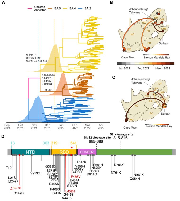
medRxiv preprint doi: https://doi.org/10.1101/2022.05.01.22274406; this version posted May 2, 2022. The copyright holder for this preprint
 (which was not certified by peer review) is the author/funder, who has granted medRxiv a license to display the preprint in perpetuity.
                                    It is made available under a CC-BY-NC-ND 4.0 International license .

  14
     Department of Medical Virology, University of Pretoria, Pretoria, South Africa
  15
     Division of Medical Virology, Faculty of Health Sciences, University of Cape Town, Cape
  Town, South Africa
  16
     National Health Laboratory, Health Services Management, Ministry of Health and Wellness,
  Gaborone, Botswana
  17
     Discipline of Virology, School of Laboratory Medicine and Medical Sciences and National
  Health Laboratory Service (NHLS), University of KwaZulu-Natal, Durban, South Africa
  18
     Department of Molecular Medicine and Haematology, Faculty of Health Science, School of
  Pathology, University of the Witwatersrand, Johannesburg, South Africa
  19
     Health Services Management, Ministry of Health and Wellness, Gaborone, Botswana
  20
     National Priority Program of the National Health Laboratory Service, Johannesburg, South
  Africa
  21
     Zoonotic Arbo and Respiratory Virus Program, Centre for Viral Zoonoses, Department of
  Medical Virology, University of Pretoria, Pretoria, South Africa
  22
     Institute of Evolutionary Biology, University of Edinburgh, Edinburgh, UK
  23
     Department of Medicine, University of Cambridge, Cambridge, UK
  24
     Department of Infectious Disease, Imperial College London, UK, W2 1PG
  25
     Biozentrum, University of Basel
  26
     Division of Virology, NHLS Groote Schuur Laboratory, Cape Town, South Africa
  27
     Wellcome Centre for Infectious Diseases Research in Africa (CIDRI-Africa), Cape Town,
  South Africa
  28
     Institute of Infectious Disease and Molecular Medicine, University of Cape Town, Cape Town,
  South Africa
  29
     Department of Zoology, University of Oxford, Oxford, UK
  30
     Department of Molecular Pathology, School of Pathology, Faculty of Health Sciences,
  University of the Witwatersrand, Johannesburg, South Africa
  31
     Department of Global Health, University of Washington, Seattle, WA, USA

  *Corresponding author: Prof Tulio de Oliveira, tulio@sun.ac.za
medRxiv preprint doi: https://doi.org/10.1101/2022.05.01.22274406; this version posted May 2, 2022. The copyright holder for this preprint
(which was not certified by peer review) is the author/funder, who has granted medRxiv a license to display the preprint in perpetuity.
It is made available under a CC-BY-NC-ND 4.0 International license .
Abstract
South Africa’s fourth COVID-19 wave was driven predominantly by three lineages (BA.1, BA.2
and BA.3) of the SARS-CoV-2 Omicron variant of concern. We have now identified two new
lineages, BA.4 and BA.5. The spike proteins of BA.4 and BA.5 are identical, and comparable to
BA.2 except for the addition of 69-70del, L452R, F486V and the wild type amino acid at Q493.
The 69-70 deletion in spike allows these lineages to be identified by the proxy marker of S-gene
target failure with the TaqPath™ COVID-19 qPCR assay. BA.4 and BA.5 have rapidly replaced
BA.2, reaching more than 50% of sequenced cases in South Africa from the first week of April
2022 onwards. Using a multinomial logistic regression model, we estimate growth advantages
for BA.4 and BA.5 of 0.08 (95% CI: 0.07 - 0.09) and 0.12 (95% CI: 0.09 - 0.15) per day
respectively over BA.2 in South Africa.
Main text
Within days of being discovered in South Africa and Botswana, on November 26, 2021, the
Omicron variant of SARS-CoV-2 was designated as a variant of concern (VOC) by the World
Health Organization1. Initially, Omicron was comprised of three sister lineages, BA.1, BA.2 and
BA.3. BA.1 caused most of the infections in South Africa’s fourth epidemic wave. However, as
that wave receded in mid-January 2022, BA.2 became the dominant South African lineage.
Despite being associated with a modest prolongation of the fourth wave, the displacement of
BA.1 by BA.2 in South Africa was not associated with a significant resurgence in cases, hospital
admissions or deaths. This pattern was not consistent worldwide, however, and in some countries
BA.2 was responsible for a greater share of cases, hospitalizations and deaths in the Omicron
wave2–7.
We recently identified two new Omicron lineages that have been designated BA.4 and BA.5 by
the Pango Network and pango-designation v1.3 (Fig. 1A)8,9. Bayesian phylogenetic methods
revealed that BA.4 and BA.5 are distinct from the other Omicron lineages. BA.4 and BA.5 are
estimated to have originated in mid-December 2021 (95% highest posterior density [HPD] 25
November 2021 to 01 January 2022) and early January 2022 (HPD 10 December 2021 to 6
February 2022) respectively (Fig. 1A). The most recent common ancestor of BA.4 and BA.5 is
estimated to have originated in mid-November 2021 (HPD 29 September 2021 to 6 December
2021) (Fig. 1A), coinciding with the emergence of the other lineages, for example BA.2 in early
November 2021 (HPD: 9 October 2021 to 29 November 2021). Phylogeographic analysis
suggests early dispersal of BA.4 from Limpopo to Gauteng, with later spread to other provinces
(Fig. 1B); and early dispersal of BA.5 from Gauteng to KwaZulu-Natal, with more limited
onward spread to other provinces (Fig. 1C).medRxiv preprint doi: https://doi.org/10.1101/2022.05.01.22274406; this version posted May 2, 2022. The copyright holder for this preprint
(which was not certified by peer review) is the author/funder, who has granted medRxiv a license to display the preprint in perpetuity.
It is made available under a CC-BY-NC-ND 4.0 International license .
BA.4 and BA.5 have identical spike proteins, most comparable to BA.2. Relative to BA.2, BA.4
and BA.5 have the additional spike mutations 69-70del, L452R, F486V and wild type amino acid
at position Q493 (Fig 1D). Outside of spike, BA.4 has the additional mutations at ORF7b:L11F
and N:P151S and a triple amino acid deletion in NSP1:141-143del whilst BA.5 has the M:D3N
mutation. Relative to BA.2, BA.5 has additional reversions at ORF6:D61 and nucleotide
positions 26858 and 27259. In addition, BA.4 and BA.5 have a nuc:G12160A synonymous
mutation in NSP8 that was present in Epsilon (B.1.429) and has arisen in BA.2 in some locations
(Extended Data Fig. 1). BA.4 and BA.5 have identical mutational patterns in the 5’ genome
region (from ORF1ab to Envelope) yet exhibit genetic divergence in the 3’ region (from M to the
3’ genome end). This suggests that BA.4 and BA.5 may be related by a recombination event,
with breakpoint between the E and M genes, prior to their emergence into the general population.
This scenario is somewhat similar to the relationship between BA.3 and BA.1/BA.2 which also
exhibit apparent ancestral recombination. Using the RASCL pipeline 10 we found no compelling
evidence of natural selection acting on the S-genes of viruses in either the BA.4 or BA.5
lineages.
It is currently unknown how differences in the mutation profiles of BA.4 and BA.5, relative to
BA.2, will impact on the phenotype. Changes at spike amino acids 452, 486 and 493 are likely to
influence human angiotensin-converting enzyme-2 (hACE2) and antibody binding. The 452
residue is in immediate proximity to the interaction interface of the hACE2 receptor. The L452R
mutation has been associated with an increased affinity for receptor binding with a resultant
increased infectivity11,12. The L452R mutation is also present in the Delta, Kappa and Epsilon
variants (and L452Q in Lambda), and mutations at this position have been associated with a
reduction in neutralization by monoclonal antibodies (particularly class 2 antibodies) and
polyclonal sera13–15. Mutations at this position (L452R/M/Q) have also arisen independently in at
least four BA.2 sublineages in different parts of the world, most notably BA.2.12.1 (L452Q)
which has become dominant in New York State.medRxiv preprint doi: https://doi.org/10.1101/2022.05.01.22274406; this version posted May 2, 2022. The copyright holder for this preprint
(which was not certified by peer review) is the author/funder, who has granted medRxiv a license to display the preprint in perpetuity.
It is made available under a CC-BY-NC-ND 4.0 International license .
Figure 1: A) Time-resolved maximum clade credibility phylogeny of the BA.2, BA.4 and BA.5 .5
lineages (n = 221, sampled between 29 December 2021 and 7 April 2022). Mutations that at
characterize the lineages are indicated on the branch at which each first emerged. The posterior
ior
distribution of the time of the most recent common ancestor (TMRCA) is also shown for BA.2, .2,
BA.4 and BA.5. B) Spatiotemporal reconstruction of the spread of the BA.4 lineage in South th
Africa. C) Spatiotemporal reconstruction of the spread of the BA.5 lineage in South Africa. In BmedRxiv preprint doi: https://doi.org/10.1101/2022.05.01.22274406; this version posted May 2, 2022. The copyright holder for this preprint
(which was not certified by peer review) is the author/funder, who has granted medRxiv a license to display the preprint in perpetuity.
It is made available under a CC-BY-NC-ND 4.0 International license .
and C, circles represent nodes of the maximum clade credibility phylogeny, coloured according
to their inferred time of occurrence (scale shown). Solid curved lines denote the links between
nodes and the directionality of movement (anti-clockwise along the curve). D) Amino acid
mutations in the spike gene of the BA.4 and BA.5 lineages. Mutations that differ from BA.2 are
denoted in red, including the wild-type amino acid at position Q493 (denoted by the red *).
Before the emergence of BA.4 and BA.5, F486V had been observed only in 54 of 10 million
publicly available genome sequences in GISAID (https://cov-
spectrum.org/explore/World/AllSamples/AllTimes/variants?aaMutations=S%3AF486V&).
Selection analyses focusing on ratios of non-synonymous and synonymous substitution rates at
individual codons have indicated that, since December 2020 S:486 has been evolving under
strong negative selection favouring the F state at this site (i.e., the amino acid that is found in
Wuhan-Hu-1) (Extended Data Fig. 2). Although rare, the F486L mutation has been observed in
some viral lineages found infecting minks and in human cases linked to mink farms and has been
shown to directly enhance entry into cells expressing mink/ferret ACE2 16. When binding to
hACE2, spike amino acid F486 interacts with hACE2 residues L79, M82, and Y83, which
collectively comprise a hotspot for ACE2 differences between mammalian species17,18.
Mutations at F486 are associated with a reduction in neutralising activity by class 1 (and some
class 2) neutralising antibodies and by polyclonal sera13–15. Deep mutational scanning suggests
that F486 is a key site for escape of vaccine- and infection-elicited RBD-targeted antibodies,
including those still able to neutralize Omicron/BA.1
19
(https://jbloomlab.github.io/SARS2_RBD_Ab_escape_maps/escape-calc/) .
The S:69-70del means BA.4 and BA.5 can again be presumptively identified (against a
background of BA.2 infection) using the proxy marker of S-gene target failure (SGTF) with the
TaqPath™ COVID-19 qPCR assay (Thermo Fisher Scientific, Waltham, MA, USA). SGTF was
successfully used to track the early spread of BA.1 (which also demonstrates SGTF), later also
enabling discrimination between BA.1 and BA.2 infections, since BA.2 viruses generally lack
the S:69-70del20. Recent data from public laboratories in South Africa suggest that the proportion
of positive PCR tests with SGTF has been increasing since early March, suggesting that BA.4
and BA.5 may be responsible for a growing share of recently confirmed cases (Fig. 2E). To
assess the validity of SGTF for identifying BA.4/BA.5, we performed qPCR with the TaqPath™
assay on 296 unselected samples submitted for sequencing to KwaZulu-Natal Research
Innovation and Sequencing Platform (KRISP) from Gauteng, Eastern Cape and KwaZulu-Natal
collected between 6 January and 3 April 2022. Of the 296 samples processed, we had a paired
valid qPCR result and sequence for 198. Of the 77 samples with SGTF on qPCR, 66 were BA.4
or BA.5, nine were BA.1, and two were BA.2. No BA.4 and BA.5 genomes were S-gene target
positive on qPCR (Extended Data Table 1). These results suggest that SGTF surveillance (wheremedRxiv preprint doi: https://doi.org/10.1101/2022.05.01.22274406; this version posted May 2, 2022. The copyright holder for this preprint
(which was not certified by peer review) is the author/funder, who has granted medRxiv a license to display the preprint in perpetuity.
It is made available under a CC-BY-NC-ND 4.0 International license .
the assay is available) may for now be a reasonable proxy to identify BA.4 and BA.5 for
countries with a low prevalence of BA.1.
At the time of writing, we have confirmed BA.4 and/or BA.5 in seven provinces in South Africa
(Eastern Cape, Gauteng, KwaZulu-Natal, Limpopo, Mpumalanga, North West and Western
Cape) in samples collected between 1 January 2022 and 20 April 2022 (Fig. 2B). In the two most
populous provinces in South Africa, Gauteng and KwaZulu-Natal, BA.4 and BA.5 have rapidly
replaced BA.2, and are responsible for approximately 60-75% of sequenced cases by the week
starting 18 April 2022 (Extended Data Fig. 3). These estimates are based on unselected sampling
for genomic surveillance (samples not selected based on SGTF or genotyping). The data suggest
geographic heterogeneity in the distribution of these two new lineages, with growth
predominantly of BA.4 in Gauteng and BA.5 in KwaZulu-Natal (Extended Data Fig. 3).
Internationally, by 20 April 2022, BA.4 had also been detected in a small number of samples in
neighbouring Botswana (estimated prevalencemedRxiv preprint doi: https://doi.org/10.1101/2022.05.01.22274406; this version posted May 2, 2022. The copyright holder for this preprint
(which was not certified by peer review) is the author/funder, who has granted medRxiv a license to display the preprint in perpetuity.
It is made available under a CC-BY-NC-ND 4.0 International license .
reported cases (Fig. 2A) and the proportions of positive qPCR and antigen tests were relatively
low (5-10%) through to early April 2022, these indicators began to rise from mid-April 2022,
and at the time of writing there are early signs of rising hospital admissions in some provinces.
Work is underway to characterise disease severity and immune escape.
There remains some uncertainty about the origin of the different Omicron lineages and
phylogenetic inference is limited by the relatively low sampling coverage in our genomic
surveillance. Whilst the Bayesian phylogenetic methods employed here suggest that BA.4 and
BA.5 are independent lineages that originated around the same time as BA.1-BA.3, other
methods suggest they could have descended from BA.2. Further sequencing (particularly
samples from Gauteng and neighbouring provinces) may help to provide more clarity.
Nevertheless, the continued discovery of genetically diverse Omicron lineages shifts the level of
support for hypotheses regarding their origin, from an unsampled location to a discrete reservoir,
such as human chronic infections (or even a network of chronic human infections) and/or animal
reservoirs, potentially contributing to further evolution and dispersal of the virus. We are actively
investigating the potential of a yet unidentified animal reservoir in the region. To date, the only
reverse zoonoses cases reported from the African region were in African lions and a puma in a
private zoo in Johannesburg, South Africa 23. Although these are unlikely species to play a role
in the emergence of new variants, it is a reminder of the susceptibility of certain wildlife species
to infections from humans. Following the emergence of Omicron, the World Organisation for
Animal Health released a statement calling for enhanced surveillance in animals to identify the
origin of new variants 24.
In conclusion, we have identified two new Omicron lineages (BA.4 and BA.5), which seem to be
associated with a resurgence in infections in South Africa approximately four months on from
the start of the Omicron wave. This once again highlights the importance of continued global
genomic surveillance and variant analysis in real-time to characterize the continuing evolution of
SARS-CoV-2.medRxiv preprint doi: https://doi.org/10.1101/2022.05.01.22274406; this version posted May 2, 2022. The copyright holder for this preprint
(which was not certified by peer review) is the author/funder, who has granted medRxiv a license to display the preprint in perpetuity.
It is made available under a CC-BY-NC-ND 4.0 International license .
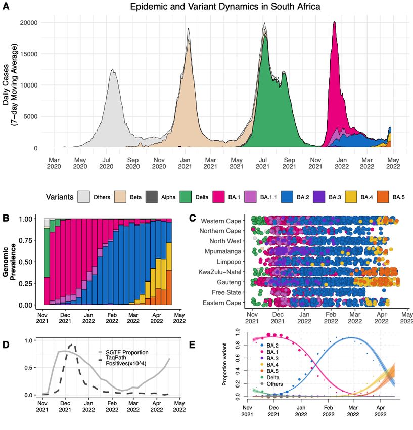
Figure 2 A) The progression of the 7-day rolling average of daily reported case numbers in
South Africa over two years of the epidemic (April 2020 – April 2022). Daily cases are colouredd
by the inferred proportion of SARS-CoV-2 variants prevalent at a particular period in the he
epidemic. B) Changes in the genomic prevalence of Omicron lineages in South Africa from m
November 2021 (when BA.1 dominated) to April 2022 (when BA.4 and BA.5 were increasing in
frequency). C) The count of Omicron lineage genomes per province of South Africa over er
November 2021 – April 2022. BA.4 and BA.5 have been detected in seven of the nine provinces. es.
D) Changes in the proportion of positive TaqPath qPCR tests exhibiting SGTF from November er
^4
2021 to April 2022. The number of TaqPath positives are to the order of 10 to the scale shown.n.
E) Modelled linear proportions of the Omicron lineages in South Africa. BA.1 rapidly lymedRxiv preprint doi: https://doi.org/10.1101/2022.05.01.22274406; this version posted May 2, 2022. The copyright holder for this preprint
(which was not certified by peer review) is the author/funder, who has granted medRxiv a license to display the preprint in perpetuity.
It is made available under a CC-BY-NC-ND 4.0 International license .
outcompeted Delta in November 2021 and was then superseded by BA.2 in early 2022. BA.4 and
BA.5 appear to be swiftly replacing BA.2 in South Africa. Model fits are based on a multinomial
logistic regression and dot size represents the weekly sample size.
References:
1. Viana, R. et al. Rapid epidemic expansion of the SARS-CoV-2 Omicron variant in southern
Africa. Nature 603, 679–686 (2022).
2. Lyngse, F. P. et al. Transmission of SARS-CoV-2 Omicron VOC subvariants BA.1 and
BA.2: Evidence from Danish Households. medRxiv (2022)
doi:10.1101/2022.01.28.22270044.
3. Rahimi, F. & Talebi Bezmin Abadi, A. The Omicron subvariant BA.2: Birth of a new
challenge during the COVID-19 pandemic. Int. J. Surg. 99, 106261 (2022).
4. Fonager, J. et al. Molecular epidemiology of the SARS-CoV-2 variant Omicron BA.2 sub-
lineage in Denmark, 29 November 2021 to 2 January 2022. Euro Surveill. 27, (2022).
5. Dai, Y. Rapid epidemic expansion of the SARS-CoV-2 Omicron BA.2 subvariant during
China’s largest outbreaks. Res. Sq. (2022) doi:10.21203/rs.3.rs-1516063/v3.
6. Chen, L.-L. et al. Contribution of low population immunity to the severe Omicron BA.2
outbreak in Hong Kong. Res. Sq. (2022) doi:10.21203/rs.3.rs-1512533/v1.
7. Hirotsu, Y. et al. SARS-CoV-2 Omicron sublineage BA.2 replaces BA.1.1: genomic
surveillance in Japan from September 2021 to March 2022. medRxiv (2022)
doi:10.1101/2022.04.05.22273483.
8. O’Toole, Á., Pybus, O. G., Abram, M. E., Kelly, E. J. & Rambaut, A. Pango lineage
designation and assignment using SARS-CoV-2 spike gene nucleotide sequences. BMC
Genomics 23, 121 (2022).medRxiv preprint doi: https://doi.org/10.1101/2022.05.01.22274406; this version posted May 2, 2022. The copyright holder for this preprint
(which was not certified by peer review) is the author/funder, who has granted medRxiv a license to display the preprint in perpetuity.
It is made available under a CC-BY-NC-ND 4.0 International license .
9. Rambaut, A. et al. A dynamic nomenclature proposal for SARS-CoV-2 lineages to assist
genomic epidemiology. Nat. Microbiol. 5, 1403–1407 (2020).
10. Lucaci, A. G. et al. RASCL: Rapid Assessment Of SARS-CoV-2 Clades Through
Molecular Sequence Analysis. BioRxiv (2022) doi:10.1101/2022.01.15.476448.
11. Motozono, C. et al. SARS-CoV-2 spike L452R variant evades cellular immunity and
increases infectivity. Cell Host Microbe 29, 1124-1136.e11 (2021).
12. Chen, J., Wang, R., Wang, M. & Wei, G.-W. Mutations Strengthened SARS-CoV-2
Infectivity. J. Mol. Biol. 432, 5212–5226 (2020).
13. Greaney, A. J. et al. Mapping mutations to the SARS-CoV-2 RBD that escape binding by
different classes of antibodies. Nat. Commun. 12, 4196 (2021).
14. Greaney, A. J. et al. Comprehensive mapping of mutations in the SARS-CoV-2 receptor-
binding domain that affect recognition by polyclonal human plasma antibodies. Cell Host
Microbe 29, 463-476.e6 (2021).
15. Greaney, A. J. et al. Complete Mapping of Mutations to the SARS-CoV-2 Spike Receptor-
Binding Domain that Escape Antibody Recognition. Cell Host Microbe 29, 44-57.e9
(2021).
16. Zhou, J. et al. Mutations that adapt SARS-CoV-2 to mink or ferret do not increase fitness in
the human airway. Cell Rep. 38, 110344 (2022).
17. Lan, J. et al. Structure of the SARS-CoV-2 spike receptor-binding domain bound to the
ACE2 receptor. Nature 581, 215–220 (2020).
18. Shang, J. et al. Structural basis of receptor recognition by SARS-CoV-2. Nature 581, 221–
224 (2020).
19. Greaney, A. J., Starr, T. N. & Bloom, J. D. An Antibody-Escape Estimator for Mutations tomedRxiv preprint doi: https://doi.org/10.1101/2022.05.01.22274406; this version posted May 2, 2022. The copyright holder for this preprint
(which was not certified by peer review) is the author/funder, who has granted medRxiv a license to display the preprint in perpetuity.
It is made available under a CC-BY-NC-ND 4.0 International license .
the SARS-CoV-2 Receptor-Binding Domain. Virus Evol. (2022) doi:10.1093/ve/veac021.
20. Scott, L. et al. Track Omicron’s spread with molecular data. Science 374, 1454–1455
(2021).
21. Sun, K. et al. Persistence of SARS-CoV-2 immunity, Omicron’s footprints, and projections
of epidemic resurgences in South African population cohorts. medRxiv (2022)
doi:10.1101/2022.02.11.22270854.
22. Madhi, S. A. et al. Population Immunity and Covid-19 Severity with Omicron Variant in
South Africa. N. Engl. J. Med. 386, 1314–1326 (2022).
23. Koeppel, K. N. et al. SARS-CoV-2 Reverse Zoonoses to Pumas and Lions, South Africa.
Viruses 14, (2022).
24. Statement from the Advisory Group on SARS-CoV-2 Evolution in Animals concerning the
origins of Omicron variant - OIE - World Organisation for Animal Health.
https://www.oie.int/en/document/statement-from-the-advisory-group-on-sars-cov-2-
evolution-in-animals-concerning-the-origins-of-omicron-variant/.
Online Methods:
Epidemiological dynamics
We analysed daily cases of SARS-CoV-2 in South Africa up to 25 April 2022 from publicly
released data provided by the National Department of Health and the National Institute for
Communicable Diseases. This was accessible through the repository of the Data Science for
Social Impact Research Group at the University of Pretoria
(https://github.com/dsfsi/covid19za)25,26. The National Department of Health releases daily
updates on the number of confirmed new cases, deaths and recoveries, with a breakdown by
province.
Sampling of SARS-CoV-2
As part of the NGS-SA27, seven sequencing hubs receive randomly selected samples for
sequencing every week according to approved protocols at each site. These samples include
remnant nucleic acid extracts or remnant nasopharyngeal and oropharyngeal swab samples frommedRxiv preprint doi: https://doi.org/10.1101/2022.05.01.22274406; this version posted May 2, 2022. The copyright holder for this preprint
(which was not certified by peer review) is the author/funder, who has granted medRxiv a license to display the preprint in perpetuity.
It is made available under a CC-BY-NC-ND 4.0 International license .
routine diagnostic SARS-CoV-2 PCR testing from public and private laboratories in South
Africa. We analysed SARS-CoV-2 genomes generated from samples collected between 1
November 2021 and 20th April 2022.
Ethical statement
The project was approved by University of KwaZulu–Natal Biomedical Research Ethics
Committee (ref. BREC/00001510/2020), the University of the Witwatersrand Human Research
Ethics Committee (HREC) (ref. M180832), Stellenbosch University HREC (ref.
N20/04/008_COVID-19) and the University of Cape Town HREC (ref. 383/2020). Individual
participant consent was not required for the genomic surveillance. This requirement was waived
by the Research Ethics Committees.
Whole-genome sequencing and genome assembly
RNA was extracted on an automated Chemagic 360 instrument, using the CMG-1049 kit (Perkin
Elmer). The RNA was stored at −80 °C before use. Libraries for whole-genome sequencing
were prepared using either the Oxford Nanopore Midnight protocol with Rapid Barcoding or the
Illumina COVIDseq Assay.
Illumina Miseq/NextSeq
For the Illumina COVIDseq assay, the libraries were prepared according to the manufacturer’s
protocol. In brief, amplicons were tagmented, followed by indexing using the Nextera UD
Indexes Set A. Sequencing libraries were pooled, normalized to 4 nM and denatured with
0.2 N sodium acetate. A 8 pM sample library was spiked with 1% PhiX (PhiX Control v3
adaptor-ligated library used as a control). We sequenced libraries using the 500-cycle v2 MiSeq
Reagent Kit on the Illumina MiSeq instrument (Illumina). On the Illumina NextSeq 550
instrument, sequencing was performed using the Illumina COVIDSeq protocol (Illumina), an
amplicon-based next-generation sequencing approach. The first-strand synthesis was performed
using random hexamers primers from Illumina and the synthesized cDNA underwent two
separate multiplex PCR reactions. The pooled PCR amplified products were processed for
tagmentation and adapter ligation using IDT for Illumina Nextera UD Indexes. Further
enrichment and clean-up was performed according to protocols provided by the manufacturer
(Illumina). Pooled samples were quantified using the Qubit 3.0 or 4.0 fluorometer (Invitrogen)
and the Qubit dsDNA High Sensitivity assay kit according to the manufacturer’s instructions.
The fragment sizes were analysed using the TapeStation 4200 (Invitrogen). The pooled libraries
were further normalized to 4 nM concentration, and 25 μl of each normalized pool containing
unique index adapter sets was combined into a new tube. The final library pool was denatured
and neutralized with 0.2 N sodium hydroxide and 200 mM Tris-HCl (pH 7), respectively.
Sample library (1.5 pM) was spiked with 2% PhiX. Libraries were loaded onto a 300-cycle
NextSeq 500/550 HighOutput Kit v2 and run on the Illumina NextSeq 550 instrument (Illumina).medRxiv preprint doi: https://doi.org/10.1101/2022.05.01.22274406; this version posted May 2, 2022. The copyright holder for this preprint
(which was not certified by peer review) is the author/funder, who has granted medRxiv a license to display the preprint in perpetuity.
It is made available under a CC-BY-NC-ND 4.0 International license .
Midnight protocol
For Oxford Nanopore sequencing, the Midnight primer kit was used as described previously54.
cDNA synthesis was performed on the extracted RNA using the LunaScript RT mastermix (New
England BioLabs) followed by gene-specific multiplex PCR using the Midnight primer pools,
which produce 1,200 bp amplicons that overlap to cover the 30 kb SARS-CoV-2 genome.
Amplicons from each pool were pooled and used neat for barcoding with the Oxford Nanopore
Rapid Barcoding kit according to the manufacturer’s protocol. Barcoded samples were pooled
and bead-purified. After the bead clean-up, the library was loaded on a prepared R9.4.1 flow-
cell. A GridION X5 or MinION sequencing run was initiated using MinKNOW software with
the base-call setting switched off.
Ion Torrent Genexus Integrated Sequencer methodology for rapid whole-genome
sequencing of SARS-CoV-2
Viral RNA was extracted using the MagNA Pure 96 DNA and Viral Nucleic Acid kit on the
automated MagNA Pure 96 system (Roche Diagnostics) according to the manufacturer’s
instructions. Extracts were then screened by quantitative PCR to acquire the mean cycle
threshold (Ct) values for the SARS-CoV-2 N and ORF1ab genes using the TaqMan 2019-nCoV
assay kit v1 (Thermo Fisher Scientific) on the ViiA7 Real-time PCR system (Thermo Fisher
Scientific) according to the manufacturer’s instructions. Extracts were sorted into batches of
n = 8 within a Ct range difference of 5 for a maximum of two batches per run. Extracts with
99% coverage of the SARS-CoV-2 genome (~30 kb) and
an additional five primer pairs targeting human expression controls. The SARS-CoV-2
amplicons range from 125 bp to 275 bp in length. TRINITY was used for de novo assembly
and the Iterative Refinement Meta-Assembler (IRMA) was used for genome assisted assembly as
well as FastQC for quality checks.
Genome assembly
We assembled paired-end and Nanopore .fastq reads using Genome Detective v.1.132
(https://www.genomedetective.com), which was updated for the accurate assembly and variant
calling of tiled primer amplicon Illumina or Oxford Nanopore reads, and the Coronavirus Typing
Tool55. For Illumina assembly, the GATK HaploTypeCaller --min-pruning 0 argument was
added to increase mutation calling sensitivity near sequencing gaps. For Nanopore, low-coverage
regions with poor alignment quality (medRxiv preprint doi: https://doi.org/10.1101/2022.05.01.22274406; this version posted May 2, 2022. The copyright holder for this preprint
(which was not certified by peer review) is the author/funder, who has granted medRxiv a license to display the preprint in perpetuity.
It is made available under a CC-BY-NC-ND 4.0 International license .
increased. We also used the wf_artic (ARTIC SARS-CoV-2) pipeline as built using the Nextflow
workflow framework56. In some instances, mutations were confirmed visually with .bam files
using Geneious v.2020.1.2 (Biomatters). The reference genome used throughout the assembly
process was NC_045512.2 (numbering equivalent to MN908947.3).
Raw reads from the Illumina COVIDSeq protocol were assembled using the Exatype NGS
SARS-CoV-2 pipeline v.1.6.1 (https://sars-cov-2.exatype.com/). This pipeline performs quality
control on reads and then maps the reads to a reference using Examap. The reference genome
used throughout the assembly process was NC_045512.2 (accession number: MN908947.3).
Several of the initial Ion Torrent genomes contained a number of frameshifts, which caused
unknown variant calls. Manual inspection revealed that these were probably sequencing errors
resulting in mis-assembled regions (probably due to the known error profile of Ion Torrent
sequencers)57. To resolve this, the raw reads from the IonTorrent platform were assembled using
the SARSCoV2 RECoVERY (Reconstruction of Coronavirus Genomes & Rapid Analysis)
pipeline implemented in the Galaxy instance ARIES (https://aries.iss.it). This pipeline fixed the
observed frameshifts, confirming that they were artefacts of mis-assembly; this subsequently
resolved the variant calls. The Exatype and RECoVERY pipelines each produce a consensus
sequence for each sample. These consensus sequences were manually inspected and polished
using Aliview v.1.27 (http://ormbunkar.se/aliview/).
All of the sequences passing internal quality control were deposited in GISAID
(https://www.gisaid.org/), and the GISAID accession identifiers are included as part of Extended
Data Table 1.
Phylogenetic analysis
We initially analysed genomes from South Africa against the global reference dataset using a
custom pipeline based on a local version of NextStrain (https://github.com/nextstrain/ncov)28.
The pipeline contains several Python scripts that manage the analysis workflow. It performs an
alignment of genomes in NextAlign 29, phylogenetic tree inference in IQ-Tree V1.6.930, tree
dating and ancestral state construction and annotation (https://github.com/nextstrain/ncov).
The initial phylogenetic analysis enabled us to identify clusters corresponding to the BA.4
(n=120) and BA.5 (n=51) lineages. We extracted these clusters and constructed a preliminary
maximum-likelihood tree with a subset of BA.2 sequences (n=52) in IQ-tree. We inspected this
maximum-likelihood tree in TempEst v.1.5.331 for the presence of a temporal or molecular clock
signal. Linear regression of root-to-tip genetic distances against sampling dates indicated that the
SARS-CoV-2 sequences evolved in a relatively strong clock-like manner (correlation coefficient
= 0.6, R2 = 0.4) (Extended Data Fig. 4).medRxiv preprint doi: https://doi.org/10.1101/2022.05.01.22274406; this version posted May 2, 2022. The copyright holder for this preprint
(which was not certified by peer review) is the author/funder, who has granted medRxiv a license to display the preprint in perpetuity.
It is made available under a CC-BY-NC-ND 4.0 International license .
We then estimated time-calibrated phylogenies using the Bayesian software package BEAST
v.1.10.432. For this analysis, we used the strict molecular clock model, the HKY+I+G, nucleotide
substitution model and the exponential growth coalescent model33. We computed Markov chain
Monte Carlo (MCMC) in duplicate runs of 20 million states each, sampling every 2,000 steps.
Convergence of MCMC chains was checked using Tracer v.1.7.134. Maximum clade credibility
trees were summarized from the MCMC samples using TreeAnnotator after discarding 10% as
burn-in. The phylogenetic trees were visualized using ggplot and ggtree35,36.
Phylogeographic analysis
To model phylogenetic diffusion of the new cluster across the country, we used a flexible relaxed
random walk diffusion model that accommodates branch-specific variation in rates of dispersal
with a Cauchy distribution37. For each sequence, latitude and longitude were attributed to the
most precise district or provincial information available and linked to the diagnostic sample.
As described in ‘Phylogenetic analysis’, MCMC chains were run in duplicate for 10 million
generations and sampled every 1,000 steps, with convergence assessed using Tracer v.1.7.1.
Maximum clade credibility trees were summarized using TreeAnnotator after discarding 10% as
burn-in. We used the R package seraphim38 to extract and map spatiotemporal information
embedded in posterior trees.
Lineage classification
We used a previously proposed dynamic lineage classification method39 from the ‘Phylogenetic
Assignment of Named Global Outbreak Lineages’ (pangolin) software suite v4.0.6 with the --
Usher option (https://github.com/cov-lineages/pangolin) 40. This is aimed at identifying the most
epidemiologically important lineages of SARS-CoV-2 at the time of analysis, enabling
researchers to monitor the epidemic in a particular geographic region. A lineage is a linear chain
of viruses in a phylogenetic tree showing connection from the ancestor to the last descendant.
Variant refers to a genetically distinct virus with different mutations to other viruses.
Selection analysis
To identify which (if any) of the observed mutations in the spike protein was most likely to
increase viral fitness, we used the natural selection analysis of SARS-CoV-2 pipeline
(https://observablehq.com/@spond/revised-sars-cov-2-analytics-page). This pipeline examines
the entire global SARS-CoV-2 nucleotide sequence dataset for evidence of: (i) polymorphisms
having arisen in multiple epidemiologically unlinked lineages that have statistical support for
non-neutral evolution (mixed effects model of evolution)41, (ii) sites at which these
polymorphisms have support for a greater-than-expected ratio of nonsynonymous-to-
synonymous nucleotide substitution rates on internal branches of the phylogenetic tree (fixed-
effects likelihood)42 and (iii) whether these polymorphisms have increased in frequency in the
regions of the world in which they have occurred.medRxiv preprint doi: https://doi.org/10.1101/2022.05.01.22274406; this version posted May 2, 2022. The copyright holder for this preprint
(which was not certified by peer review) is the author/funder, who has granted medRxiv a license to display the preprint in perpetuity.
It is made available under a CC-BY-NC-ND 4.0 International license .
Estimating transmission advantage
We analysed 12,528 SARS-CoV-2 sequences from South Africa generated in this study and
uploaded to GISAID with sample collection dates from 1 November 2021 to 20 April 2022 43.
We used a multinomial logistic regression model to estimate the growth advantage of Omicron
BA.2 lineage compared with BA.1, BA.4 and BA.5 lineages at the time point at which the
proportion of Omicron BA.4 and BA.5 collectively reached 50% 44,45. We fitted the model using
the multinom function of the nnet package and estimated the growth advantage using the
package emmeans in R 46.
S-Gene Target Failure Monitoring
SGTF monitoring is performed through analysing SARS-CoV-2 laboratory test results from
nasopharyngeal specimens received from the public health sector and referred for PCR testing
undertaken by the National Health Laboratory Service (NHLS) in South Africa. The NHLS has a
single laboratory information system connecting laboratory testing platforms to a corporate data
warehouse, where data can be mined in near real-time. The TaqPath™ COVID-19 [Thermo
Fisher Scientific, Waltham, MA, USA] assay accounts for around 20% of NHLS PCR tests
performed, with around half of those performed in Gauteng. The TaqPath assay targets three
gene regions, ORF1ab, N and S, with the lack of probe fluorescence of the latter culminating in
S-gene target failure (SGTF). In Fig 2D, we analysed and plotted the weekly number of TaqPath
positive tests as well as the proportion of the positive tests with SGTF (defined as samples with
non-detectable S gene target and either N or ORF1ab gene positive with CT valuemedRxiv preprint doi: https://doi.org/10.1101/2022.05.01.22274406; this version posted May 2, 2022. The copyright holder for this preprint
(which was not certified by peer review) is the author/funder, who has granted medRxiv a license to display the preprint in perpetuity.
It is made available under a CC-BY-NC-ND 4.0 International license .
(2020) doi:10.5281/zenodo.3819126.
27. Msomi, N., Mlisana, K., de Oliveira, T. & Network for Genomic Surveillance in
South Africa writing group. A genomics network established to respond rapidly to public
health threats in South Africa. Lancet Microbe 1, e229–e230 (2020).
28. Hadfield, J. et al. Nextstrain: real-time tracking of pathogen evolution.
Bioinformatics 34, 4121–4123 (2018).
29. GitHub - neherlab/nextalign: Viral genome reference alignment.
https://github.com/neherlab/nextalign.
30. Nguyen, L.-T., Schmidt, H. A., von Haeseler, A. & Minh, B. Q. IQ-TREE: a fast
and effective stochastic algorithm for estimating maximum-likelihood phylogenies. Mol.
Biol. Evol. 32, 268–274 (2015).
31. Rambaut, A., Lam, T. T., Max Carvalho, L. & Pybus, O. G. Exploring the
temporal structure of heterochronous sequences using TempEst (formerly Path-O-Gen).
Virus Evol. 2, vew007 (2016).
32. Suchard, M. A. et al. Bayesian phylogenetic and phylodynamic data integration
using BEAST 1.10. Virus Evol. 4, vey016 (2018).
33. Griffiths, R. C. & Tavaré, S. Sampling theory for neutral alleles in a varying
environment. Philos. Trans. R. Soc. Lond. B Biol. Sci. 344, 403–410 (1994).
34. Rambaut, A., Drummond, A. J., Xie, D., Baele, G. & Suchard, M. A. Posterior
summarization in bayesian phylogenetics using tracer 1.7. Syst. Biol. 67, 901–904 (2018).
35. Wickham, H. ggplot2. WIREs Comp Stat 3, 180–185 (2011).
36. Yu, G. Using ggtree to Visualize Data on Tree-Like Structures. Curr. Protoc.
Bioinformatics 69, e96 (2020).medRxiv preprint doi: https://doi.org/10.1101/2022.05.01.22274406; this version posted May 2, 2022. The copyright holder for this preprint
(which was not certified by peer review) is the author/funder, who has granted medRxiv a license to display the preprint in perpetuity.
It is made available under a CC-BY-NC-ND 4.0 International license .
37. Lemey, P., Rambaut, A., Welch, J. J. & Suchard, M. A. Phylogeography takes a
relaxed random walk in continuous space and time. Mol. Biol. Evol. 27, 1877–1885 (2010).
38. Dellicour, S., Rose, R., Faria, N. R., Lemey, P. & Pybus, O. G. SERAPHIM:
studying environmental rasters and phylogenetically informed movements. Bioinformatics
32, 3204–3206 (2016).
39. Rambaut, A. et al. A dynamic nomenclature proposal for SARS-CoV-2 to assist
genomic epidemiology. BioRxiv (2020) doi:10.1101/2020.04.17.046086.
40. O’Toole, Á. et al. Assignment of epidemiological lineages in an emerging
pandemic using the pangolin tool. Virus Evol. 7, veab064 (2021).
41. Murrell, B. et al. Detecting individual sites subject to episodic diversifying
selection. PLoS Genet. 8, e1002764 (2012).
42. Kosakovsky Pond, S. L. & Frost, S. D. W. Not so different after all: a comparison
of methods for detecting amino acid sites under selection. Mol. Biol. Evol. 22, 1208–1222
(2005).
43. Shu, Y. & McCauley, J. GISAID: Global initiative on sharing all influenza data -
from vision to reality. Euro Surveill. 22, 30494 (2017).
44. Davies, N. G. et al. Estimated transmissibility and impact of SARS-CoV-2
lineage B.1.1.7 in England. Science 372, (2021).
45. Campbell, F. et al. Increased transmissibility and global spread of SARS-CoV-2
variants of concern as at June 2021. Euro Surveill. 26, (2021).
46. Lenth RV. emmeans: Estimated Marginal Means, aka Least-Squares Means,R
package version 1.6.1. (2021).medRxiv preprint doi: https://doi.org/10.1101/2022.05.01.22274406; this version posted May 2, 2022. The copyright holder for this preprint
(which was not certified by peer review) is the author/funder, who has granted medRxiv a license to display the preprint in perpetuity.
It is made available under a CC-BY-NC-ND 4.0 International license .
NGS-SA consortium author list
Phillip A. Bester, Maciej F. Boni, Mohammed Chand, Kutlo Macheke, Rachel Colquhoun,
Michaela Davids, Koen Deforche, Deelan Doolabh, Louis du Plessis, Susan Engelbrecht, Diana
Hardie, Verity Hill, Nei-Yuan Hsiao, Arash Iranzadeh, Arshad Ismail, Charity Joseph, Rageema
Joseph, Legodile Koopile, Faith Hungwe, Nokuthula Ndlovu, Lesego Kuate-Lere, Oluwakemi
Laguda-Akingba, Onalethatha Lesetedi-Mafoko, Shahin Lockman, Nkhensani Mtileni, Ashlyn
S.C. Davis Makama, Annabel Enoch, Luicer Olubayo, Arisha Maharaj, Boitshoko Mahlangu,
Kamela Mahlakwane, Gert van Zyl, Mathilda Claassen, Shannon Wilson, Zinhle Makatini, Gert
Marais, Koleka Mlisana, Anele Mnguni, Thabo Mohale, Kgomotso Moruisi, Mosepele
Mosepele, Kereng V. Masupu, Gerald Motsatsi, Modisa S. Motswaledi, Thongbotho
Mphoyakgosi, Noxolo Ntuli, Martin Nyaga, Lucier Olubayo, Botshelo Radibe, Yajna Ramphal,
Upasana Ramphal, Roger Shapiro, Naume Tebeila, Wilhelmina Strasheim, Joseph Tsui,
Stephanie van Wyk, Steven Weaver, Nicole Wolter, Alexander E. Zarebski, Boitumelo Zuze,
Dominique Goedhals, Armand (Phillip) Bester, Martin Nyaga and Peter Mwangi.
Funding Information
This research was supported by the South African Medical Research Council (SAMRC) with
funds received from the National Department of Health. Sequencing activities for NICD are
supported by a conditional grant from the South African National Department of Health as part
of the emergency COVID-19 response; a cooperative agreement between the National Institute
for Communicable Diseases of the National Health Laboratory Service and the United States
Centers for Disease Control and Prevention (CDC)(grant number 5 U01IP001048-05-00; 1
NU51IP000930-01-00); the African Society of Laboratory Medicine (ASLM) and Africa Centers
for Disease Control and Prevention through a sub-award from the Bill and Melinda Gates
Foundation grant number INV-018978; the UK Foreign, Commonwealth and Development
Office and Wellcome (Grant no 221003/Z/20/Z); and the UK Department of Health and Social
Care and managed by the Fleming Fund and performed under the auspices of the SEQAFRICA
project. This research was also supported by The Coronavirus Aid, Relief, and Economic
Security Act (CARES ACT) through the CDC and the COVID International Task Force (ITF)
funds through the CDC under the terms of a subcontract with the African Field Epidemiology
Network (AFENET) AF-NICD-001/2021. Sequencing activities at KRISP and CERI are
supported in part by grants from the World Health Organization, the Abbott Pandemic Defense
Coalition (APDC), the National Institute of Health USA (U01 AI151698) for the United World
Antivirus Research Network (UWARN) and the INFORM Africa project through IHVN (U54
TW012041), and the South African Department of Science and Innovation (SA DSI) and the
SAMRC under the BRICS JAF #2020/049.
Acknowledgements
We thank additional members from originating and sequencing laboratories in South Africa,
listed as part of the NGS-SA consortium authors, that helped to generate and made public themedRxiv preprint doi: https://doi.org/10.1101/2022.05.01.22274406; this version posted May 2, 2022. The copyright holder for this preprint
(which was not certified by peer review) is the author/funder, who has granted medRxiv a license to display the preprint in perpetuity.
It is made available under a CC-BY-NC-ND 4.0 International license .
SARS-CoV-2 sequences (through GISAID) used as reference dataset in this study (a complete
list of individual contributors of sequences is provided in Supplementary Table S1).
Data Availability Statement
All of the SARS-CoV-2 genomes generated and presented in this manuscript are publicly
accessible through the GISAID platform (https://www.gisaid.org/). The GISAID accession
identifiers of the sequences analysed in this study are provided as part of Supplementary Table
S1. Other raw data for this study are provided as a supplementary dataset at
https://github.com/krisp-kwazulu-natal/SARSCoV2_South_Africa_Omicron_BA4_BA5. The
reference SARS-CoV-2 genome (MN908947.3) was downloaded from the NCBI database
(https://www.ncbi.nlm.nih.gov/).
Code Availability Statement
All custom scripts to reproduce the analyses and figures presented in this Article are available at
https://github.com/krisp-kwazulu-natal/SARSCoV2_South_Africa_Omicron_BA4_BA5.
Author Contributions
Genomic or Diagnostics data generation: H.T., M.Moir, J.E., C.S., K.S., S.Moyo, D.G.A.,
U.J.A., D.K., R.V., J.G., T.M., D.M., W.C., M.Matshaba, S.Mayaphi, N.Mbhele, N.B.M., Y.N.,
S.P., T.J.S., J.E.S, L.Scott, L.Singh, N.A.M., P.S.L., W.S., G.D., D.T., N.W., W.P., F.K.T, C.W.,
J.B.
Sample collection and metadata curation: N.Msomi, M.V., K.S., F.K.T., M.D., G.C., A.M.,
C.M.,
Data analysis: H.T., M.Moir, M.G., E.W., J.E., D.G.A., K.S., A.OT., C.R., T.P.P., C.R., O.G.P,
D.P.M., A.R.
Study design and data interpretation: H.T., M.Moir, E.W., C.L.A, R.J.L., C.W., O.G.P, J.B.,
A.G., D.P.M., A.R., S.G., A.vG., T.dO
Manuscript writing: H.T., M.Moir, M.G., E.W., C.B., R.J.L., T.dO
All of the authors reviewed the manuscript.A B
Johannesburg/
LP
Tshwane
GP
MP
NW
KZN
FS
NC
15 EC Durban
WC
10
N: P151S
ORF7b: L11F Cape Town Nelson
2022.00
Mandela
2022.10
Bay
2022.20
5
NSP1: Del 141-143
medRxiv preprint doi: https://doi.org/10.1101/2022.05.01.22274406; this version posted May 2, 2022. The copyright holder for this preprint
(which was not certified by peer review) is the author/funder, who has granted medRxiv a license to display the preprint in perpetuity.
It is made available under a CC-BY-NC-ND 4.0 International license .
2022.00
Jan 2022 2022.10
Feb 2022 2022.20
March 2022
0 15
09/13/21 10/19/21 S:Del
11/25/2169-70 01/01/22 02/06/22 03/15/22 04/20/22
10
S:L452R date C Johannesburg/
S:F486V
Tshwane
density
S:R493Q LP
5
GP
M:D3N
MP
0
NW
09/13/21 10/19/21 11/25/21 01/01/22 02/06/22 03/15/22 04/20/22
date KZN
9 FS
NC
density
6
3
EC Durban
WC
0
09/13/21 10/19/21 11/25/21 01/01/22 02/06/22 03/15/22 04/20/22
Nelson Mandela Bay
Cape Town
2022.00 2022.10 2022.20
date
DA Epidemic and Variant Dynamics in South Africa
20000
(7−day Moving Average)
15000
Daily Cases
10000
5000
0
Mar May Jul Sep Nov Jan Mar May Jul Sep Nov Jan Mar May
2020 2020 2020 2020 2020 2021 2021 2021 2021 2021 2021 2022 2022 2022
Variants Others Beta Alpha Delta BA.1 BA.1.1 BA.2 BA.3 BA.4 BA.5
B 1.00 C Western Cape
Northern Cape
0.75 North West
Prevalence
Genomic
Mpumalanga
0.50 Limpopo
KwaZulu−Natal
0.25 Gauteng
Free State
0.00 Eastern Cape
Nov Dec Jan Feb Mar Apr May Nov Dec Jan Feb Mar Apr May
2021 2021 2022 2022 2022 2022 2022 2021 2021 2022 2022 2022 2022 2022
1.0
1.0
D E
0.8
BA.2
Proportion variant
SGTF Proportion BA.1
0.6
TaqPath BA.3
Positives(x10^4) BA.4
0.5
0.4
BA.5
Delta
0.2
Others
0.0
0.0
Nov Dec Jan Feb Mar Apr May
2021 2021 2022 2022 2022 2022 2022 Nov Dec Jan Feb Mar Apr
2021 2021 2022 2022 2022 2022You can also read