Corporate Medical Policy - Blue Cross NC
←
→
Page content transcription
If your browser does not render page correctly, please read the page content below
Corporate Medical Policy
General Inflammation Testing AHS – G2155
File Name:                     general_inflammation_testing
Origination:                   01/01/2019
Last CAP Review:               02/2021
Next CAP Review:               02/2022
Last Review:                   07/2021

Description of Procedure or Service
    Inflammatory response can occur due to tissue injury and/or various disorders, including arthritis,
    lupus, and infection. Acute phase reactants, such as serum C-reactive protein (CRP), are released in
    the acute phase response during inflammation and can be used to monitor inflammation.
    Inflammation may also be measured using the simple laboratory technique of erythrocyte
    sedimentation rate (ESR) (Kushner, 2021).

    Conditions Associated with Acute Inflammatory Responses
    Diseases most associated with an acute inflammatory response measured by C-reactive protein (CRP)
    and/or erythrocyte sedimentation rate (ESR) include arthritis, especially rheumatoid arthritis (RA),
    polymyalgia rheumatica (PMR), giant cell arteritis (GCA), systemic lupus erythematosus (SLE),
    cardiovascular disease (CVD) (Kushner, 2021), and Hodgkin lymphoma (HL) (NCCN, 2021a). RA is
    a systemic polyarthritis that can lead to joint loss as well as tendon and ligament deformation to the
    point of affecting day-to-day living. The diagnosis of RA can be made in a patient “with
    inflammatory arthritis involving three or more joints, positive RF [rheumatoid factor] and/or anti-
    citrullinated peptide/protein antibody, disease duration of more than six weeks, and elevated CRP or
    ESR, but without evidence of diseases with similar clinical features” (Venables & Maini, 2021). PMR
    “is an inflammatory rheumatic condition characterized clinically by aching and morning stiffness
    about the shoulders, hip girdle, and neck (Docken, 2021).” PMR is frequently associated with GCA
    (also known as Horton disease), which is vasculitis of medium-to-large blood vessels and can include
    the aorta and cranial arteries. Cranial arteritis can lead to permanent vision loss. An estimated 40-
    50% of patients with GCA also suffer from PMR whereas 15% of all PMR patients are also diagnosed
    with GCA. Due to inflammation of the aorta and aortic branches, aortic aneurysm and aortic
    dissection can occur in patients with GCA (Docken & Rosenbaum, 2019). In both PMR and GCA,
    ESR and CRP levels are typically elevated. SLE “is a complex autoimmune disease with chronic
    relapsing-remitting course and variable manifestations leading a spectrum from mild mucocutaneous
    to devastating, life-threatening illness… Epigenetic modifications mediate the effect of the
    environment on immunologic responses, eventually leading to an inflammatory, autoimmune, multi-
    systemic disease characterized by autoantibody production and tissue injury (Gergianaki & Bertsias,
    2018).” Since patients with SLE can be prone to infection, ESR and CRP may be used in monitoring
    inflammation (Kushner, 2021). CVD is a very common inflammatory disorder in the United States.
    Although serum CRP is a non-specific inflammatory marker and is not a causative agent of CVD,
    serum CRP can be used as a biomarker for CVD (Black, Kushner, & Samols, 2004; Kushner, 2021).
    Hodgkin lymphoma accounts for 10% of lymphomas and is characterized as a B-cell lymphoma
    “containing a minority of neoplastic cells (Reed-Sternberg cells and their variants) in an
    inflammatory background” (Aster & Pozdnyakova, 2020). ESR is elevated in HL, and an ESR ≥50 is
    considered as an “early-stage unfavorable factor” (NCCN, 2021a).

    Erythrocyte Sedimentation Rate (ESR)

                                                                                                 Page 1 of 20
                         An Independent Licensee of the Blue Cross and Blue Shield Association
General Inflammation Testing AHS – G2155
Erythrocyte sedimentation rate (ESR) is a common laboratory method used to monitor general
inflammation. ESR is used to analyze many different conditions, including RA, SLE, arteritis, PMR
(Kushner, 2021; Wu et al., 2010). The simple Westergren method of ESR consists of measuring the
distance a blood sample travels in a tube within one hour. The International Council for
Standardization in Hematology (ICSH) established a calibration reference to this method using
citrate-diluted samples. Automated ESR methods have been established; however, some of these
analyzers use different dilution solutions, such as EDTA, rather than citrate. EDTA is commonly used
as an anticoagulant in hematology measurements whereas the use of citrate is less prevalent. Horsti,
Rontu, and Collings (2010) compared blood samples from 200 patients using the traditional
Westergren method versus an EDTA-based method. Their data has an R2 value of only 0.72 and 55
subjects had a difference of over 30%, clearly indicating that ESR is significantly affected by sample
preparation methods (Horsti et al., 2010). ESR can also be affected by red blood cell morphology,
ambient conditions (such as high room temperature or tilting of the ESR tube), anemia, renal disease,
obesity, heart failure, and hypofibrinogenemia (Kushner, 2021; Taylor & Maini, 2020).
More, ESR may be affected by noninflammatory factors, thus reducing its specificity for
inflammatory processes. Noninflammatory biological factors and environmental conditions can
increase a sample’s observed ESR. If the serum sample contains elevated concentrations of ions or
charged proteins, an elevated ESR may occur; for example, an increase in positively charged plasma
proteins could result in agglutination of erythrocytes within a sample for rapid sedimentation (Hale,
Ricotta, & Freed, 2019).
The ICSH established a Working Group to investigate the ESR methodology used in laboratories; the
findings of this working group were published in 2017. Data from over 6000 laboratories on four
different continents was examined. Of the laboratories included in the study, only 28% used the “gold
standard” Westergren method exclusively (i.e. the method with the established validation by the
ICSH) “while 72% of sites used modified or alternate methods.” The data obtained from the new
methodologies could deviate from the Westergren method by up to 142% and could differ “from each
other of up to 42%.” The ICSH released recommendations based up the results of these studies. One
such recommendation for labs using the non-Westergren method of ESR is to “consider adding an
interpretative comment to every result stating that ‘This result was obtained with an ESR instrument
that is not based on the standard Westergren method. The sensitivity and specificity of this method
for various disease states may be different from the standard Westergren method’” (Kratz et al.,
2017).
C-reactive Protein (CRP)
C-reactive protein (CRP) was first discovered in the early twentieth century when it was isolated in a
co-precipitation reaction with the pneumococcal C polysaccharide. The polysaccharide component
bound by CRP later was identified to be phosphocholine. Since then, studies have shown that CRP
can bind a number of ligands other than bacterial cell wall components. During an acute
inflammatory response, hepatocytes can upregulate CRP synthesis more than 1000-fold. The increase
in serum CRP “after tissue injury or infection suggests that it contributes to host defense and that it is
part of the innate immune response” (Black et al., 2004). Determining CRP concentration and
fluctuations in plasma CRP can be useful in monitoring inflammatory response; however, what
dictates “normal” CRP levels is of debate since CRP concentrations can vary considerably between
individuals, people groups, and laboratory testing methodology. The units used to denote CRP
concentrations also vary between laboratories (Kushner, 2021).
Clinical Validity and Utility of CRP and ESR in Measuring Inflammatory Processes
Both CRP and ESR have been used to monitor RA. Elevated CRP and ESR does correlate to observed
radiologic damage in RA. Unlike ESR, CRP can be evaluated in stored serum. This could be
advantageous due to the time constraints of ESR testing (Taylor & Maini, 2020). A 2009 study by
Crowson, Rahman, and Matteson (2009) show that the use of both ESR and CRP testing in the case of
RA is not warranted. Data from three randomized trials of 1247 RA patients was examined. “Where
available, the CRP alone may be preferred for disease activity assessment as a simple, validated,
Page 2 of 20
An Independent Licensee of the Blue Cross and Blue Shield AssociationGeneral Inflammation Testing AHS – G2155
reproducible, non age-dependent test” (Crowson et al., 2009). Since both ESR and CRP have been
incorporated into composite scoring for RA, the elimination of one or the other will not hinder the
quantitative evaluation of the patient using a composite scoring system such as DAS (Disease
Activity Score) or SDAI (Simplified Disease Activity Index). A 2015 Danish study clearly shows that
the data obtained in DAS using either ESR or CRP “are interchangeable when assessing RA patients
and the two versions of DAS28 are comparable” (Nielung et al., 2015). This study compared the
baseline data and one-year follow-up of 109 different patients with RA using the DAS28-ESR and
DAS28-CRP. Using the EULAR (European League Against Rheumatism) response criteria, only 14
patients show a divergence between using the ESR and CRP methods. Of those 14, “12 showed a
better response (in terms of responder category) using DAS28-CRP, while two patients showed a
better response using DAS28-ESR.” However, a 2006 study by Fransen and van Riel (2006) show
that it is still possible for a patient to have a high number of swollen joints and yet receive a low
DAS28-ESR score within the remission range due to a low ESR value since ESR has a significant
weight on the DAS28-ESR algorithm (Fransen & van Riel, 2006). This study did not include CRP
measurements to compare its validity to that of the DAS28-ESR. Another study released in 2010
(Hensor, Emery, Bingham, & Conaghan, 2010) shows that the DAS28-CRP could also underestimate
RA remission rates since those values are usually lower than the corresponding DAS28-ESR values,
but the discrepancy is not significant if age and gender are added as factors into the DAS28-CRP
methodology. To confound issues, “newer biologic agents that target specific inflammatory cytokines
are differentially reflected in the ESR and CRP and may therefore disproportionately deflate the
composite score (Anderson et al., 2012).”
ESR cannot be used to predict RA as a screening method. Suarez-Almazor and colleagues
investigated the predictive value of ESR for connective tissue diseases (CTD) and RA. Their review
of 711 records by more than 300 different primary care physicians in Alberta show that ESR
positively predicted 35% for CTD and only 17% for cases of RA. For SLE, the positive predictive
value for ESR was even lower at only 3%. CRP testing was not included in this study. The authors
note that “most tests were negative, and were often requested in patients without CTD, resulting in
low positive predictive values and questionable clinical utility” (Suarez-Almazor et al., 1998). A
study by Keenan, Swearingen, and Yazici (2008) compared the utilization of ESR and CRP in RA,
SLE, and osteoarthritis. The data showed that for the 188 patients with RA, the number of patients
with both ESR and CRP elevated were statistically the same as those with normal test levels or those
with only one test elevated. Conclusions stated “that another look at the role of ESR and CRP as
markers of inflammation in RA patients seen in routine care may be in order (Keenan et al., 2008).”
Bitik et al. (2015) researched the use of elevated ESR and CRP levels in distinguishing the definitive
diagnosis of a rheumatic disorder from patients with nonspecific inflammation. In their study of 112
patients, 47 had a previously diagnosed rheumatic disorder and 65 had no history of a rheumatism.
Of the 65 patients with no history of a rheumatic disorder, 52.3% were diagnosed with a new
rheumatic disorder with PMR/GCA comprising 38.2%, while 47.7% had a non-rheumatic diagnosis.
Within this latter group, only the “CRP levels were significantly higher in infections when compared
with new onset RD (rheumatic disease) or malignancies (p < 0.05) (Bitik et al., 2015).” The ESR
levels between the three groups were statistically insignificant. This indicates that CRP is more
sensitive to acute infections than ESR. The authors state that “although ESR and CRP levels have a
very low specificity in differentiating between these conditions, in cases of unusually high levels of
CRP (especially above 200), more consideration should be given to infections or malignancies.”
A 2014 study of 60 different PMR patients compared the efficacy of ESR and CRP in assessing
disease activity versus patient-reported outcomes and plasma fibrinogen. In this study, the VASDA
(Visual analog scale disease activity) and VASQOL (VAS quality of life), two patient-reported
outcome methods, were the most responsive to changes in disease activity. Of the serum biomarkers,
fibrinogen, ESR, and CRP, fibrinogen was the most accurate with a correlation coefficient of 1.63
whereas 1.2 and 1.05 were the correlation coefficients of ESR and CRP, respectively. These data
suggest that plasma fibrinogen would be a more sensitive measure of PMR disease activity as
compared to either ESR or CRP (McCarthy et al., 2014).
Page 3 of 20
An Independent Licensee of the Blue Cross and Blue Shield AssociationGeneral Inflammation Testing AHS – G2155 A two-year retrospective study released in 2010 (Ernst, Weiss, Tracy, & Weiss, 2010) researched the validity of using either ESR and/or CRP in assessing septic joints. This study consisted of 163 patients and included both genders as well as patients with alcohol or drug histories. The mean ESR value for the 119 control non-septic joints was 46 while the septic joint mean ESR value was 57, which was however, the mean CRP value was 13 in the septic joints and 8.5 in the non-septic joints. The conclusion of the authors is that “CRP is helpful in determining the presence of a septic joint; ESR is not (Ernst et al., 2010).” ESR is used in determining the algorithm to follow in the treatment of Hodgkin lymphoma (CHL). For example, in stage 1A CHL, a patient with an ESR
General Inflammation Testing AHS – G2155
   included in this study would not have been diagnosed with PJI according to the American Association
   of Orthopaedic Surgeons (AAOS) guidelines or the Musculoskeletal Infection Society definition
   (Perez-Prieto et al., 2017).

   Kheir, Tan, Shohat, Foltz, and Parvizi (2018) evaluated the accuracy of inflammatory markers in
   diagnosis periprosthetic joint infections (PJI). A total of “549 periprosthetic joint infection cases and
   653 aseptic total joint arthroplasty revisions” were reviewed. The sensitivity of ESR to diagnose PJI
   was 0.85 and 0.88 for CRP. ESR was also elevated in antibiotic-resistant strains of bacteria compared
   to culture-negative cases. For CRP, gram-negative species had higher levels of CRP than culture-
   negative cases. Overall, the authors concluded that both ESR and CRP had higher false-negative
   levels than previously reported (Kheir et al., 2018).

   Hamann et al. (2019) compared the DAS28-ESR and DAS28-CRP to determine the impact on disease
   activity stratification in RA. A total of 31,074 paired data sets were included in this study and were
   obtained from the British Society for Rheumatology Biologics Register for RA. Results showed that
   “DAS28-CRP scores were ∼0.3 lower than DAS28-ESR overall, with greatest differences for women
   (-0.35) and patients over 50 years old (-0.34). Mean male DAS28-CRP scores were 0.15 less than
   corresponding DAS28-ESR scores (Hamann et al., 2019).” When DAS28-CRP data is adjusted by
   gender, significant agreement (P
General Inflammation Testing AHS – G2155
State and Federal Regulations, as applicable
Testing of serum acute phase reactants and ESR is performed in laboratories meeting Clinical
Laboratory Improvement Act (CLIA) quality standards. The FDA has approved multiple tests for
human CRP, including assays for conventional CRP, high sensitivity CRP (hsCRP), and cardiac CRP
(cCRP). On September 22, 2005, the FDA issued guidelines concerning the assessment of CRP (FDA,
2005). A search of the FDA Medical Devices database (FDA, 2018) on April 20, 2021, shows that the
FDA has approved ESR systems from multiple companies, incuding the ESR Control -M Hematology
Erythrocyte Sedimentation system (K972172) and the ESR Control -HC Hematology Erythrocyte
Sedimentation system (K972170) by R & D Systems, the Seditainer Erythrocyte Sedimentation Rate
System (K953994) from Becton Dickinson Vacutainer Systems, the Westergren Dispette for ESR
(K831195) by Ulster Scientific, and the Dade ESR Kit (K823368) from American Dade.
***Note: This Medical Policy is complex and technical. For questions concerning the technical
language and/or specific clinical indications for its use, please consult your physician.
Policy
BCBSNC will provide coverage for general inflammation testing when it is determined the medical
criteria or reimbursement guidelines below are met.
Benefits Application
This medical policy relates only to the services or supplies described herein. Please refer to the Member's
Benefit Booklet for availability of benefits. Member's benefits may vary according to benefit design;
therefore member benefit language should be reviewed before applying the terms of this medical policy.
When general inflammation testing is covered
Reimbursement for measurement of erythrocyte sedimentation rate (ESR) for patients with Hodgkin
Lymphoma is allowed.
Reimbursement for measurement of either C-Reactive Protein (CRP) or ESR in the diagnosis,
assessment and monitoring of inflammatory disorders, and/or undiagnosed conditions, and/or to
detect acute phase inflammation is allowed. (*please see Note 1).
*NOTE 1: For policy regarding the use of CRP as a cardiac biomarker, please see policy AHS-
G2150 Cardiac Biomarkers for Myocardial Infarction. For policy regarding ANA/ENA Testing for
systemic autoimmune rheumatic diseases and idiopathic inflammatory myopathies, please see policy
AHS-G2022 ANA/ENA Testing.
When general inflammation testing is not covered
Reimbursement is not allowed for the measurement of both CRP and ESR, at the same visit, in the
diagnosis, assessment and monitoring of inflammatory disorders, and/or undiagnosed conditions,
and/or to detect acute phase inflammation.
Reimbursement is not allowed for the measurement of either CRP and/or ESR during general exam
without abnormal findings.
Policy Guidelines
Page 6 of 20
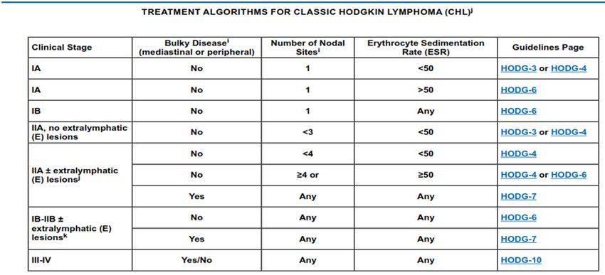
An Independent Licensee of the Blue Cross and Blue Shield AssociationGeneral Inflammation Testing AHS – G2155 Guidelines and Recommendations World Health Organization (WHO) (WHO, 2018, 2019) On May 16, 2018, the WHO released their first edition of the Model List of Essential In Vitro Diagnostics (EDL) “to advance universal health coverage, address health emergencies, and promote healthier populations.” This list of in vitro diagnostics (IVD) is to be used as a reference of the essential diagnostic tools for laboratories to complement their Model List of Essential Medicines. With respect to the diagnostic tool “to detect inflammation as an indicator of various conditions,” the WHO recommends CRP either in an EIA (enzyme immunoassay) or RDT (rapid diagnostic test) assay format. The specimen type can be venous whole blood, serum, or plasma. In 2019, the WHO released the Second WHO Model List of Essential In Vitro Diagnostics. In a table titled General IVDs for Use in Clinical Laboratories, CRP is once again listed. The WHO now recommends CRP in an RDT, latex agglutination assay or immunoassay format (WHO, 2019). National Comprehensive Cancer Network (NCCN) (NCCN, 2020, 2021a, 2021b) The 2020 NCCN guidelines concerning Hodgkin Lymphoma (NCCN, 2019b, 2020b) uses ESR as a diagnostic tool in characterizing the type of Classic Hodgkin Lymphoma (CHL) as well as the primary treatment of the disease. In the diagnosis/workup of Hodgkin Lymphoma in adults (age ≥18 years) (recommendation 2A), they list erythrocyte sedimentation rate (ESR) as “essential” and that ESR should be tested within 6 months of diagnosis; in fact, ESR is used extensively in the treatment algorithm for CHL as depicted in the table below (NCCN, 2020b). In the guidelines concerning follow-up after completion of treatment, the NCCN (2021a) states that “interim physical examinations and blood tests (CBC [complete blood count], platelets, and ESR is elevated at initial diagnosis and chemistry profile) are performed every 3 to 6 months for 1 to 2 years and then every 6 to 12 months for the next 3 years and then annually.” ESR is also used in determining the dosage of involved-site radiation therapy (ISRT). “A dose of 20 Gy following ABVD X 2 is sufficient if the patient has non-bulky stage I-IIA disease with an ESR
General Inflammation Testing AHS – G2155
   Regarding diagnostic criteria for idiopathic MCD (Multicentric Castleman Disease), minor diagnostic
   criteria include elevated CRP (>10 mg/L) or ESR (>15 mm/h) where an “Elevation of CRP is
   mandatory and tracking CRP levels is highly recommended, but ESR will be accepted if CRP is not
   available (NCCN, 2021b).”

   In the NCCN guidelines concerning the T-cell lymphomas, they state that the “evaluation of
   serological markers such as rheumatoid factor (RF), antinuclear antibodies (ANA), and erythrocyte
   sedimentation rate (ESR) is useful in patients with autoimmune disease”(NCCN, 2020). [Please note
   that the Avalon policy AHS-G2022 covers ANA testing.] The guidelines concerning T-cell
   lymphomas do not mention the diagnostic or prognostic use of CRP.

   2015 American Society for Clinical Pathology (ASCP) (Pathology, 2015)
   In the Choosing Wisely site of the ABIM Foundation, the ASCP released the recommendation to not
   “order an erythrocyte sedimentation rate (ESR) to look for inflammation in patients with undiagnosed
   conditions. Order a C-reactive protein (CRP) to detect acute phase inflammation” due to the
   sensitivity and specificity of CRP for acute phase of inflammation. “In the first 24 hours of a disease
   process, the CRP will be elevated, while the ESR may be normal. If the source of inflammation is
   removed, the CRP will return to normal within a day or so, while the ESR will remain elevated for
   several days until excess fibrinogen is removed from the serum.”

   European League Against Rheumatism (EULAR) (Colebatch et al., 2013; Combe et al., 2017;
   Dejaco et al., 2018; Dejaco et al., 2015; Mukhtyar et al., 2009)
   In 2009, EULAR issued their recommendations concerning the management of large vessel
   vasculitis. With a “Level of Evidence 3, Strength of recommendation C”, they recommend
   “monitoring of therapy for large vessel vasculitis should be clinical and supported by measurement of
   inflammatory markers…. For patients with giant cell arteritis, a relapse is usually associated with a
   rise in ESR and CRP” (Mukhtyar et al., 2009). In this paper, no mention of the frequency of ESR
   and/or CRP testing is mentioned.

   In 2013 in EULAR recommendations for the use of imaging of the joints in the clinical management
   of rheumatoid arthritis (Colebatch et al., 2013), they state that “baseline inflammatory disease
   measured by scintigraphy appears to be associated with radiographic progression. In addition,
   multiple regression analysis has demonstrated that progression of radiographic joint destruction was
   primarily predicted by 99mTc-IgG scintigraphy; joint swelling, ESR and IgM RF (Rheumatoid Factor)
   were not predictive. This suggests that scintigraphy may be superior to conventional clinical and
   laboratory measurements in the prediction of joint destruction.” This set of guidelines did not include
   any mention concerning CRP or the frequency of ESR testing.

   In 2015, EULAR and the American College of Rheumatology (ACR) issued joint recommendations
   concerning the management of polymyalgia rheumatica (PMR) (Dejaco et al., 2015). Within their
   recommendations, they list assessments that “every case of PMR should have…prior to the
   prescription of therapy (primary or secondary care).” They include a basic laboratory workup “to
   exclude mimicking conditions and establish a baseline for monitoring of therapy”, and they state that
   this includes “rheumatoid factor and/or anti-cyclic citrullinated peptide antibodies (ACPA), C-
   reactive protein and/or erythrocyte sedimentation rate (ESR), blood count, glucose, creatinine, liver
   function tests, bone profile (including calcium, alkaline phosphatase) and dipstick urinalysis.” They
   do not state a specific preference of either CRP or ESR nor do they state the frequency of testing.

   EULAR in 2016 updated their 2007 recommendations concerning the management of early arthritis
   (Combe et al., 2017). The 2016 updates included the following recommendation: “Monitoring of
   disease activity should include tender and swollen joint counts, patient and physician global
   assessments, ESR and CRP, usually by applying a composite measure. Arthritis activity should be
   assessed at 1-month to 3-month intervals until the treatment target has been reached.” The
   recommendation concerning including both ESR and CRP did not change between the 2016 and 2007
   recommendations. Within the discussion of the recommendations, they state, “In every patient with
   active arthritis, closely monitoring disease activity is now considered of particular importance in the
                                                                                                Page 8 of 20
                        An Independent Licensee of the Blue Cross and Blue Shield Association
General Inflammation Testing AHS – G2155
therapeutic strategy to provide a good outcome…. Monitoring disease activity should be as frequent
as the level of disease activity mandates, usually every 1-3 months, then potentially less frequently
(such as every 6-12 months) once the treatment target has been achieved. Nevertheless, three changes
were proposed to this item…. First, a composite measure was recommended as the method of choice
to monitor disease activity; second, a specific time frame for monitoring structural damage was
deliberately left out and third, patient-reported outcomes were expanded beyond functional
assessments” (Combe et al., 2017).
In 2018, EULAR issued EULAR recommendations for the use of imaging in large vessel vasculitis in
clinical practice (Dejaco et al., 2018). They make no recommendation concerning the preference of
ESR or CRP nor do they state the frequency of testing; they do state “in patients with a high clinical
suspicion of GCA (>50%), for example, in case of new-onset headache, visual symptoms, jaw
claudication and elevated erythrocyte sedimentation rate (ESR) and C reactive protein, a positive
ultrasound would result in a post-test probability of >95%.”
American College of Rheumatology (ACR) (Anderson et al., 2012; Barber et al., 2019; England
et al., 2019; National Guideline, 2016; Singh et al., 2015; Ward et al., 2016)
In 2012, ACR released their recommendations concerning the clinical practice of using disease
activity measures of rheumatoid arthritis (RA) (Anderson et al., 2012). The recommend using the
Disease Activity Score with 28-joint counts (DAS28), the Clinical Disease Activity Index, the Patient
Activity Scale (PAS), the PAS-II, the Simplified Disease Activity Index (SDAI), and Routine
Assessment of Patient Index Data with 3 measures. The DAS28 is a composite test that can use either
CRP or ESR data. The ACR states that both the CRP or ESR used in the DAS28 have been validated
in RA. Of the six activity measures recommended by the ACR, only DAS28 received “excellent”
recommendations for all three psychometric properties—reliability, validity, and responsiveness.
Within the guidelines, the ACR also issued the scores corresponding to remission, low/minimal,
moderate, and high/severe RA for all of the disease activity measures, including the DAS28, as well
as the mathematical formula using either CRP or ESR data to determine the DAS28. CRP is also
used in the SDAI; however, the SDAI is rated as “good” for reliability because they state that “test-
retest reliability for composite has not been evaluated” for the SDAI. No mention of frequency of
testing is made. They do note that the “inclusion of acute-phase reactants in the DAS28 and SDAI
complicates the logistics and timing using these measures in point-of-care clinical decision making.
Although these measures have traditionally been used in clinical trials, academic medical centers, and
large multispecialty clinics, logistical barriers have likely delayed their widespread adoption in
smaller practice settings (Anderson et al., 2012).”
The ACR in 2015 (Singh et al., 2015) issued guidelines for the treatment of RA. While not specifying
a preference of either CRP or ESR in diagnosing or predicting the prognosis of RA, they do state in
their “Key provisos and principles” that “functional status assessment using a standardized, validated
measure should be performed routinely for RA patients, at least once per year, but more frequently if
disease is active.” They also state that disease activity be measured using ACR-validated scales,
including the aforementioned DAS28 and/or SDAI. Moreover, they define RA remission as “a tender
joint count, swollen joint count, C-reactive protein level (mg/dl), and patient global assessment of ≤1
each or a Simplified DAS of ≤3.3, 1 of 6 ACR-endorsed disease activity measures”.
Also, in 2015 (but published in 2016), the ACR and the Spondylitis Association of America (SAA)
issued their joint recommendations concerning the treatment of ankylosing spondylitis (AS) and
nonradiographic axial spondyloarthritis (National Guideline, 2016; Ward et al., 2016). Regarding
“the treatment of patients with either active or stable AS…we conditionally recommend regular-
interval use and monitoring of the CRP concentrations or erythrocyte sedimentation rate (ESR) over
usual care without regular CRP or ESR monitoring.” This received a “very low-quality evidence;
vote 100% agreement” rating. They do make note that as of the time of publication “no studies
addressed the effect of routine monitoring of a disease activity measure” but that “the panel thought
that monitoring would be most helpful in patients with active symptoms as a guide to treatment.”
Testing is not required for every clinic visit.
Page 9 of 20
An Independent Licensee of the Blue Cross and Blue Shield AssociationGeneral Inflammation Testing AHS – G2155 In 2019, updated recommendations by the RA disease activity measures working group were published by England et al. (2019). Recommended tests include the Clinical Disease Activity Index (CDAI), the Simplified Disease Activity Index (SDAI), the Routine Assessment of Patient Index Data 3 (RAPID3), and the 28-Joint Disease Activity Score (DAS28). As noted above, the DAS28 is a composite test that can use either CRP or ESR data. The ACR states that both the CRP or ESR used in the DAS28 have been validated in RA. American Academy of Family Physicians (AAFP) (Caylor & Perkins, 2013; Wasserman, 2018) In 2013, the AAFP released Recognition and Management of Polymyalgia Rheumatica and Giant Cell Arteritis. For polymyalgia rheumatica (PMR), they note that “a normal ESR is found in 6% to 20% of persons with [PMR], although in those cases C-reactive protein level is elevated. ESR predicts relapse more reliably, but C-reactive protein is more sensitive, and is less affected by age and other factors (Caylor & Perkins, 2013).” For giant cell arteritis (GCA), ESR is elevated in up to 89% of patients, but the sensitivity and specificity increase to 99% and 97%, respectively, if both ESR and CRP are tested. Regardless of using either ESR or CRP testing, the AAFP recommends that either ESR or CRP is tested at each clinic visit for patients with either PMR or GCA. American College of Radiology (ACR) (Ha et al., 2014; National Guideline, 2014) The ACR released their updated guidelines concerning the follow-up of Hodgkin lymphoma in 2014. They state that “limited data are available on the role of routine blood work in detecting relapses.” ESR is listed as one of the tests conducted as routine blood work in follow-up of Hodgkin lymphoma. They summarize their findings as the following: “In general a majority of recurrences can be detected initially by history and physical examination rather than by routine imaging studies or blood tests such as ESR, CBC, and chemistry (Ha et al., 2014).” Four of the five variants they reviewed had ESR tests conducted 1 – 2 times per year, and the ACR rated the use of ESR as a 3, 5, 5, and 7 in these four variants where a “3” indicates “usually not appropriate,” a “5” is “may be appropriate”, and a “7” falls in the “usually appropriate” category. The ACR released guidelines concerning management of multi-system inflammatory syndrome in children and devised a two-tier algorithm for diagnosis. ACR recommends routine lab tests as tier 1 testing which include complete blood count, comprehensive metabolic panel, erythrocyte sedimentation rate (ESR), and CRP. If tier 1 lab results include CRP ≥5 or ESR≥40 and one suggestive lab feature such as neutrophilia or platelet count
General Inflammation Testing AHS – G2155

   The British Society for Rheumatology (BSR) (Gordon et al., 2018; Mackie et al., 2020)
   The BSR alone issued their guidelines for the management of systemic lupus erythematosus (SLE) in
   2018 (Gordon et al., 2018). For the statement “CRP low or normal unless infection,” the BSR gives
   an overall level of evidence of 2++ with a B grade of recommendation whereas they grade the
   statement “ESR correlates with active lupus” a 2+ and only a C grade of recommendation. “ESR is
   often raised in active SLE, but can also reflect persistent polyclonal hypergammaglobulinaemia, and
   is not a reliable marker of disease activity…. A significantly raised CRP is more likely to indicate
   infection, and patients with raised CRP will need therefore to be thoroughly screened for infection,
   given that infection is the commonest cause of death in lupus patients. In contrast, a raised ESR does
   not discriminate between active lupus and infection.” They recommend that CRP is tested at initial
   diagnosis and then every 1-3 months during active disease states. Once stabilized, then testing
   frequency can be every 6-12 months. They also state that CRP testing should be conducted on
   mothers with SLE during pregnancy, but they do not state the frequency of the testing during
   pregnancy.

   The BSR has also published guidelines on the diagnosis and treatment of giant cell arteritis (GCA).
   Regarding which evaluations should be performed when starting treatment, the BSR states that
   “When starting glucocorticoids for suspected GCA, diagnostically relevant symptoms and signs
   should be documented. Blood should be taken for full blood count, CRP and ESR before or
   immediately after commencing high-dose glucocorticoids. If GCA is strongly suspected, the first
   dose of glucocorticoid can be given without waiting for laboratory results (Mackie et al., 2020).”
   Further, the BSR provides a list of clinical assessments which should be carried out at or near a GCA
   diagnosis. These lists includes “Measures of activity of GCA: laboratory markers of inflammation
   (CRP for all patients, plus either ESR or plasma viscosity) and full blood count (platelet count may
   be elevated in GCA).” Finally, regarding follow-up visits, “Each follow-up visit should include at
   least a full history, targeted physical examination and measurement of at least a full blood count,
   ESR and/or CRP, plus follow-up of any abnormalities relevant to the individual patient as well as
   drug-specific screening for toxicity (Mackie et al., 2020).”

   Canadian Rheumatology Association (CRA) (Bykerk et al., 2012; Keeling et al., 2018)
   The 2012 guidelines by the CRA titled Canadian Rheumatology Association Recommendations for
   Pharmacological Management of Rheumatoid Arthritis with Traditional and Biologic Disease-
   modifying Antirheumatic Drugs recommends (with Level II and Strength B) “the presence of the
   following poor prognostic features should be assessed at baseline and considered when making
   treatment decisions: RF positivity, anti-CCP positivity, functional limitation, high number of swollen
   and tender joints, early erosions, extraarticular features, high ESR or CRP.” They also recommend
   (with Level I and Strength A) “RA care providers should monitor disease activity as frequently as
   every 1 to 3 months in patients with active RA.” The disease activity should be monitored by a
   validated method, such as DAS28 or SDAI.

   In 2018, CRA released guidelines on assessment and monitoring of Systemic Lupus Erythematosus.
   Regarding diagnosis, CRA recommends that best clinical practice includes a complete history and physical
   examination at baseline with laboratory monitoring which could possibly include (but is not limited to) the
   following tests: “complete blood count (CBC), liver enzymes, creatine kinase, creatinine and estimated
   glomerular filtration rate (eGFR), urine routine/microscopic (urinalysis), urine protein-creatinine ratio, C-
   reactive protein (CRP), erythrocyte sedimentation rate (ESR), complements (C3, C4), anti-dsDNA,
   antinuclear antibodies, antibodies to extractable nuclear antigens, antiphospholipid antibodies (aPL), lupus
   anticoagulant (LAC), anticardiolipin (aCL), anti-β2-glycoprotein I (anti-β2-GPI), and lipid profile.
   Follow up laboratory monitoring will depend on the patient’s clinical status and may include CBC, eGFR,
   urinalysis, urine protein-creatinine ratio, CRP, and/or ESR, C3, C4, and anti-dsDNA antibodies (Keeling et
   al., 2018).”

   The National Collaborating Centre for Chronic Conditions (NCC-CC) (Conditions, 2009)
   The NCC-CC produced extensive guidelines for RA on behalf of the National Health Service of the
   UK in 2009. They state in their guidelines that “in people with recent-onset active RA, measure C-
                                                                                            Page 11 of 20
                         An Independent Licensee of the Blue Cross and Blue Shield Association
General Inflammation Testing AHS – G2155
reactive protein (CRP) and key components of disease activity (using a composite score such as
DAS28) monthly until treatment has controlled the disease to a level previously agreed with the
person with RA [Recommendation 35].” Regarding using CRP for prognostication, they state that
“baseline CRP is a poor predictor of who will go on to develop RA.” Another recommendation
[Recommendation 34] within the guidelines says to “measure CRP and key components of disease
activity (using a composite score such as DAS28) regularly in people with RA to inform decision-
making about increasing treatment to control disease [and] cautiously decreasing treatment when
disease is controlled.”
The Rheumatoid Arthritis Working Group of The Royal Australian College of General
Practitioners (RACGP) (March et al., 2009)
The RACGP released guidelines concerning the diagnosis and management of early rheumatoid
arthritis for the National Health and Medical Research Council of Australia in 2009. They
recommend (Grade A) the use of ESR and/or CRP. “For patients presenting with painful and swollen
joints, GPs should support clinical examination with appropriate tests to exclude other forms of
arthritis and other differential diagnoses, and to predict patients likely to progress to erosive disease.
Base investigations should include erythrocyte sedimentation (ESR) and/or C-reactive protein
(CRP).” Prior to beginning treatment with an antirheumatic drug therapy, they also recommend CRP
testing as good practice. ESR/CRP testing should be a part of basic therapy “to monitor for
continuing efficacy” (Grade A). With a Grade B recommendation, “general practitioners should be
involved in monitoring disease progression, response to treatment and comorbidities in conjunction
with the treating rheumatologist and other members of the multidisciplinary team.... Arthritis activity
should be assessed at least three times per year. Treatment should be adjusted to keep the swollen and
tender joint count, and the CRP levels, as low as possible.”
Joint Task Force on Practice Parameters (JTFPP) of the Academy of Allergy, Asthma &
Immunology (AAAAI); the American College of Allergy, Asthma & Immunology (ACAAI); and the
Joint Council of Allergy, Asthma & Immunology (Bernstein et al., 2014)
The JTFPP within their guidelines concerning the diagnosis and management of acute and chronic urticaria
state, “Targeted laboratory testing based on history or physical examination findings is appropriate, and
limited laboratory testing can be obtained. Limited laboratory testing includes a CBC with differential,
sedimentation rate, and/or C-reactive protein, liver enzyme, and thyroid-stimulating hormone (TSH)
measurement… Targeted laboratory testing based on history and/or physical examination (eg, obtaining
TSH in a patient with weight gain, heat/cold intolerance, and thyromegaly) is recommended (Bernstein et
al., 2014).”
National Institute for Health and Care Excellence (NICE) (National Guideline, 2015; NICE, 2017,
2020)
NICE first issued the guidelines concerning irritable bowel syndrome (IBS) in 2008 with updates in 2015
and 2017. After initial assessment for IBS, they recommend ESR and CRP along with full blood count and
antibody testing for celiac disease or tissue transglutaminase to exclude other possible diagnoses. They do
not state anything concerning follow-up testing of either ESR or CRP.
In 2020, NICE issues guidelines concerning management of rheumatoid arthritis (RA). In adults with active
RA, they recommend measuring CRP and disease activity monthly in specialist care until remission or low
disease activity is achieved (NICE, 2020).
Musculoskeletal Infection Society (MSIS) (Javad Parvizi et al., 2018; J. Parvizi et al., 2011)
The MSIS published a set of diagnostic criteria for PJI, including “major” and “minor” criteria. Elevated
CRP and ESR were considered one “minor” criterion. Four out of six minor criteria were sufficient for a PJI
diagnosis according to the guideline (J. Parvizi et al., 2011).
The MSIS published an updated definition in 2018. In the update, a point scale was added to the diagnostic
criteria. ≥6 points were considered an infection, 2-5 points were considered “possibly” infected, and 0-1
point was considered “not” infected. Elevated CRP or D-dimer was given a value of two points, and
elevated ESR was given a value of one point (Javad Parvizi et al., 2018).
Page 12 of 20
An Independent Licensee of the Blue Cross and Blue Shield AssociationGeneral Inflammation Testing AHS – G2155
American Academy of Orthopaedic Surgeons (AAOS) (AAOS, 2019)
The AAOS notes that “Strong evidence supports the use of [ESR and CRP] to aid in the preoperative
diagnosis of prosthetic joint infection (PJI).” However, the AAOS remarks that neither biomarker is
perfectly accurate for PJI diagnosis and should not be used as sole tests for diagnosis. Critically, neither
marker informs clinicians of the microbiology of the PJI.
These guidelines were endorsed by IDSA, the American College of Radiology, and the Society of Nuclear
Medicine and Molecular Imaging (AAOS, 2019).
Table Summarizing Guidelines and Recommendations
Society Year Condition Test Frequency of Recommendations
Prefer- Testing
ence (if stated)
(if
stated)
WHO 2018, General CRP NS CRP in an RDT, latex
2019 Inflammation agglutination assay or
immunoassay is an
essential diagnostic tool
NCCN 2019, Hodgkin ESR Every 3 to 6 Can be used in evaluating
2020 Lymphoma months for 1 to therapy
2 years and
then every 6 to
12 months for
the next 3 years
and then
annually
NCCN 2019, Castleman’s CRP and NS “Essential” tests but does
2020 Disease ESR not explicitly state to use
both
NCCN 2020 T-cell ESR NS “Useful” but does not
lymphomas state as requirement
ASCP 2015 General CRP NS Specifically recommends to
(Choosing Wisely) Inflammation NOT use ESR
EULAR 2009 Large Vessel CRP and NS Level of evidence is 3 with
Vasculitis ESR only a “C” strength of
recommendation
EULAR 2013 Rheumatoid NS NS ESR is not useful in disease
Arthritis progression prediction
EULAR/ACR 2015 Polymyalgia CRP NS At initial workup prior to
(Rheumatology) Rheumatica and/or prescription of therapy
ESR
EULAR 2016 Arthritis CRP and 1-3 months Composite measure is best
ESR initially; 6-12 recommendation for
months later monitoring disease
EULAR 2018 Large Vessel CRP or NS With respect to the use of
Vasculitis ESR imaging techniques, they
recommend doing so in case
of elevated CRP or ESR levels
ACR 2012, Rheumatoid CRP or NS To be used as part of
(Rheumatology) 2019 Arthritis ESR composite (such as DAS28,
CDAI, SDAI and RAPID3)
ACR 2015 Rheumatoid CRP or At least once per Preference not specifically
(Rheumatology) Arthritis ESR year or more stated, but CRP specifically
frequently for mentioned in RA remission
active disease
ACR 2015 Ankylosing CRP or Regular-interval “Very low-quality evidence”
(Rheumatology)/SAA Spondylitis ESR use
AAFP 2013 Polymyalgia CRP or Follow-up lab For either PMR or GCA, CRP
Rheumatica & ESR with each clinic or ESR levels should be
visit checked at each clinical visit
Page 13 of 20
An Independent Licensee of the Blue Cross and Blue Shield AssociationGeneral Inflammation Testing AHS – G2155
Giant Cell
Arteritis
ACR (Radiology) 2014 Hodgkin ESR 1-2 times per Does not mention CRP;
Lymphoma year, depending limited data
on variant
BSR/BHPR 2010 Giant Cell CRP Follow-up lab Their recommendation is a
Arteritis and/or with each clinic “consensus statement, level
ESR visit C”
BSR/BHPR 2010 Polymyalgia CRP At initial Generic recommendation
Rheumatica and/or diagnosis; every 3 (level B) of vigilant
ESR months during monitoring
long-term steroid
therapy
BSR 2018 Systemic Lupus CRP At initial The frequency of CRP during
Erythematosus assessment; every pregnancy is not specified
1-3 months during
active disease;
every 6-12
months during
stable disease;
during pregnancy
BSR 2020 Giant Cell ESR At or near Measures of activity of GCA:
Arteritis and/or diagnosis of GCA laboratory markers of
CRP and during inflammation (CRP for all
follow-up visits patients, plus either ESR or
plasma viscosity) and full
blood count (platelet count
may be elevated in GCA)
CRA 2012 Rheumatoid CRP or At initial During active disease,
Arthritis ESR assessment prior CRP/ESR monitoring is part
to treatment; of composite testing, such as
every 1-3 months DAS28 or SDAI
during active
disease
NCC-CC 2009 Rheumatoid CRP At initial Recommendation 34: regular
Arthritis assessment; use of CRP and DAS28 to
monthly until inform decision-making
disease is Recommendation 35: use of
controlled CRP/DAS28 for initial
assessment and then monthly
until disease is controlled
RACGP 2009 Rheumatoid CRP At initial For initial assessment, CRP
Arthritis and/or assessment; to and/or ESR should be used for
ESR monitor therapy diagnosing/assessing RA;
efficacy; CRP however, in Recommendation
testing at least 29, only CRP testing is
every 4 months specifically mentioned.
Recommendation 29 is
concerning disease
monitoring.
NICE 2015 Irritable Bowel CRP and NS Only at initial assessment for
Disorders ESR exclusionary purposes
JTFPP 2014 Acute and CRP NS Can be used in diagnosis and
Chronic Urticaria and/or management of disease
ESR
MSIS 2011, Periprosthetic CRP NS Included as minor criteria to
2018 Joint Infections and/or support diagnosis of PJI.
(PJI) ESR Guidelines note these markers
may be elevated for unrelated
reasons after surgery
AAOS 2019 Periprosthetic CRP NS May provide supporting
Joint Infections and/or evidence for pre-operativ e
ESR diagnosis of PJI. Guidelines
note these markers may be
elevated for unrelated reasons
after surgery
Page 14 of 20
An Independent Licensee of the Blue Cross and Blue Shield AssociationGeneral Inflammation Testing AHS – G2155
NS = Not specified; NA = Not applicable; EIA = enzyme immunoassay; RDT = rapid diagnostic test; DAS = Disease Activity
Score; SDAI = Simplified Disease Activity Index
Billing/Coding/Physician Documentation Information
This policy may apply to the following codes. Inclusion of a code in this section does not guarantee that it
will be reimbursed. For further information on reimbursement guidelines, please see Administrative
Policies on the Blue Cross Blue Shield of North Carolina web site at www.bcbsnc.com. They are listed in
the Category Search on the Medical Policy search page.
Applicable service codes: 85651, 85652, 86140
BCBSNC may request medical records for determination of medical necessity. When medical records are requested, letters of
support and/or explanation are often useful, but are not sufficient documentation unless all specific information needed to
make a medical necessity determination is included.
Scientific Background and Reference Sources
AAOS. (2019). Diagnosis and Prevention of Periprosthetic Joint Infections Clinical Practice Guideline.
Retrieved from https://www.aaos.org/pjiguideline
Anderson, J., Caplan, L., Yazdany, J., Robbins Mark, L., Neogi, T., Michaud, K., . . . Kazi, S. (2012).
Rheumatoid arthritis disease activity measures: American College of Rheumatology
recommendations for use in clinical practice. Arthritis Care & Research, 64(5), 640-647.
doi:10.1002/acr.21649
Aster, J. C., & Pozdnyakova, O. (2018, 06/11/2018). Epidemiology, pathologic features, and diagnosis of
classic Hodgkin lymphoma. UpToDate. Retrieved from
https://www.uptodate.com/contents/epidemiology-pathologic-features-and-diagnosis-of-classic-
hodgkin-lymphoma
Barber, C. E. H., Zell, J., Yazdany, J., Davis, A. M., Cappelli, L., Ehrlich-Jones, L., . . . Michaud, K.
(2019). 2019 American College of Rheumatology Recommended Patient-Reported Functional
Status Assessment Measures in Rheumatoid Arthritis. Arthritis Care Res (Hoboken), 71(12), 1531-
1539. doi:10.1002/acr.24040
Berbari, E., Baddour, Larry, Chen, Antonia. (2019). Prosthetic joint infection: Epidemiology, microbiology,
clinical manifestations, and diagnosis. Retrieved from
Page 15 of 20
An Independent Licensee of the Blue Cross and Blue Shield AssociationGeneral Inflammation Testing AHS – G2155
https://www.uptodate.com/contents/prosthetic-joint-infection-epidemiology-microbiology-clinical-
manifestations-and-
diagnosis?search=periprosthetic%20joint%20infection&source=search_result&selectedTitle=1~15
0&usage_type=default&display_rank=1#H2957426242
Berbari, E., Mabry, T., Tsaras, G., Spangehl, M., Erwin, P. J., Murad, M. H., . . . Osmon, D. (2010).
Inflammatory blood laboratory levels as markers of prosthetic joint infection: a systematic review
and meta-analysis. J Bone Joint Surg Am, 92(11), 2102-2109. doi:10.2106/jbjs.I.01199
Bernstein, J. A., Lang, D. M., Khan, D. A., Craig, T., Dreyfus, D., Hsieh, F., . . . Wallace, D. (2014). The
diagnosis and management of acute and chronic urticaria: 2014 update. J Allergy Clin Immunol,
133(5), 1270-1277. doi:10.1016/j.jaci.2014.02.036
Bingham, J. S., Hassebrock, J. D., Christensen, A. L., Beauchamp, C. P., Clarke, H. D., & Spangehl, M. J.
(2019). Screening for Periprosthetic Joint Infections With ESR and CRP: The Ideal Cutoffs. J
Arthroplasty. doi:10.1016/j.arth.2019.11.040
Bitik, B., Mercan, R., Tufan, A., Tezcan, E., Küçük, H., İlhan, M., . . . Göker, B. (2015). Differential
diagnosis of elevated erythrocyte sedimentation rate and C-reactive protein levels: a rheumatology
perspective. European Journal of Rheumatology, 2(4), 131-134. doi:10.5152/eurjrheum.2015.0113
Black, S., Kushner, I., & Samols, D. (2004). C-reactive Protein. J Biol Chem, 279(47), 48487-48490.
doi:10.1074/jbc.R400025200
Bykerk, V. P., Akhavan, P., Hazlewood, G. S., Schieir, O., Dooley, A., Haraoui, B., . . . Bombardier, C.
(2012). Canadian Rheumatology Association Recommendations for Pharmacological Management
of Rheumatoid Arthritis with Traditional and Biologic Disease-modifying Antirheumatic Drugs.
The Journal of Rheumatology, 39(8), 1559. Retrieved from
http://www.jrheum.org/content/39/8/1559.abstract
Caylor, T. L., & Perkins, A. (2013). Recognition and management of polymyalgia rheumatica and giant cell
arteritis. Am Fam Physician, 88(10), 676-684.
Colebatch, A. N., Edwards, C. J., Østergaard, M., van der Heijde, D., Balint, P. V., Agostino, M.-A., . . .
Conaghan, P. G. (2013). EULAR recommendations for the use of imaging of the joints in the
clinical management of rheumatoid arthritis. Annals of the Rheumatic Diseases, 72(6), 804.
Retrieved from http://ard.bmj.com/content/72/6/804.abstract
Combe, B., Landewe, R., Daien, C. I., Hua, C., Aletaha, D., Álvaro-Gracia, J. M., . . . van Vollenhoven, R.
(2017). 2016 update of the EULAR recommendations for the management of early arthritis.
Annals of the Rheumatic Diseases, 76(6), 948. Retrieved from
http://ard.bmj.com/content/76/6/948.abstract
Conditions, N. C. C. f. C. (2009). National Institute for Health and Clinical Excellence: Guidance. In
Rheumatoid Arthritis: National Clinical Guideline for Management and Treatment in Adults.
London: Royal College of Physicians (UK) Royal College of Physicians of London.
Crowson, C. S., Rahman, M. U., & Matteson, E. L. (2009). Which Measure of Inflammation to Use? A
Comparison of Erythrocyte Sedimentation Rate and C-Reactive Protein Measurements from
Randomized Clinical Trials of Golimumab in Rheumatoid Arthritis. The Journal of Rheumatology,
36(8), 1606. Retrieved from http://www.jrheum.org/content/36/8/1606.abstract
Dasgupta, B. (2010). Concise guidance: diagnosis and management of giant cell arteritis. Clin Med (Lond),
10(4), 381-386.
Dasgupta, B., Borg, F. A., Hassan, N., Alexander, L., Barraclough, K., Bourke, B., . . . Samanta, A. (2010).
BSR and BHPR guidelines for the management of giant cell arteritis. Rheumatology (Oxford),
49(8), 1594-1597. doi:10.1093/rheumatology/keq039a
Dasgupta, B., Borg, F. A., Hassan, N., Barraclough, K., Bourke, B., Fulcher, J., . . . Samanta, A. (2010).
BSR and BHPR guidelines for the management of polymyalgia rheumatica. Rheumatology
(Oxford), 49(1), 186-190. doi:10.1093/rheumatology/kep303a
Dejaco, C., Ramiro, S., Duftner, C., Besson, F. L., Bley, T. A., Blockmans, D., . . . Schmidt, W. A. (2018).
EULAR recommendations for the use of imaging in large vessel vasculitis in clinical practice.
Page 16 of 20
An Independent Licensee of the Blue Cross and Blue Shield AssociationGeneral Inflammation Testing AHS – G2155
Annals of the Rheumatic Diseases, 77(5), 636. Retrieved from
http://ard.bmj.com/content/77/5/636.abstract
Dejaco, C., Singh Yogesh, P., Perel, P., Hutchings, A., Camellino, D., Mackie, S., . . . Dasgupta, B. (2015).
2015 Recommendations for the Management of Polymyalgia Rheumatica: A European League
Against Rheumatism/American College of Rheumatology Collaborative Initiative. Arthritis &
Rheumatology, 67(10), 2569-2580. doi:10.1002/art.39333
Docken, W. P. (2017, 11/02/2017). Clinical manifestations and diagnosis of polymyalgia rheumatica.
UpToDate. Retrieved from https://www.uptodate.com/contents/clinical-manifestations-and-
diagnosis-of-polymyalgia-rheumatica
Docken, W. P., & Rosenbaum, J. T. (2017, 12/08/2017). Clinical manifestations of giant cell arteritis.
UpToDate. Retrieved from https://www.uptodate.com/contents/clinical-manifestations-of-giant-
cell-arteritis
England, B. R., Tiong, B. K., Bergman, M. J., Curtis, J. R., Kazi, S., Mikuls, T. R., . . . Michaud, K. (2019).
2019 Update of the American College of Rheumatology Recommended Rheumatoid Arthritis
Disease Activity Measures. Arthritis Care Res (Hoboken), 71(12), 1540-1555.
doi:10.1002/acr.24042
Ernst, A. A., Weiss, S. J., Tracy, L. A., & Weiss, N. R. (2010). Usefulness of CRP and ESR in predicting
septic joints. South Med J, 103(6), 522-526. doi:10.1097/SMJ.0b013e3181ddd246
FDA. (2005). Review Criteria for Assessment of C-Reactive Protein (CRP), High Sensitivity C-Reactive
Protein (hsCRP)k and Cardiac C-Reactive Protein (cCRP) Assays. Rockville, MD: U.S.
Department of Health and Human Services Retrieved from
https://www.fda.gov/downloads/medicaldevices/deviceregulationandguidance/guidancedocuments
/ucm071017.pdf
FDA. (2018). Devices@FDA. Retrieved from
https://www.accessdata.fda.gov/scripts/cdrh/devicesatfda/index.cfm
Fransen, J., & van Riel, P. L. (2006). DAS remission cut points. Clin Exp Rheumatol, 24(6 Suppl 43), S-29-
32.
Gaitonde, S., Samols, D., & Kushner, I. (2008). C‐reactive protein and systemic lupus erythematosus.
Arthritis Care & Research, 59(12), 1814-1820. doi:10.1002/art.24316
Gergianaki, I., & Bertsias, G. (2018). Systemic Lupus Erythematosus in Primary Care: An Update and
Practical Messages for the General Practitioner. Frontiers in Medicine, 5, 161.
doi:10.3389/fmed.2018.00161
Gordon, C., Amissah-Arthur, M.-B., Gayed, M., Brown, S., Bruce, I. N., D’Cruz, D., . . . Guidelines
Working, G. (2018). The British Society for Rheumatology guideline for the management of
systemic lupus erythematosus in adults. Rheumatology, 57(1), e1-e45.
doi:10.1093/rheumatology/kex286
Ha, C. S., Hodgson, D. C., Advani, R., Dabaja, B. S., Dhakal, S., Flowers, C. R., . . . Constine, L. S. (2014,
2014). Follow-up of Hodgkin lymphoma. American College of Radiology ACR Appropriateness
Criteria. Retrieved from https://acsearch.acr.org/docs/69388/Narrative/
Hale, A. J., Ricotta, D. N., & Freed, J. A. (2019). Evaluating the Erythrocyte Sedimentation Rate. Jama,
321(14), 1404-1405. doi:10.1001/jama.2019.1178
Hamann, P. D. H., Shaddick, G., Hyrich, K., Green, A., McHugh, N., & Pauling, J. D. (2019). Gender
stratified adjustment of the DAS28-CRP improves inter-score agreement with the DAS28-ESR in
rheumatoid arthritis. Rheumatology (Oxford), 58(5), 831-835. doi:10.1093/rheumatology/key374
Henderson, L. A., Canna, S. W., Friedman, K. G., Gorelik, M., Lapidus, S. K., Bassiri, H., . . . Mehta, J. J.
(2020). American College of Rheumatology Clinical Guidance for Multisystem Inflammatory
Syndrome in Children Associated With SARS-CoV-2 and Hyperinflammation in Pediatric
COVID-19: Version 1. Arthritis Rheumatol, 72(11), 1791-1805. doi:10.1002/art.41454
Hensor, E. M. A., Emery, P., Bingham, S. J., & Conaghan, P. G. (2010). Discrepancies in categorizing
rheumatoid arthritis patients by DAS-28(ESR) and DAS-28(CRP): can they be reduced?
Rheumatology, 49(8), 1521-1529. doi:10.1093/rheumatology/keq117
Page 17 of 20
An Independent Licensee of the Blue Cross and Blue Shield AssociationYou can also read