COVID-19 triggering mucormycosis in a susceptible patient: a new phenomenon in the developing world?
←
→
Page content transcription
If your browser does not render page correctly, please read the page content below
Case report
BMJ Case Rep: first published as 10.1136/bcr-2021-241663 on 27 April 2021. Downloaded from http://casereports.bmj.com/ on December 3, 2021 by guest. Protected by copyright.
COVID-19 triggering mucormycosis in a susceptible
patient: a new phenomenon in the developing world?
Shweta Mallikarjun Revannavar , Supriya P S, Laxminarayana Samaga, Vineeth V K
General Medicine, Nitte SUMMARY eye direct light reflex and left eye visual acuity of
University K S Hegde Medical A middle-aged woman with diabetes presented with 6/36 (figure 1). The right eye movements and vision
Academy, Mangalore, left-sided facial pain, complete ptosis and fever of short were normal. The rest of the neurological exam-
Karnataka, India
duration. On presentation, she had hyperglycaemia ination was within normal limits. The patient was
without ketosis. There was total ophthalmoplegia of the afebrile on presentation. Pulse was 78 bpm, blood
Correspondence to
Dr Shweta Mallikarjun left eye with a visual acuity of 6/36. She incidentally pressure was 124/80 mm Hg and oxygen saturation
Revannavar; tested positive for COVID-19. CT paranasal sinus and was 98% on room air. Systemic examination was
shweta.connect2@g mail.com MRI brain revealed left-sided pansinusitis with acute unremarkable, and there was no clinical evidence
infarct in the left parieto-occipital region without of ketoacidosis.
Accepted 15 April 2021 angioinvasion. An emergency functional endoscopic sinus
procedure was done, which confirmed mucormycosis
on histopathological examination. After 1 week of INVESTIGATIONS
conventional amphotericin B and antibiotics, repeat Admission glucose was 378 mg/dL without ketosis.
CT brain showed improvement in mucosal thickening Serum glycated haemoglobin was 12.39%. A
and sinusitis. This case is a rare presentation of CT paranasal sinus showed total opacification
mucormycosis associated with rapid progression to of the left ethmoid, maxillary and frontal sinuses
orbital apex syndrome with brain infarction in a patient (figure 2). An MRI brain showed acute infarct in
with non-ketotic diabetes and COVID-19. Early diagnosis the left parieto-occipital lobe with a subperiosteal
and treatment are essential to prevent further end- abscess in the superomedial extraconal aspect of
organ damage. It is also interesting that there was no the left orbit (figure 3) with thickening and peri-
angioinvasion and transient periarterial inflammation neural enhancement of the left optic nerve. There
was attributed to brain infarction. was no thrombus in the left internal carotid artery
(ICA); however, there was some periarterial inflam-
mation of the left ICA (figure 4). The total leuco-
cyte count was 10.6 × 10⁹/L with a lymphocyte
BACKGROUND count of 1696 cells/uL. Serial monitoring showed
Mucormycosis is an angioinvasive disease caused by
a falling trend of lymphocyte count from 1696
fungi of the order Mucorales like Rhizopus, Mucor,
(day 1), to 1246 (day 5), to 924 (day 10), to 666
Rhizomucor, Cunninghamella and Absidia. The
(day 14) cells/uL. However, CD4 or CD8 testing
prevalence of mucormycosis in India is approxi-
could not be done. Serum liver biochemistry was
mately 0.14 cases per 1000 population, about
within normal limits. Blood urea was 12.3 mg/dL,
80 times the prevalence in developed countries.1
and serum creatinine was 0.44 mg/dL. The serum
COVID-19 infection has been associated with
electrolytes were within normal limits. She had a
fungal infections. Mucormycosis is more often seen
B-positive blood type. Inflammatory markers were
in immunocompromised individuals, and complica-
C reactive protein, 68.35 mg/L (reference range:
tions of orbital and cerebral involvement are likely
≤6 mg/L); procalcitonin, 0.069 ng/mL (reference
in diabetic ketoacidosis and with concomitant use
range: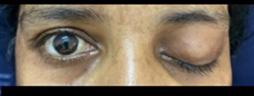
Case report
BMJ Case Rep: first published as 10.1136/bcr-2021-241663 on 27 April 2021. Downloaded from http://casereports.bmj.com/ on December 3, 2021 by guest. Protected by copyright.
Figure 1 Patient presented with left total ophthalmoplegia.
showed fungal colonies of broad aseptate hyphae at an obtuse
angle with periodic acid–Schiff stain, which was consistent with Figure 3 Cross-sectional image of MRI brain (DWI and ADC
mucormycosis. Fungal culture of the sample obtained after mismatch) showing acute infarct of the parieto-occipital lobe. ADC,
sinus debridement in FESS confirmed mucormycosis (Rhizopus apparent diffusion coefficient; DWI, diffusion weighted image.
species). However, serological fungal markers and fungal culture
sensitivity were not tested. The patient was hospitalised for 17 protons by transferrin in diabetic ketoacidosis and fungal heme
days. Her sugars were well controlled after the initiation of oxygenase, which promotes iron absorption for its metabolism.
insulin therapy. However, there was no resolution of ophthal- In a case described of severe COVID-19 associated with fungal
moplegia or ptosis until she was discharged. coinfection, cell counts revealed that there was a progressive
increase in white blood cell count and neutrophils while lympho-
TREATMENT cytes progressively decreased.9 It is hypothesised that SARS-CoV-2
She was initiated on conventional amphotericin B (given for infection may affect CD4+ and CD8+ T-cells, which are highly
11 days) and aspirin for acute cerebral infarct. Post FESS, CT involved in the pathological process of COVID-19 infection. It
paranasal sinus imaging was done after 1 week of treatment has been shown that in severe COVID-19 cases, there is a reduc-
with antifungal therapy and showed a reduction in the diffuse tion in the absolute number of lymphocytes and T-cells, which
opacification of the left ethmoid, frontal and maxillary sinuses is associated with the worst outcomes. Mucorales-specific T-cells
(figure 6). (CD4+ and CD8+) produce cytokines such as interleukin (IL)
4, IL-10, IL-17 and interferon-gamma (IFN-γ) that damage the
DISCUSSION fungal hyphae. Such specific T-cells were seen only in patients
An active search of literature reviewed few reported rhino- affected by invasive mucormycosis, and they concluded that
orbitary cases associated with COVID-19.3 5 6 Diabetes mellitus they could be a useful surrogate diagnostic marker of an invasive
is an independent risk factor for rhino-orbital–cerebral mucor- fungal disease. It might be speculated that lymphopenia could
mycosis in a meta-analysis of 600 series with 851 cases. The most increase the risk of developing invasive mucormycosis, while
common species isolated was Rhizopus species, with an overall the recovery of lymphocyte count could improve the adaptive
mortality of 46%.7 immune system and induce the production of Mucorales-specific
A case of COVID-19 with rhino-orbital mucormycosis coin- T-cells, which might have a role in controlling the invasive
fection associated with ketoacidosis was reported in a patient infection.
with recent- onset diabetes mellitus.8 Pathogenic mechanisms
involved in fungal aggressiveness include decreased phagocytic
activity, accessible amounts of iron due to the displacement of
Figure 2 Coronal section of CT paranasal sinus showing left ethmoid Figure 4 MRI brain showing periarterial inflammation of internal
sinusitis encroaching on the lamina papyracea. carotid artery (red arrowhead).
2 Revannavar SM, et al. BMJ Case Rep 2021;14:e241663. doi:10.1136/bcr-2021-241663Case report
BMJ Case Rep: first published as 10.1136/bcr-2021-241663 on 27 April 2021. Downloaded from http://casereports.bmj.com/ on December 3, 2021 by guest. Protected by copyright.
endothelial inflammation and vasoconstriction associated
with endothelial dysfunction put individuals with diabetes
at a greater risk for endotheliitis in several organs. Change
of vascular tone towards more vasoconstriction can lead to
subsequent organ ischaemia, tissue oedema and a procoagu-
lant state. Finally, dysregulated immune cell populations and
activity observed in individuals with diabetes play a critical
role in aggravating the severity.10
A case series in the Indian subcontinent reported six cases
of rhino- orbital–cerebral mucormycosis following COVID-19
infections.11 The mean duration between the diagnosis of
COVID-19 and the development of symptoms of mucormycosis
was 15.6±9.6 days. Control of hyperglycaemia, early treatment
with liposomal amphotericin B and surgery are essential for the
successful management of mucormycosis. Thus, the use of gluco-
corticoids in mild COVID-19 cases (without hypoxaemia) or the
utilisation of higher doses of glucocorticoids should be avoided.
Further, in the absence of a clear benefit, drugs targeting
immune pathways such as tocilizumab should be discouraged.
For successful management of mucormycosis, a high index of
Figure 5 Chest X-ray (PA view). PA, posteroanterior. clinical suspicion, low threshold for diagnosis in patients with
risk factors, neuroimaging and specific diagnostic tests with a
coordinated effort from a multidisciplinary team including
There are a significant number of reports showing alter-
ophthalmology, otorhinolaryngology, infectious diseases, neuro-
ations in cell-mediated immunity, such as chemotaxis, phago-
surgery, critical care, microbiology and pathology department
cytosis and cytokine secretion in both type 1 and type 2
are crucial. A delay of even 6 days in initiating treatment doubles
diabetics. Individuals with diabetes have been described
to have alterations in innate immune system components. the 30-day mortality from 35% to 66%.11
Natural killer cell activity is reduced in individuals with Simple tests like vision, pupil, ocular motility and sinus
diabetes, and more pro- inflammatory M1 macrophages tenderness can be part of routine physical evaluation of a patient
are present. Furthermore, T-cell activity is skewed. Disease with COVID-19 hospitalised with moderate to severe infection
severity in patients is due to not only the viral infection or diabetics with COVID-19 or those receiving systemic cortico-
but also the host response. Elevated glucose levels may also steroids. Visual prognosis, however, continues to remain poor.
suppress the antiviral response. In the context of COVID- Thus, it is important to have a high index of suspicion for
19, severe disease progression is described by a delay in fungal coinfection in patients with COVID-19 presenting with
IFN-γ response with a prolonged hyperinflammatory state comorbidities. Further, they should undergo immediate imaging
and lower CD4 and CD8 cell numbers. Regardless of the studies with an emphasis on the requirement of surgical inter-
involvement of the endothelial cells, the initial delay in IFN-γ vention. There is a need to stress on the judicious use of steroids
response together with the hyperinflammatory response in to avoid flaring up of the fungal infection.
individuals with diabetes may exacerbate the ‘cytokine storm’
and increase COVID-19 severity. Increased vascular lesions, Learning points
►► This case is an unusual presentation of rapidly developing
fungal infection in a patient with non-ketotic diabetes in the
background of COVID-19. Severe disease progression in the
absence of use of immunosuppressants makes it a rare case.
►► An alteration in the T-cell population in COVID-19 infection is
linked to the pathogenesis of fungal infection.
►► Early diagnosis and treatment of mucormycosis that involve
antifungal therapy and surgical debridement are necessary to
reduce mortality and prevent end-organ damage.
►► Judicial use of immunosuppressive therapy in COVID-19
infection should be considered particularly in regard to
treatment of fungal coinfections.
Twitter Shweta Mallikarjun Revannavar @ShwetaMR
Contributors All four authors were involved in patient care directly. Writing the
initial manuscript was done by SMR. Selecting appropriate image templates was
done by VVK. The necessary corrections and final outcome of the article were done
under the guidance of SPS and LS.
Funding The authors have not declared a specific grant for this research from any
funding agency in the public, commercial or not-for-profit sectors.
Figure 6 Coronal section of CT paranasal sinus showing reduced Competing interests None declared.
opacification of the left ethmoid sinus. Patient consent for publication Consent obtained from parent(s)/guardian(s).
Revannavar SM, et al. BMJ Case Rep 2021;14:e241663. doi:10.1136/bcr-2021-241663 3Case report
BMJ Case Rep: first published as 10.1136/bcr-2021-241663 on 27 April 2021. Downloaded from http://casereports.bmj.com/ on December 3, 2021 by guest. Protected by copyright.
Provenance and peer review Not commissioned; externally peer reviewed. 4 Radulesco T, Verillaud B, Béquignon E, et al. COVID-19 and rhinology, from the
consultation room to the operating theatre. Eur Ann Otorhinolaryngol Head Neck Dis
This article is made freely available for use in accordance with BMJ’s website
2020;137:309–14.
terms and conditions for the duration of the covid-19 pandemic or until otherwise
5 Mekonnen ZK, Ashraf DC, Jankowski T, et al. Acute invasive rhino-orbital
determined by BMJ. You may use, download and print the article for any lawful,
mucormycosis in a patient with COVID-19-associated acute respiratory distress
non-commercial purpose (including text and data mining) provided that all copyright
syndrome. Ophthalmic Plast Reconstr Surg 2021;37:e40–80.
notices and trade marks are retained.
6 Werthman-Ehrenreich A. Mucormycosis with orbital compartment syndrome in a
ORCID iD patient with COVID-19. Am J Emerg Med. In Press 2021;42:264.e5–264.e8.
Shweta Mallikarjun Revannavar http://orcid.org/0000-0003-1574-9586 7 Jeong W, Keighley C, Wolfe R, et al. The epidemiology and clinical manifestations of
mucormycosis: a systematic review and meta-analysis of case reports. Clin Microbiol
Infect 2019;25:26–34.
8 Waizel-Haiat S, Guerrero-Paz JA, Sanchez-Hurtado L, et al. A case of fatal rhino-orbital
REFERENCES mucormycosis associated with new onset diabetic ketoacidosis and COVID-19. Cureus
1 Skiada A, Pavleas I, Drogari-Apiranthitou M. Epidemiology and diagnosis of 2021;13:e13163.
mucormycosis: an update. J Fungi 2020;6:265. 9 Pasero D, Sanna S, Liperi C, et al. A challenging complication following SARS-CoV-2
2 Prakash H, Chakrabarti A. Global epidemiology of mucormycosis. J Fungi infection: a case of pulmonary mucormycosis. Infection 2020:1–6.
2019;5:26. 10 Erener S. Diabetes, infection risk and COVID-19; molecular metabolism 39, 2020.
3 Mehta S, Pandey A. Rhino-Orbital mucormycosis associated with COVID-19. Cureus 11 Sen M, Lahane S, Lahane TP, et al. Mucor in a viral land: a tale of two pathogens.
2020;12:e10726. Indian J Ophthalmol 2021;69:244–52.
Copyright 2021 BMJ Publishing Group. All rights reserved. For permission to reuse any of this content visit
https://www.bmj.com/company/products-services/rights-and-licensing/permissions/
BMJ Case Report Fellows may re-use this article for personal use and teaching without any further permission.
Become a Fellow of BMJ Case Reports today and you can:
►► Submit as many cases as you like
►► Enjoy fast sympathetic peer review and rapid publication of accepted articles
►► Access all the published articles
►► Re-use any of the published material for personal use and teaching without further permission
Customer Service
If you have any further queries about your subscription, please contact our customer services team on +44 (0) 207111 1105 or via email at support@bmj.com.
Visit casereports.bmj.com for more articles like this and to become a Fellow
4 Revannavar SM, et al. BMJ Case Rep 2021;14:e241663. doi:10.1136/bcr-2021-241663You can also read