Cryo-SEM & Cryo-FIB/SEM Preparation - TECHNIQUES & APPLICATIONS www.emsdiasum.com - Electron Microscopy ...
←
→
Page content transcription
If your browser does not render page correctly, please read the page content below
Cryo-SEM &
        Cryo-FIB/SEM
          Preparation
                 TECHNIQUES & APPLICATIONS
          featuring the PP3010 Cryo-SEM/Cryo-FIB/SEM Preparation System

www.emsdiasum.com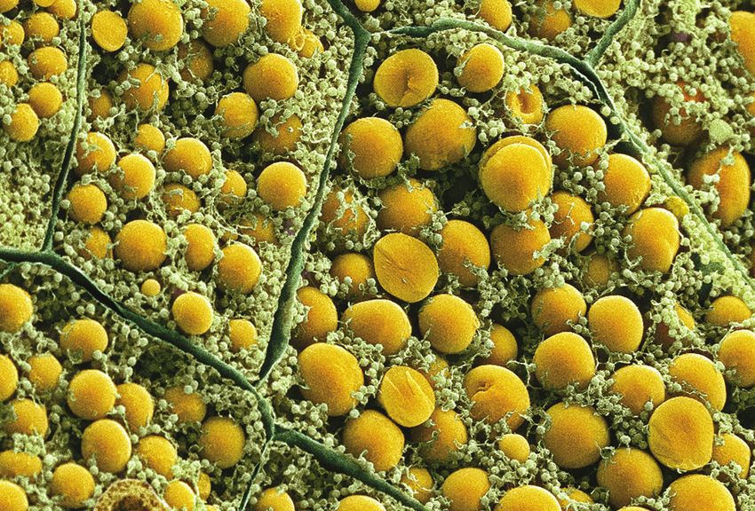
What is...                                                                         CRYO-SEM & CRYO-FIB/SEM PREPARATION EDITION II

Cryogenic Specimen                                 Techniques and Applications
Preparation?                                       Cryo-SEM — the advantages
In this instance we are referring to frozen
                                                   The Scanning Electron Microscopist is faced with           Why choose cryo-SEM?
hydrated bulk specimens for Scanning
                                                   the inescapable fact that liquid is a fundamental
Electron Microscopy (SEM), commonly                part of practically all lifesciences – and many
                                                                                                              The limitations of conventional ‘wet processing’
termed Cryo-SEM. When biological                                                                              include:
                                                   materials – specimens. Since water occupies up
specimens are prepared by alternative              to 98% of some animal and plant tissues it                 • Shrinkage and distortion
methods, such as critical point drying, they       represents a most formidable specimen problem              • Extraction of soluble materials
may collapse and distort due to the removal        to most Scanning Electron Microscopists.                   • Relocation of highly diffusible elements
of their water content. In addition diffusible     Cryo-SEM is a quick, reliable and effective way to         • Mechanical damage (fragile specimens can be
elements are often removed or relocated,           over come these not inconsiderable SEM                       damaged during conventional processing)
affecting the validity of subsequent X-ray         preparation problems. Additionally the technique           • Slow (24 hours or longer)
microanalysis.                                     is widely used for observing ‘difficult’ samples,
                                                                                                              • Toxic reagents are required (fixatives, buffers
                                                   such as those with greater beam sensitivity and
Cryo-SEM offers the best solution to this and                                                                   etc)
                                                   of an unstable nature. An important application,
in addition allows observation and analysis of     often overlooked, is the ability to use cryo-SEM to        Advantages of cryo-SEM:
liquid, semi-liquid and beam-sensitive             study dynamic processes (industrial or otherwise)          • Specimen viewed in fully hydrated state
specimens, such as emulsions, suspensions          by using a series of time resolved samples.                • Soluble materials are retained
and foams. Cryo preparation is increasingly        Naturally the advent of various “higher pressure”          • Less relocation of highly diffusible elements
being used with FIB/SEM instruments for a          modes, such as VP, LV and ESEM has allowed
                                                   such samples examined in SEM without resorting             • Little or no mechanical damage
wide range of specimen types, including
                                                   to freezing or drying methods. However, cryo-              • Time lapse experiments and evaluating
some materials where low temperature
                                                   SEM is still by far the most effective method of             industrial processes at timed intervals
milling conditions are desirable.
                                                   preventing sample water loss, which will in fact           • Usually no exposure to toxic reagents
For biological and other “wet” specimens           occur at any vacuum level –even with Peltier               • Rapid process
rapid freezing is essential in order to reduce     stages fitted to the SEM and the careful addition          • High resolution capability (compared to low-
morphological distortion, a key consideration      of water vapor in the SEM chamber. Cryo-SEM                  vacuum techniques)
for structural observation.                        also a number of a additional advantages,
                                                   including the ability to fracture and selectively          • Extra information obtained by low-temperature
The aim of fast freezing is to reduce the size     remove surface water (ice) by controlled                     fracturing (compared with conventional and
of ice crystals within specimens by reaching       specimen sublimation.                                        low-vacuum methods)
as quickly as possible the point at which                                                                     • Good for liquid, semi-liquids and beam
recrystallization takes place (for pure water                                                                   sensitive specimens
this is in the order of -130°C) and maintaining                                                               • Ability to selectively etch (sublimate to reveal
the specimen below this temperature during                                                                      information)
transfer, preparation and observation.                                                                        • Ability to ‘rework’ specimen (eg re-fracture and
                                                                                                                coat)
For larger specimens commonly used in SEM
and FIB/SEM rapid freezing is normally done
by plunging into liquid nitrogen in its ‘slushy’
form at -210°C. This is the standard method
supplied with the PP3010, but it is also fitted
with an “advanced specimen handling”               Learn how to do it...
system which allows specimens that have            Our EMS Microscopy Academy offers a Cryo SEM
been frozen by alternative (faster) freezing       Workshop for individuals who are new to the field of
                                                   cryo SEM, desire a technical refresher to maintain
methods to be manipulated and loaded
                                                   current skills, or just want to see and learn all of the
under liquid nitrogen and then transferred         possibilities of the technology.
under vacuum into the PP3010 for                   This course will cover the process of rapid freezing,
subsequent processing and observation.             fracturing, coating, and imaging of a variety of
                                                   samples.
                                                                                                                ...ENDLESS POSSIBILITIES
Cover Photos:                                      Main Curriculum                                             Our new academy is now open!
                                                   • Theory and overview of cryo SEM
                                                                                                               Located minutes from Philadelphia, next to our
                                                   • Mounting and adhering
                                                                                                               extensive warehouse in Hatfield, PA, we have
                                                   • Freezing, loading and fracturing
                                                                                                               many offerings led by our certified faculty,
                                                   • In situ coating
                                                                                                               including: ten educational workshops, corporate
                                                   • Operation of SEM
                                                                                                               training, group training, private training, and
                                                   • Cryo face-off Leica UC 7 Crion
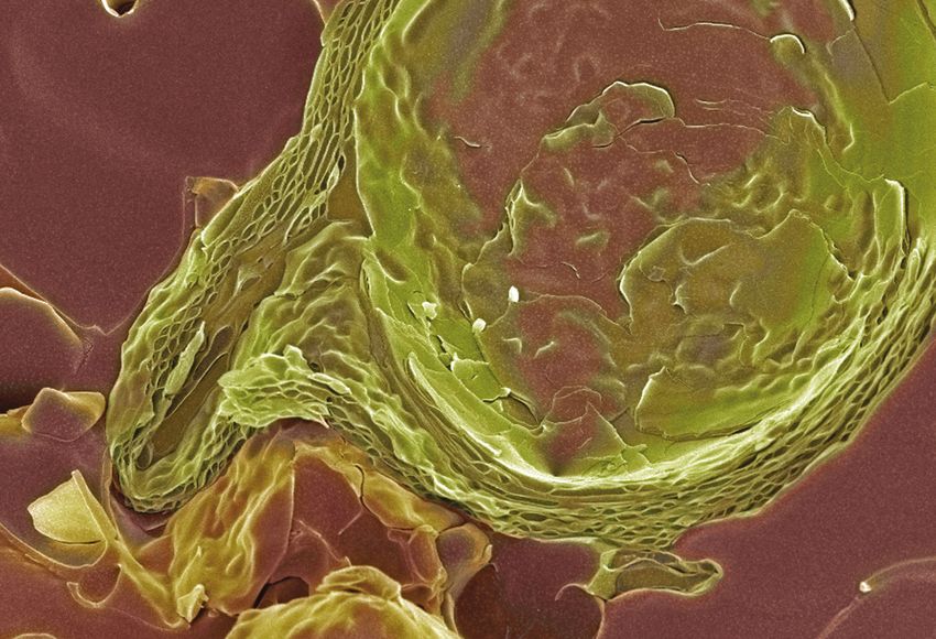
Hand Cream      Latex            Soya Bean Leaf    • Specific techniques of Cryo SEM imaging
                                                                                                               www.emsmicroscopyacademy.com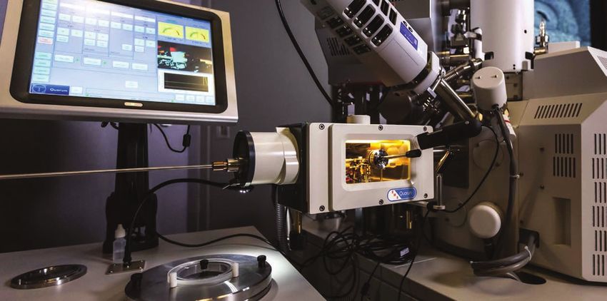
CRYO-SEM & CRYO-FIB/SEM PREPARATION EDITION II

Techniques and Applications
A summary of the cryo-SEM preparation technique
Cryo preparation techniques for scanning electron       phase products, such as ice cream, confectionery       Rivet mounting
microscopy (SEM) have become essential for the          and dairy products.                                    For liquids and for when
observation of wet or ‘beam sensitive’                  Botanical: Cryo-SEM is the perfect method for          specimens need to be frozen off
specimens. Using such techniques removes the            highly hydrated botanical material.                    the stub to achieve fast freezing
need for conventional preparation techniques,                                                                  rates. The rivet is placed in the
such as critical point drying or freeze-drying, and     Some specimen mounting
                                                                                                               hole and filled with liquid prior to
allows observation of the specimen in its ‘natural’     techniques for cryo-SEM
                                                                                                               freezing. If the specimen needs to
hydrated state.                                         Surface mounting                                       be frozen away from the stub, two
The specimen is rapidly cooled and transferred          This technique is used for                             liquid-filled rivets are held together
under vacuum to the cold stage of the                   leaf specimens etc. Roughen                            and then frozen prior to transfer
preparation chamber, which is mounted onto the          stub surface with fine emery                           onto the stub.
SEM chamber. The preparation chamber is                 paper. Specimen is laid on
pumped either with a rotary pump (PP2000) or by         top of mounting media.
a specially designed turbomolecular pumping                                                                    Alternative rivet mounting method
                                                        Edge mounting
system (PP2000T). The specimen can be
fractured, sublimated (‘etched’) to reveal greater      This technique is used for                              Pipette liquid sample into         Place metal rivet or small piece
                                                                                                                  hole in sample stub              of plastic tubing on top of hole
detail, and coated with metal by sputtering or          edge observation and                                                                          (containing liquid sample).
with carbon by thermal evaporation.                     fracture. Roughen surface of                                                              Note: Small drop of “Super Glue”
                                                        stub with fine emery paper.                                                               can be used to hold tube to stub.
Finally, the specimen can be moved under vacuum         Specimen is placed on its
into the SEM chamber where it is easily located on      edge in a machined slot and
a cold stage specifically tailored to the SEM. At all   secured with mounting
stages of the procedure the specimen is                 media.
maintained at a ‘safe’ temperature of typically lower
than -140°C.                                            Film emulsion mounting
                                                        This technique is useful when
Typical applications
                                                        a small specimen would be
Biological sciences including botany, mycology,         obscured by the Tissue-Tek
zoology, biotechnology and biomedical – plus            mounting media, or when specimens need to be                                                               Pipette liquid sample
economically import agricultural sciences.              recovered. Specimens need to be slightly damp                    Freeze & transfer onto                          into tube
                                                                                                                          preparation chamber
More recently cryo-SEM is becoming an essential         to use this method (good for nemotode worms).                          cold stage
tool for pharmaceutical, cosmetics and healthcare       The specimen is laid on surface so that its
industries, where it is used in basic applied           dampness slightly dissolves the film emulsion
research and for routine QA of many products,           allowing the specimen to adhere to the film
such as creams, cosmetics and drug delivery             surface. Exposed unused film with the emulsion
systems.                                                side uppermost is secured to the stub with
Cryo-SEM has long been a standard preparation           mounting media. It may be useful to scrape off                                       Result: clean surface fracture
method in the food industry. Of interest are multi-     the protective coating of the film emulsion first to
                                                        assist conductivity.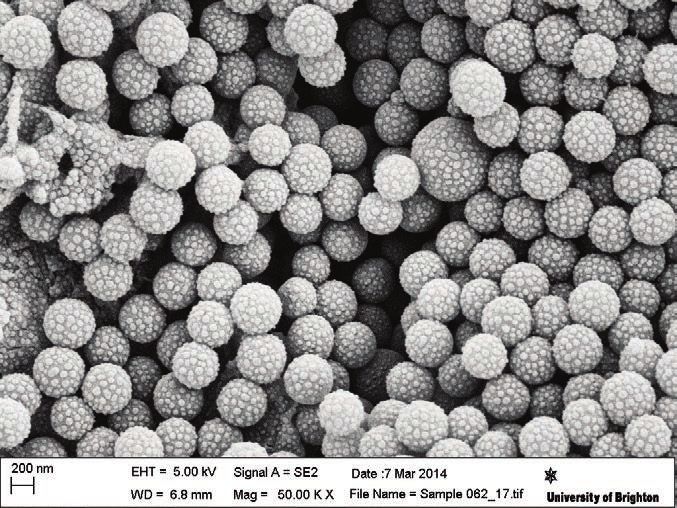
CRYO-SEM & CRYO-FIB/SEM PREPARATION EDITION II

                                              zzz PP3010 Cryo-SEM/Cryo-FIB/SEM Preparation System
                                              KEY FEATURES
                                              z High resolution performance on SEM, FE-SEM and FIB/SEMs
                                              z Totally gas cooled, including cryo preparation chamber – no boiling nitrogen on the SEM
                                              z Efficient cooling (down to at least -190°C)
                                              z 24 hours plus run times on one fill of LN2 are typical – allowing unattended overnight
                                                operation (at typical operating temperatures)
                                              z Large recipe driven touch-screen interface
                                              z Automated sublimation, coating and system start up
                                              z Superb specimen visibility (including preparation chamber CCD camera)
OVERVIEW                                      z Fully compatible with SEM beam deceleration/stage bias modes up to 5kV
The PP3010 is a highly automated, easy-       z Off column cooling and pumping systems – minimum mass on the SEM
to-use, column-mounted, gas-cooled cryo       z On-screen data logging and diagnostics
preparation system suitable for most makes    z Pumped storage of the cryo transfer device
and models of SEM, FE-SEM and FIB/SEM.        z Prepdek® workstation – self contained work area, extra bench space not required
The PP3010 has all the facilities needed to   z Cryo workflow options
rapidly freeze, process and transfer          z Specialist support and three-year warranty
specimens. The cryo preparation chamber       PRODUCT FEATURES
is turbomolecular pumped and includes         Mounting, Freezing and Transferring Specimens —
tools for cold fracturing, controlled         easy with the Prepdek® Workstation
automatic sublimation and sputter coating.    The PP3010 Prepdek® workstation is fitted with a slushy
After processing, the specimen is             nitrogen freezing station, connected to the pumping system.
transferred from the cryo preparation         Rapid freezing reduces ice crystal damage and results in
chamber onto a highly stable SEM cold         improved specimen preservation. For handling pre-frozen
stage for observation. Cold trapping in the   material the Prepdek® freezing system allows specimens that
cryo preparation chamber and SEM              have been frozen by alternative freezing methods (or stored field
                                              specimens) to be manipulated — in or just above liquid
chamber ensures the whole process is
                                              nitrogen — and then transferred under vacuum into the
frost-free. Specimen process times are        PP3010 preparation chamber for subsequent processing and                Slushy nitrogen freezing station
typically between five and ten minutes.       observation.
                                              Additionally the TEM Prep Slusher and Glove Box Interface/Airlock options allow workflow amongst a
                                              range of other platforms, including cryo-TEM, cryoultramicrome, XPS and glove box.
CRYO-SEM & CRYO-FIB/SEM PREPARATION EDITION II

zzz PP3010 Cryo-SEM/Cryo-FIB/SEM Preparation System (continued)
Cryo Transfer Device – Including Vacuum Storage
The vacuum transfer device is compact (fits easily into one hand), reliably
vacuum-tight and has a bayonet connection to the specimen shuttle to
ensure rapid pick up and transfer.
Set into the Prepdek® work surface is a pumped storage tube for the cryo
transfer device (see Prepdek® workstation section below).
Specimen Stubs, Shuttles
The PP3010 is supplied with universal 10 mm specimen stubs with surface
slots, holes and a flat area – useful for most specimen types. Blank and
slotted stubs are also included. In addition a range of optional holders is
available, including shuttles for large specimens and top-loading holders for
high pressure freezing, TEM Autogridstm (for cryo-FIB/SEM applications)
and clamping shuttles for hard specimens.
Cryo Preparation Chamber                                                                                           Specimen transfer device
The cryo preparation chamber is connected directly to the microscope and
includes a highly efficient nitrogen gas cold stage, extensive cold trapping
and facilities to fracture, sublimate and sputter coat specimens. The
chamber is fitted with two fully integrated and interlocked gate valves. The
outer load-lock valve includes a pumped airlock which accepts the cryo
transfer device – the inner SEM valve ensures rapid high-vacuum to high-
vacuum specimen exchange.
Highly Efficient Gas Cooled Stage and Cold Traps
At the heart of the cryo preparation chamber is a nitrogen gas cooled
specimen stage. The stage has a dovetail fitting to accept a cryo shuttle and
can be precisely controlled over a temperature range from 100°C to -190°C
or lower. Large gas cooled cold traps located above, below and behind the
specimen stage ensure clean, high vacuum conditions in the chamber.
Both the cold stage and cold traps are cooled with the fully integrated
CHE3010 off-column cooling system (see below), which at normal
operating temperatures give typical hold times of at up to 24 hours
between fills (provided the nitrogen gas is dry).
High Visibility – Plus CCD Camera
There is superb visibility into the preparation chamber. In addition to the
large front window (75 x 150 mm), there are two top viewing ports. The
chamber is lit by three LEDS and a CCD camera allows the specimen cold                                         View during specimen transfer
stage area to be viewed on the control screen and the images saved.
An optional stereo microscope can be fitted to the cryo preparation chamber.
Cold Fracturing
Twin fracturing tools manipulators (actively cooled) are available and allow
a range of specimen types to be cold fractured.
The PP3010 is fitted as standard with a front mounted fracturing and manip-
ulation device. The ball-jointed mount offers flexible movement of the blade
which can be used both as a surface pick (probe) and a fracturing knife.
An optional micrometer-advanced fracturing tool with rigid blade is
available, in addition to the standard front-mounted tool.
Fractured fragments are captured in the large cold trap located below the
specimen stage.
Automatic Sublimation and Sputtering
Sublimation temperatures and times can be preset and stored for easy
retrieval. The process is fully automatic and graphically displayed on the
control screen, showing the actual verses the predicted temperature curves.
The high resolution sputter coater is based on the market leading series of
bench top coaters. The coating system will give fine grain films essential            Front-mounted fracturing and specimen manipulation tool
for FE-SEM applications. A platinium target is fitted as standard – optional
metals include gold, gold/palladium, chromium and iridium. An optional
fully integrated carbon fiber evaporation head can also be fitted.
An optional terminating film thickness monitor is available.
CRYO-SEM & CRYO-FIB/SEM PREPARATION EDITION II

                                                         zzz PP3010 Cryo-SEM/Cryo-FIB/SEM Preparation System (continued)
                                                         Turbomolecular Pumping – High Vacuum
                                                         Performance
                                                         The preparation chamber is pumped by a
                                                         remotely-positioned 70 L/s turbomolecular
                                                         pumping system. Typical preparation chamber
                                                         vacuums when cold are in the region of 10-7mbar
                                                         or better. Positioning the turbomolecular pump
                                                         away from the SEM ensures total elimination of
                                                         mechanical vibration and has the advantage of
Mayonnaise                                               significantly reducing the total cryo system mass
Image courtesy of FEI Company.                           connected to the SEM. A vacuum buffer tank
                                                         (remotely located in the Prepdek®) is
                                                         automatically pumped when required. The
                                                         pumping system is connected to the preparation
                                                         chamber by flexible stainless-steel bellows, which
                                                         allows flexible positioning of the pumping system.
                                                         A 5 m3/hr rotary vacuum pump is required to "back"
                                                         the turbomolecular pump and for slushing and
                                                         rough pumping operations. The rotary pump can be
                                                         located up to five meters from the system, allowing
                                                         remote location if required. Dry pumping alternatives
                                                         are available – see Ordering Information.
Shaving Cream                                            Prepdek® Workstation
Fractured at -140ºC, sublimated at -90ºC for 3 minutes   The Prepdek® workstation has been designed to
and coated with 5nm of platinum.                         allow specimen mounting, freezing (plus pre-
                                                         frozen specimen manipulation) and transfer
                                                         device storage on one ergonomically designed
                                                         work surface. The control electronics are
                                                         mounted in a sealed but accessible cabinet
                                                                                                                 Remotely mounted turbomolecular pumping system
                                                         beneath the Prepdek®.
                                                         Set into the work surface is a pumped storage
                                                         tube which allows the cryo transfer device to be
                                                         stored under vacuum conditions when not in use.
                                                         Panel PC Touch Screen User Interface
                                                         The PP3010 is controlled using a large touch
                                                         screen panel PC, mounted on the Prepdek®
                                                         workstation. User-defined 'recipes' can be
Face Cream                                               entered and stored for instant future access. The
                                                         screen can be set to suit operator preferences;
Anti-aging face cream. Specimen rapidly frozen in
                                                         for example, vacuum measurement can be
slushy nitrogen, fractured at -140°C and sputter
coated with 5nm of platinum                              displayed in millibar, Pascal or Torr.
                                                         Many of the key steps in the specimen preparation
                                                         process are automated (system set up, gas flow
                                                         control, sublimation and sputter coating).              When not in use, the cryo transfer device can be
                                                                                                                 stored under vacuum in the pumped storage tube,
                                                         SEM Cold Stage, Cold Trap and Cooling System            located on the Prepdek® work surface
                                                         A highly stable, thermally isolated, nitrogen gas-
                                                         cooled stage attaches to the SEM stage. The
                                                         SEM stage and cold trap are cooled by separate
                                                         cold gas circuits – both capable of reaching
                                                         temperatures of -190°C or lower. This
                                                         configuration allows the operator to select stage
                                                         and cold trap temperatures that are optimized for
                                                         specific specimens. For example, for some non-
                                                         biological materials, it is useful to hold the
                                                         specimen at very low temperatures – for
                                                         example, a cold stage temperature of -175°C.
                                                         This is possible with the PP3010, as cold trap
                                                         temperatures of -190°C or lower can be
                                                         selected, but not possible with conduction cooled       Typical screen view during operation (with camera
                                                                                                                 image minimized)
CRYO-SEM & CRYO-FIB/SEM PREPARATION EDITION II

zzz PP3010 Cryo-SEM/Cryo-FIB/SEM Preparation System (continued)

                                                     OPTIONS AND ACCESSORIES
                                                     Rapid Warming Tower for the CHE3010
                                                     This device is meant to facilitate a rapid warming of the CHE3010
                                                     (Cryo-Heat Exchanger) associated with the PP3010 Cryo-SEM
                                                     preparation system. The Rapid Warming Tower in conjunction with a
                                                     1 liter/min Nitrogen gas flow rate dramatically reduces the lag time
                                                     before the SEM or Prep chamber components return to room
                                                     temperature so venting can occur. A process that normally takes
                                                     hours can be completed in minutes, allowing maintenance or
Gas-cooled SEM cold trap (temperatures down to
                                                     reconfiguring the SEM back to room temperature operation. Accepts
-190°C). Tailor-made to suit each SEM                many common, locally obtained hair dryers. (Hair Dryer not included).
                                                                                                                                         Rapid Warming Tower
                                                     Specimen shuttles and stubs
systems. The SEM cold stage has a temperature        The PP3010 is supplied with a selection of holders, and a range of
range of down to -190°C and a temperature            additional specimen shuttles and stubs is also available. (See
stability of < 0.5°C.                                Ordering Information for details).
Compatibility with SEM Stage Bias Mode               Carbon Evaporation and Film Thickness Monitor
The PP3010 cold stage is fully compatible with       A carbon evaporation attachment and a terminating film thickness
SEM stage bias/beam deceleration modes of up         monitor can be fitted. Both are fully integrated (no external control                       Specimen shuttle
to 5kV.                                              boxes required).
CHE3010 Off-Column Cooling                           Pressurized LN2 Dewar
The CHE3010 is a fully integrated, remotely          The PP7450/60L is a highly recommended option that generates dry
mounted cooling system which comes as                nitrogen gas used for cooling the SEM cold stage & cold trap and cryo
standard with every PP3010. The CHE3010 is           preparation chamber and cold shields. In addition, LN2 can be
used to cool the SEM stage, SEM cold trap and        decanted for slushing (freezing). During normal operation the
cryo preparation chamber cold stage and cold         PP7450/60L will generate dry nitrogen gas for up to eight days usage.
traps and will typically reach temperatures down     If the PP7450/60L is not included, appropriate, locally sourced,                        Gas cooling dewar
to -90°C or lower.                                   nitrogen gas cylinders can be used. It is important to ensure that it                     and turbo pump
The CHE3010 is remotely positioned (typically on     has low moisture content – if in doubt, please contact us.
the floor behind the microscope) and at normal       Cryoflow Work Options
operating temperatures can run for up to 24
hours between fills. This greatly simplifies the     Glove Box Valve/Airlock Interface
                                                                                                                                 Glove box
cryo process (no more checking on dewar status       The airlock is connected to a glove box using a generic NW fitting
                                                                                                                                 valve/airlock
and topping off), but also allows overnight,         and for most applications requires a suitable pumping system (rotary        interface
unattended operation – particularly useful for       pump or turbomolecular plus diaphragm pumping system).
some automated FIB/SEM "slice and view"              The airlock is designed to accept the PP3010 vacuum transfer device.
protocols.                                           For full details of this and other accessories please see Ordering
Single Port Interface to the SEM or FIB/SEM          Information.
Where SEM geometry allows, both the cryo             TEM Prep Slusher Option
preparation chamber and the SEM cooling              Offers an easy, convenient way of transporting pre-frozen specimens to
system can be fitted to a single chamber port        and from the PP3010.
(the minimum port diameter is 38 mm). This                                                                                       Cryo preparation chamber with
gives a tidy installation and frees up a valuable    • Conveniently locates into the Prepdek® workstation of the PP3010
                                                                                                                                      cryo transfer device fitted
chamber port.                                        • Freely transportable, e.g. between high pressure freezer,
                                                       cryoultramicrotome and the PP3010
                            Rotary Cold Stage for    • Ideal for loading/unloading TEM grids and grid holders
                          continuous 360 degree
                                                     • Tilting holder for cryo shuttles (holders). Allows the easy transfer of
                           rotation below -165˚C
                                                       specimens from external freezers (e.g. high pressure, jet, slam etc)
                                                     • Option for PP3010 and previous PP3000T and PP2000/PP2000T
                                                       models
                                                     Pre-Frozen Sample Loading Jig for PP3010                                    Pre-Frozen Sample Loading Jig
                                                     This loading jig allows you to comfortably load pre-frozen samples, ie:
                                                     HPF or Rivets, into the PP3010 specimen shuttles (not included). The
                                                     loaded shuttles can then be transferred to the PP3010 slushing pot
                                                     on the Prepdek®. Includes the LN2 container, Lexan cover, loading jig,
                                                     and a short transfer rod.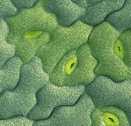
CRYO-SEM & CRYO-FIB/SEM PREPARATION EDITION II

                                              SPECIFICATIONS                                                                                     Standard?
                                              Cryo Preparation Chamber (column-mounted)
                                              Gas cooled preparation chamber with a twenty-four hour run time between fills                         Yes
                                              Two integral gate valves (loading and SEM) with appropriate electrical interlocks                     Yes
                                              Variable temperature gas-cooled specimen stage                                                        Yes
                                              Large cold shield above, below, behind the cold stage                                                 Yes
                                              Robust micrometer-fed fracturing knife (actively cooled)                                            Option
                                              Side-mounted surface knife/probe (actively cooled). A range of
                                              scalpel blades can be fitted to suit different specimen requirements                                    Yes
Cross-section of oil/water/rock.              Automatic sublimation (controlled and viewed on the touch screen)                                       Yes
                                              Fully automatic, high resolution sputter coater with platinum (Pt) target.
                                              (Other targets, including gold (Au), gold/palladium (Au/Pd), chromium (Cr) and iridium (Ir),
                                              are available as options.) Sputtering controlled and viewed on the user touch screen                  Yes
                                              Carbon fiber evaporation head and power supply                                                      Option
                                              Large front viewing window (150 x 78mm) plus top viewing ports                                        Yes
                                              Preparation chamber camera (CCD)                                                                      Yes
                                              Vacuum transfer device                                                                                Yes
                                              Chamber illumination — three LEDs                                                                     Yes
                                              Pumping System and Controls
                                              Remotely-mounted turbomolecular pumping system (70L/s). Includes: vacuum buffer tank,
                                              vacuum valves and stainless-steel bellows connection to the preparation chamber.
                                              Typical preparation chamber vacuum when cold: 10-7 mbar                                           Yes
                                              Single 50L/m rotary pump required                                                     Order separately
                                              SEM Cooling Dewar, SEM Cold Stage and Cold Trap
Cryo prepared image of blue stilton cheese    (anticontaminator)
(Penicillium roqueforti).                     Gas-cooled nitrogen cold stage assembly (-190°C). Temperature stability of >0.5°C                       Yes
                                              Separate gas-cooling circuits for SEM stage and SEM anti-contaminator                                   Yes
                                              21L capacity, off-column cooling dewar with run time between fills of up to 24 hours                    Yes
                                              SEM CCD camera-fitted when space allows                                                                 Yes
                                              LED lighting (interlocked)                                                                              Yes
                                              System Control and Specimen Handling
                                              Control via a color user touch screen monitor (15") mounted on the Prepdek®                             Yes
                                                 • Multi-ability user interface screen
                                                 • Quick, easy overview of system status
                                                 • User-definable “recipes” can be stored
                                                 • Quick access to videos outlining preparation techniques and system maintenance
                                                 • Fully automatic sputtering
                                                 • Automatic sublimation
                                                 • Quick, easy overview of system status
                                                 • CCD camera image of preparation chamber
Cross-section through plant palisade cells.   Twin liquid nitrogen slushing and specimen handling system — ideal for handling pre-frozen specimens.
                                              Mounted on the Prepdek®                                                                                 Yes
                                              System electronics stored in a ventilated, sealed unit under the Prepdek®                               Yes
                                              Specimen shuttles (x2). E7449-9 multi-specimen stubs (pack of 10) and E7402 blank aluminium (Al)
                                              stubs (pack of 10). Other shuttles and stubs available – see Ordering Information                       Yes
                                              Specimen Shuttles and Stubs (Others available — see Ordering Information)
                                                 • (2) Specimen shuttles (to hold 10 mm diameter cryo stubs)
                                                 • Blank 10 mm stubs – pack of 10
                                                 • Multi-stubs 7 mm high (with holes and slots) – pack of 5
                                                 • Multi-stubs 5 mm high (with holes and slots) – pack of 5
                                                 • Dovetail holder shuttle
                                                 • Brass rivets for fracturing liquids – pack of 100
                                                 • Copper (Cu) stub with 3 mm x 3 mm slot – pack of 5
                                                 • Copper (Cu) stub with 1 mm x 3 mm slot – pack of 5                                                 Yes
                                              Installation and Training
                                              Installation and training at the customer site                                                 Contact EMS
Cross-section image through sunscreen
                                              Support and Other Information
                                              Comprehensive start-up kit with key spares                                                              Yes
                                              Three-year warranty                                                                                     Yes
                                              SEM column interfaces and SEM stage adaptor (tailored to each microscope)                               Yes
                                              Some Options and Accessories (see Ordering Information for full list)
                                              Terminating film thickness monitor (FTM)                                                            Option
                                              Self-pressurizing LN2 dewar and regulator (for storage and venting)                                 Option
                                              Carbon fiber evaporation head                                                                       Option
                                              Wide range of specimen holders and specimen stubs                                                   Option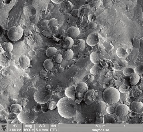
CRYO-SEM & CRYO-FIB/SEM PREPARATION EDITION II
zzz PP3010 Cryo-SEM/Cryo-FIB/SEM Preparation System (continued)
ORDERING INFORMATION
For a full quotation, including on-site installation and customer training, please contact us.
PP3010 Turbo Pumped Cryo System for SEM, FE-SEM and FIB/SEM. 25276-Q Shuttle for clamping hard flat specimens
Includes 13297 Sircal gas dryer. (clamps for upright and flat mounting) each
For a full detailed description please contact us each 10245 Cryo shuttle for high pressure freezer
(“Balzer”) planchette-style holders each
Pumping 13054 Shuttle for clamping hard, flat specimens.
PP3010 has an integrated turbomolecular pump, but also requires a Suitable for flat specimens (front of shuttle shown with
13034 rotary pump or 20063 oil-free scroll pump clamp lever) and cross-fracturing (sprung-loaded vice) each
13034 Pfeiffer DUO-6 5m3/hr rotary vacuum pump 20529 Top loading specimen holder shuttle (similar to
with oil mist filter each AL200077B but stub clamping mechanism is located
20063 Edward NXDS6i oil-free scroll pump on the top – useful for handling pre-frozen specimens
(replaces 13034 rotary pump) each mounted on a stub). One included with PP3010 each
Shipping 13419 Tilt-rotate shuttle. Tilt and rotation can be altered
on the preparation chamber cold stage using
13064 Wooden crate and packing for export each system knife/probe each
Installation and Training 20530 Top loading, vice style specimen shuttle for
directly clamping two 328116510 fracturing rivets each
PP3010-I Installation and training -
12406 TEM grid holder cryo shuttle, including cryo shield,
Four day on site installation and customer training
for cryo-FIB/SEM applications. Takes two TEM “Autogrids” each
PP3010-T Engineer costs - Travel from PA to customer site,
12922 Cryo shuttle for FEI Polara™ cartridge each
including local hotels
13359 STEM shuttle for 3mm TEM grid,
PP3010 Options and Accessories includes grid location tool. For STEM detectors
PP7450 60 liter pressurized liquid nitrogen dewar. mounted below the SEM cold stage each
Used for generating dry nitrogen gas required to cooling Specimen Holder Stubs (10mm dia.) For use with appropriate shuttles
the SEM stage and cold trap plus preparation chamber
E7433 10mm diameter specimen stub for freeze fracture rivets
cold stage and cold traps. If not ordered, cylinder gas
(fits into 10246) each
can be used (check purity requirement with EMS) each
E7402 Specimen stub, aluminum (10mm dia. X 5mm high) blank,
11000 Rotary Cold Stage: standard stage alternative, tilts up to 52°,
pkt of 10. One packet included with PP3010 each
rotates 360°, maintains desired cryotemperatures each
E7449-5 Multi-purpose specimen stub with slots and holes,
10996 Glove Box Interface/Airlock –
10 mm dia. X 7 mm high (pack of five).
for vacuum or inert gas transfer each
One pack included with PP3010 each
10997 TEM Prep Slusher option for transporting
11541 1 x multi-purpose specimen stub with slots and holes
pre-frozen specimens to and from the PP3010 each
(10 mm dia. X 5 mm high) blank.
PP3050-SLJ Pre-Frozen Sample Loading Jig for PP3010 each
Five included with PP3010 each
13297 Sircal nitrogen gas dryer 220-240V
E7402 Aluminum stubs, 10 mm dia. x 5 mm, high, blank,
13298 (100-110V part: 13298).
pkt of 10. One packet included with PP3010 each
Note: useful if quality of the nitrogen gas source
E7403 Specimen stub, copper (10mm dia. X 5mm high) blank,
is unknown. Included with PP3010 each
pkt of 10. each
PP7424 Binocular stereo microscope with fitting to the PP3010
E7405 Specimen stub for clamping thin, flat specimens x 1 each
cryo preparation chamber. Note: the PP3010 preparation
E7406 Specimen stub, copper with 3mm wide x 3mm deep slot,
chamber is fitted with a CCD camera as standard.
pkt of 5 each
Both PP7424 and CCD can fitted together each
E7407 Specimen stub, copper with 1mm wide x 3mm deep slot,
20996 Carbon fiber evaporation head and power supply,
pkt of 5 each
including 100cm of high purity carbon fiber. each
328116510 Rivets for liquid fracture (pkt 100).
12147 Terminating film thicknesss monitor (FTM) each
One packet included with PP3010 each
12145 Micrometer controlled fracturing device
with tooled steel blade. each Spares Kit
PP3050-RWT CHE3010 Rapid Warming Tower each 13061 Two-years consumable and basic spares kit for PP3010 each
12340-SPARE Cryo transfer device (spare) - one included with PP3010 each
Sputtering Targets and Carbon Fiber
Specimen Shuttles (dovetail). Some will accept 10mm dia.
(All targets 24.5 mm in diameter) - all other target types available upon request
AL200077B Standard specimen shuttle with 10m hole for cryo stub. E7400-314A Gold (Au) sputtering target, 24mm dia. X 0.2mm thick each
Two included with PP3010 each E7400-314B Gold/Palladium (Au/Pd) sputtering target,
12434 Blank specimen shuttle for large specimens. 24mm dia. X 0.2mm thick each
Total area: 290 mm2 each E7400-314C Platinum (Pt) sputtering target, 24mm dia. X 0.2mm thick.
20718 Blank specimen shuttle with extended length One included with the PP3010 each
for large specimens. Total area: 350 mm2 each E7400-314IR Iridium (Ir) sputtering target, 24mm dia. X 0.3mm thick each
20720 Shuttle for large, flat specimens E7400-314CR Chromium (Cr) target 0.3 mm thick each
(with light microscope-style stage clips) each 91047-1 Carbon fiber cord - standard grade (1M) each
13524 Shuttle for clamping hard, flat specimens. Suitable for 91047-5 Carbon fiber cord - standard grade (10M) each
flat specimens (front of shuttle with clamp lever) and
cross-fracturing (sprung-loaded vice at rear of shuttle). each
12013 For clamping, larger flat specimens
(handle removed after loading specimen).
Useful for loading pre-frozen specimens each
CRYO-SEM & CRYO-FIB/SEM PREPARATION EDITION II

zzz Cryo Transfer Systems
Building on the success of the PP3010 cryo-SEM/FIB/SEM preparation system, we are pleased to announce three new
related products for ambient and cryo temperature transfer.

                                               PP3004 QuickLok
                                               Ambient temperature airlock for SEM, FIB/SEM, beamline and vacuum platforms
                                               Quick Overview
                                               The QuickLok provides a rapid way of transferring ambient temperature specimens into SEM,
                                               FIB/SEM or other suitable vacuum systems. A key feature of the QuickLok is the ability to vacuum
                                               transfer specimens that are sensitive to normal environmental conditions. The transfer device uses a
                                               sealed vacuum chamber which can be interfaced to a glove box for inert gas transfer or allow
                                               vacuum transfer from a wide range of platforms.
                                               Key Features
                                               z Rapid specimen exchange
                                               z Vacuum and inert gas transfer
                                               z Field-retrofittable to most systems
                                               z Upgrade path to CoolLok
                                               z Custom designed holders available
PP3004 QuickLok                                                                                  Specimen Holder Examples
                                               z 3 year warranty

                                               Components
                                               Mounted onto a suitable vacuum chamber
                                               port, the QuickLok consists of a loading
                                               chamber body with integrated controls for
                                               pumping, venting and transfer. A custom-
                                               designed interface flange and connections to
                                               the pumping system are included (see
                                               Pumping below).
                                               The compact vacuum transfer device has an
                                               easy-release bayonet fitting to a dovetail-
                                               profile specimen holder (shuttle). Standard
                                               shuttles are included, but optional holders
                                               allow a range of specimen types to be
                                               handled.
                                               Inside the microscope is a stage to accept
                                                                                                 Simple controls for specimen exchange
                                               the specimen shuttle. To aid specimen
                                               exchange an interlocked LED chamber light
                                               is mounted to the inside of the QuickLok
                                               interface.
                                               Use
                                               The specimen is mounted on a suitable
                                               holder and the transfer device fitted onto the
                                               QuickLok. The airlock and transfer device
                                               are then evacuated to a pre-set vacuum and
                                               the gate valve opened. The specimen is then
                                               guided onto the microscope stage.
                                               For transfer from other vacuum systems, or
                                               a glove box, additional interface flanges are
                                               available on request.
                                               Pumping
                                               The QuickLok requires either a rotary pump
                                               or oil-free vacuum turbomolecular pumping         QuickLok specimen stage and adaptor to SEM
                                               station (see Options).
QuickLok and specimen transfer device
CRYO-SEM & CRYO-FIB/SEM PREPARATION EDITION II zzz Cryo Transfer Systems (continued) PP3005 SEMCool Non-airlock cryo cooling for SEM, FIB/SEM, beamline and vacuum platforms Quick Overview The SEMCool is based on the PP3006 CoolLok but without the PP3004 QuickLok components. It is designed for cryogenic applications where airlock exchange of specimens into the microscope is not required. Key Features z Temperature range down to -190°C, with z Independent cooling of cold stage and stability better than 0.5°C cold trap z Off-column cooling with all-day runtime z Upgrade path to CoolLok between fills z 3 year warranty Components PP3005 SEMCool Specimen holders and transfer device: The compact vacuum transfer device has an easy- release bayonet fitting to a dovetail-profile specimen holder (shuttle). Standard shuttles are included, but optional holders allow a range of different specimen types to be handled. Cold stage and cold trap: A highly stable, thermally isolated, nitrogen gas-cooled cold stage attaches to the microscope stage. The location and shape of the cold trap is tailored to suit the internal geometry of the microscope. Both cold stage and cold trap are capable of reaching temperatures down to -190°C with a stability of
CRYO-SEM & CRYO-FIB/SEM PREPARATION EDITION II

                                                           zzz Cryo Transfer Systems (continued)
                                                           PP3006 CoolLok
                                                           Cryo transfer systems for SEM, FIB/SEM, beamline and vacuum platforms
                                                           Quick Overview
                                                           The CoolLok offers rapid transfer and cryo temperature observation of specimens for SEM, FIB/SEM,
                                                           beamline or other vacuum systems. Applications include thermal protection of beam-sensitive
                                                           specimens and low temperature observation of materials such as plastics, polymers low-K dielectrics
                                                           and hard-soft mixtures. The system can also be used for inert gas transfer of ambient temperature
                                                           specimens from a glove box.
                                                           Please Note: The PP3006 is not a replacement for the PP3010, which is a full cryo preparation
                                                           system. The PP3006 does not have a cryo preparation chamber and is designed for materials
                                                           applications where cold fracturing and sputtering are not required.
PP3006 CoolLok
                                                           Key Features
                                                           z Rapid specimen exchange
                                                           z Temperature range down to -190°C with stability better than 0.5°C
                                                           z Off-column cooling with all-day runtime between fills
                                                           z Independent cooling of cold stage and cold trap
                                                           z Vacuum or inert gas transfer
                                                           z Rapid specimen freezing option
                                                           z 3 year warranty

                                                           Components
                                                           Vacuum airlock
                                                           cold gas feedthrough
                                                           Mounted onto a suitable
                                                           vacuum chamber port,
                                                           the CoolLok consists of a
                                                           loading chamber body
                                                           with built-in controls for
                                                           pumping, venting and
                                                           transfer. A custom-
                                                           designed interface flange
                                                           to the vacuum chamber
                                                           and connections and
                                                           fittings to the pumping
                                                           system are included (see
                                                           Pumping below). The                                                        PP3006 installation example
On-microscope components: airlock, cold stage, cold trap
plus cryo transfer device                                  interface has cold
                                                           nitrogen gas feeds to and from the microscope
                                                           cold stage and cold trap.
                                                           Specimen holders and transfer device
                                                           The compact vacuum transfer device has an
                                                           easy-release bayonet fitting to a dovetail-
                                                           profile specimen holder (shuttle). Standard
                                                           shuttles are included, but optional holders
                                                           allow a range of different specimen types to
                                                           be handled.
                                                           Cold stage and cold trap A highly stable,
                                                           thermally isolated, nitrogen gas-cooled cold
                                                           stage attaches to the microscope stage. The
                                                           location and shape of the cold trap is tailored
                                                           to suit the internal geometry of the
                                                           microscope. Both cold stage and cold trap are
Load lock with vacuum isolated gas cooling lines           capable of reaching temperatures down to -
CRYO-SEM & CRYO-FIB/SEM PREPARATION EDITION II zzz Cryo Transfer Systems (continued) PP3006 CoolLok (continued) 190°C with a stability of
CRYO-SEM & CRYO-FIB/SEM PREPARATION EDITION II

zzz Cryo Transfer Systems (continued)

SPECIFICATIONS for PP3004, PP3005, PP3006
                                            PP3004                                 PP3005                                         PP3006
Temperature                                 Ambient                               RT to -190°C                                RT to -190°C
Cooling Runtime                             N/A                                   Up to 24 hours                              Up to 24 hours
LN2 Dewar Capacity                          N/A                                   21 liters                                   21 liters
Cool-Down Time to -190°C                    N/A                                   Typically
CRYO-SEM & CRYO-FIB/SEM PREPARATION EDITION II
Wax crystals in gas oil
When cooled to a temperature below about 2°C, the
waxes in fuel oils such as this tend to crystallize out. Wax
crystal size and shape can be varied by altering the rate at
which the oil is cooled.
Stable emulsion of a hydrophobic polymer
This image illustrates a stable emulsion of a synthetic
liquid polymer dispersed in an aqueous continuous
phase.
Latex
Latex particles are very electron beam sensitive, so cryo-SEM is an ideal method for their observation
Dendritic Ice Crystals
If it is cooled slowly, water forms dendritic ice crystals.
These can have a variety of branching patterns — the
complexity of which depends upon cooling rate. Arms
extend from the main body of the crystal at an angle of
60°. Some, such as the one illustrated, resemble the
arms of a snowflake. Bar: 2umCryo-SEM Applications
Zoological Botanical
Frozen hydrated aphid Pollen of cactus Zygocactus truncatus
In comparison with the critical point dried aphid, Germinating pollen grains of Zygocactus truncatus.
this image shows that there is no distortion of the
abdomen nor any other parts of the aphid following
freeze drying.
Fungi Geological
Maize root starch granules
Baker’s yeast (Saccharomyces cerevisiae) Wax crystals in gas oil
The specimen was rapidly frozen in nitrogen slush, When cooled to a temperature below about 2°C, the
fractured and coated with 4nm of platinium (Pt). waxes in fuel oils such as this tend to crystallize out.
10nm yeast cell transmembrane particles (in Wax crystal size and shape can be varied by altering
hexagonal arrays) can be observed. the rate at which the oil is cooled.
Cryo-DualBeam Polymers
Arabidopsis plant Stable emulsion of a hydrophobic polymer
Cryo-FIB/SEM. Image courtesy of Hannah Edwards This image illustrates a stable emulsion of a
and Arabidopsis plants provided by Darren Wells, synthetic liquid polymer dispersed in an aqueous Predatory Mite, courtesy of USDA Beltsville,
Centre for Plant Integrative Biology, School of continuous phase. Dr Gary Buchan
Biosciences, University of Nottingham, UK.
P.O. Box 550 • 1560 Industry Rd.
TO REQUEST A COPY OF OUR CATALOG:
Hatfield, Pa 19440
www.emsdiasum.com/requests/catalog
Tel: (215) 412-8400
Fax: (215) 412-8450 TO VIEW OUR DIGITAL CATALOG:
email: info@emsdiasum.com catalog.emsdiasum.com
or stacie@ems-secure.com
OUR MAIN INTERACTIVE WEBSITE: ...OR SCAN OUR QR CODE...
www.emsdiasum.comYou can also read


