Diagonal Earlobe Crease (Frank's Sign) for Diagnosis of Coronary Artery Disease: A Systematic Review of Diagnostic Test Accuracy Studies - MDPI
←
→
Page content transcription
If your browser does not render page correctly, please read the page content below
Journal of
Clinical Medicine
Review
Diagonal Earlobe Crease (Frank’s Sign) for Diagnosis of
Coronary Artery Disease: A Systematic Review of Diagnostic
Test Accuracy Studies
Krzysztof Wi˛eckowski 1, *, Tomasz Gallina 1 , Andrzej Surdacki 2 and Bernadeta Chyrchel 2
1 Students’ Scientific Group at Second Department of Cardiology, Institute of Cardiology, Faculty of Medicine,
Jagiellonian University Medical College, 2 Jakubowskiego Street, 30-688 Cracow, Poland;
tomasz.gallina@student.uj.edu.pl
2 Second Department of Cardiology, Institute of Cardiology, Faculty of Medicine, Jagiellonian University
Medical College, 2 Jakubowskiego, 30-688 Cracow, Poland; andrzej.surdacki@uj.edu.pl (A.S.);
bernadeta.chyrchel@uj.edu.pl (B.C.)
* Correspondence: k.wieckowski@student.uj.edu.pl
Abstract: Coronary artery disease is a global challenge for healthcare systems. Early diagnosis
is a key issue to improve quality of life and reduce morbidity and mortality. Diagonal earlobe
crease, a wrinkle extending obliquely across the earlobe, was linked by many authors to various
atherosclerotic diseases. This systematic review aimed at summarizing the diagnostic accuracy of
diagonal earlobe crease for diagnosis of chronic and acute coronary syndromes in adults. Cochrane’s
recommendations for systematic reviews of diagnostic test accuracy studies were followed. The
protocol was registered on PROSPERO. Seven electronic databases were searched up to April 2021.
The risk of bias and applicability were assessed using the QUADAS-2 tool. Meta-analysis was
Citation: Wi˛eckowski, K.; Gallina, T.; not performed. Finally, 13 cross-sectional studies evaluating 3951 patients were analyzed, all of
Surdacki, A.; Chyrchel, B. Diagonal which focused on chronic coronary syndromes defined as anatomically significant coronary stenosis.
Earlobe Crease (Frank’s Sign) for Invasive coronary angiography was used as a reference in most studies, except one which utilized
Diagnosis of Coronary Artery computed tomography angiography. Sensitivity ranged from 26% to 90%, and specificity from 32%
Disease: A Systematic Review of
to 96%. Positive likelihood ratios varied from 1.11 to 7.03, but most results were below 2. Negative
Diagnostic Test Accuracy Studies. J.
likelihood ratios were from 0.84 to 0.30, but most values exceeded 0.5. Diagnostic accuracy of diagonal
Clin. Med. 2021, 10, 2799. https://
earlobe crease for the detection of chronic coronary syndromes is insufficient. It only slightly changes
doi.org/10.3390/jcm10132799
pre-test probability, and its mere presence or absence should not affect the clinical management of
Academic Editor: Michael Henein the patients. However, for its feasibility and easy interpretation, Frank’s sign could be considered as
a part of physical examination.
Received: 11 May 2021
Accepted: 22 June 2021 Keywords: coronary artery disease; coronary syndromes; diagonal earlobe crease; Frank’s sign
Published: 25 June 2021
Publisher’s Note: MDPI stays neutral
with regard to jurisdictional claims in 1. Introduction
published maps and institutional affil- Coronary artery disease (CAD) is one of the most common cardiovascular disor-
iations.
ders and one of the main global causes of death [1]. It usually develops as a result of
atherosclerotic obstruction of epicardial coronary arteries [2]. CAD leads to myocardial
ischemia and presents as chronic coronary syndromes (CCS) or acute coronary syndromes
(ACS) [3]. Early diagnosis is essential to provide an adequate treatment that reduces
Copyright: © 2021 by the authors. mortality, eliminates symptoms, and increases the quality of life [4].
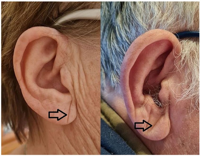
Licensee MDPI, Basel, Switzerland. Diagonal earlobe crease (DELC) is a wrinkle extending obliquely from the tragus
This article is an open access article towards the border of the earlobe, as seen in Figure 1. It was firstly described by Frank
distributed under the terms and in 1973 in the New England Journal of Medicine in a case series of patients with CAD [5].
conditions of the Creative Commons
Since then, there have been many reports published regarding its association mainly
Attribution (CC BY) license (https://
with atherosclerosis, especially CAD [6]. However, it is barely known and rarely used,
creativecommons.org/licenses/by/
likely because its clinical application has not been well established. Noteworthily, the
4.0/).
J. Clin. Med. 2021, 10, 2799. https://doi.org/10.3390/jcm10132799 https://www.mdpi.com/journal/jcm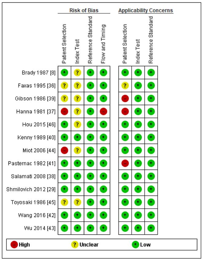
J. Clin. Med. 2021, 10, 2799                                                                                                  2 of 12

                                examination of DELC is effortless, non-invasive, and easy to interpret. It could be used in
                                primary care or emergency departments if its diagnostic accuracy is sufficient to support
                                decision-making.

       Figure 1. Diagonal earlobe crease in a woman without coronary artery disease and a man with coronary artery disease.

                                     In 2020, Stoyanov et al. published research investigating earlobes along with cardiac
                                samples in the autopsy study [7]. Histopathological examination of DELC-positive earlobes
                                showed myoelastofibrosis in the arterial vessel located at the base of the earlobe, fibrosis,
                                and Wallerian-like degeneration with eosinophilic inclusions in the peripheral nerves. The
                                authors stated that this location is a line of merging of preformed structures prenatally, and
                                thus it may be susceptible to chronic hypoxia–reoxygenation injury due to atherosclerosis.
                                Moreover, they revealed increased cardiac weight and left and right ventricular thickness
                                in DELC-positive patients, and no difference in age between groups. This study supports
                                the hypothesis that DELC is not a random finding but is directly linked to atherosclerosis.
                                Previous studies on the pathophysiology of DELC raised some other mechanisms of its
                                formation, including skin aging [8], collagen degeneration [9], or telomere shortening [10].
                                     We aimed at summarizing the diagnostic accuracy of the DELC for the diagnosis of
                                CCS and ACS in adults.

                                2. Materials and Methods
                                     This systematic review was reported according to the Preferred Reporting Items for
                                Systematic Review and Meta-Analysis [11]. Cochrane’s recommendations for systematic
                                reviews of diagnostic accuracy studies were followed [12]. The protocol was registered on
                                PROSPERO (CRD42021229551).
                                     We focused on two adult populations—patients with suspected CCS referred to further
                                cardiologic evaluation, and patients with suspected ACS who presented in the emergency
                                department. DELC was defined as a wrinkle extending obliquely from the tragus towards
                                the border of the earlobe, and studies that evaluated DELC as an index test were included
                                in the analysis. Reference standards had to agree with the present recommendations of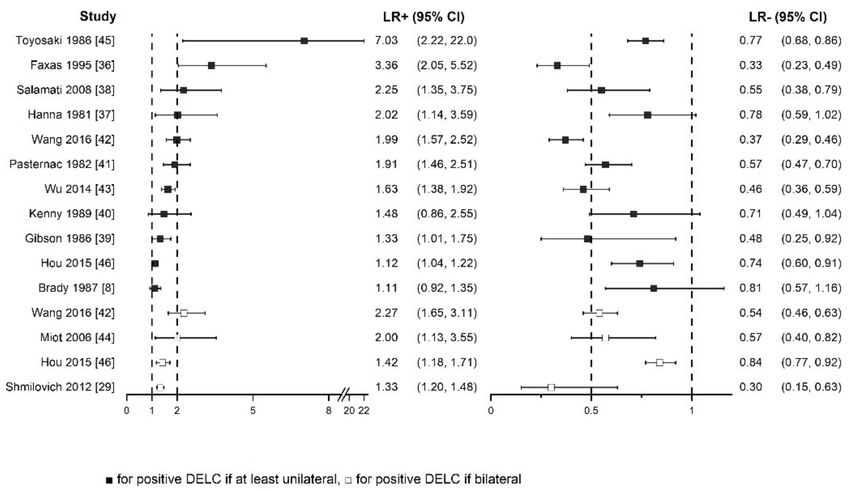
J. Clin. Med. 2021, 10, 2799                                                                                            3 of 12

                               The European Society of Cardiology [3,13] for diagnosis of CCS and ACS. Cross-sectional
                               studies were considered for inclusion, whereas case-control ones were excluded because of
                               the evaluation of nonrepresentative populations due to separate sampling of patients and
                               overestimation of diagnostic accuracy [12]. Only full-text publications were accepted.
                                    Seven electronic databases, including Pubmed MEDLINE, Embase, Web of Science,
                               Cochrane CENTRAL, CINAHL, Clinicaltrials.gov, and Scopus, were searched up to
                               8 January 2021 without any restrictions on the language or date of publication. The search
                               included the terms “Frank’s sign” and “earlobe crease” with variants. The full search
                               strategy is reported in Supplementary Materials File 1. Additionally, we manually screened
                               the references of all papers regarding DELC. The search was repeated on 13 April 2021,
                               and no new eligible papers were found.
                                    All titles and abstracts were screened, and potentially eligible papers were chosen
                               for full-text assessment, which resulted in the inclusion of studies according to the criteria
                               mentioned above. All relevant data, including study design, population, index test, refer-
                               ence standard characteristics, and study results were extracted. The Quality Assessment of
                               Diagnostic Accuracy Studies 2 (QUADAS-2) tool was used to assess the methodological
                               quality [14]. Every stage was preceded by a calibration round to ensure understanding of
                               the criteria and was performed by two reviewers (K.W., T.G.) independently. Any disagree-
                               ments were resolved by discussion and help from the third reviewer (A.S.) if necessary.
                                    Diagnostic accuracy of each study presented as sensitivity, specificity, and positive
                               and negative likelihood ratios (LR+ and LR−) with 95% confidence intervals (95% CI)
                               calculated from true positive (TP), false positive (FP), false negative (FN), and true negative
                               (TN) values. Analyses were carried out in Review Manager Version 5.4. (Copenhagen, The
                               Nordic Cochrane Centre, The Cochrane Collaboration, 2020) and OriginPro Version 2021
                               (OriginLab Corporation, Northampton, MA, USA).
                                    Separate analyses of diagnostic accuracy depending on some variables, including
                               sex, age, location, DELC definition, reference standards, and methodological quality were
                               planned to be done if relevant data were available. Due to the preliminary search as well
                               as analysis of previous reviews, we expected considerable heterogeneity between studies,
                               and the decision was made not to perform a meta-analysis.

                               3. Results
                                     The study selection flowchart is presented in Figure 2. From the 421 unique records
                               identified through database search, 34 full texts were assessed, and 13 studies were
                               finally included.
                                     Reasons for exclusion of full texts were as follows: not relevant population [9], ref-
                               erence standard [15–21], target condition [22], and case-control design [23–25]. Many
                               probable errors in methodology description and data presentation were present in the
                               research by Bawaskar et al., making it not possible to interpret [26]. Two records were
                               letters [27,28] and one was a subgroup analysis from another study included in our anal-
                               ysis [29,30]. Five studies published in the years 1979–1990 were not accessible [31–35].
                               Detailed explanations are presented in Supplementary Materials File 3.
                                     Characteristics of included studies are detailed in Table 1. The total number of patients
                               was 3,951. Six studies were conducted in North America, five in Asia, one in Europe, and
                               one in South America. Most of them were carried out on both sexes, whereas three included
                               only men. Various descriptions of the age of the population were used, and available data
                               are presented in Table 1. The total prevalence of DELC was 60.5% and varied from 17.0%
                               to 73.0%. The overall prevalence of CAD was 53.9% and ranged from 16.5% to 80.8%.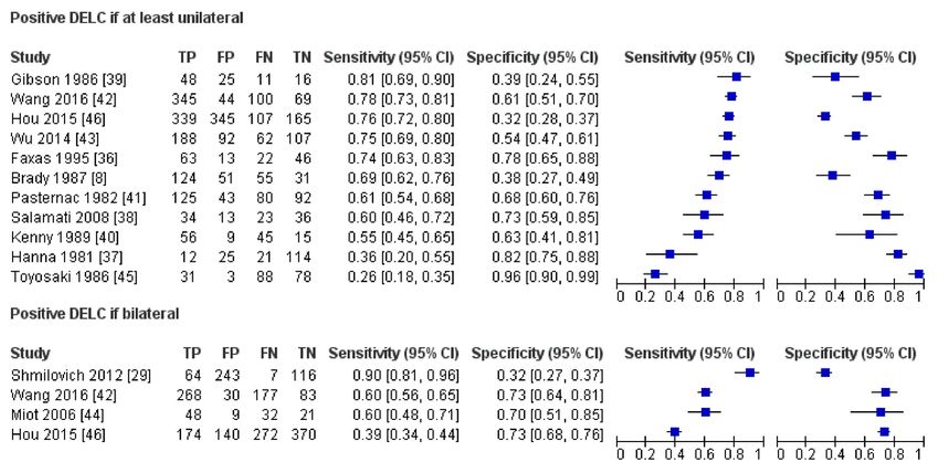
J. Clin. Med. 2021, 10, 2799                                                                                                           4 of 12

                                                      Figure 2. Flowchart of studies selection.

                                                     Table 1. Characteristics of included studies.

                                          Sample                                  DELC            CAD                              Reference
      Author (Year)            Country                Males        Age 1                                    DELC Definition
                                          Size (n)                              Prevalence     Prevalence                          Standard
                                                                 DELC+ 60
     Brady (1987) [8]           USA         261       100%                         67%               69%    At least unilateral      ICA
                                                                 DELC− 52
                                                                  Males 65
    Gibson (1986) [39]          USA         100        68%                         73%               59%    At least unilateral      ICA
                                                                 Females 70
     Faxas (1995) [36]          Cuba        144        NA           NA             53%               59%    At least unilateral      ICA
    Hanna (1981) [37]           USA         172       100%      94% below 50       22%               19%    At least unilateral      ICA
                                                                CAD+ 55 ± 9                                 At least unilateral,
      Hou (2015) [46]           China       956        57%                       72%, 33%            47%                             ICA
                                                                CAD− 51 ± 8                                      bilateral
     Kenny (1989) [40]         Ireland      125       90%       Range 35–90        52%               81%    At least unilateral      ICA
     Miot (2006) [44]           Brazil      110       100%        58 ± 12          52%               73%         Bilateral           ICA
   Pasternac (1982) [41]       Canada       340       74%          50 ± 8          49%               60%    At least unilateral      ICA
    Salamati (2008) [38]        Iran        106       66%         50 ± 14          44%               54%    At least unilateral      ICA
  Shmilovich (2012) [29]        USA         430       61%         61 ± 13          71%               17%         Bilateral           CTA
   Toyosaki (1986) [45]         Japan       200       76%       72% above 50       17%               60%    At least unilateral      ICA
                                                                                                            At least unilateral,
     Wang (2016) [42]           China       558       72%            64          70%, 53%            80%                             ICA
                                                                                                                 bilateral
      Wu (2014) [43]            China       449       62%         63 ± 12          62%               56%    At least unilateral      ICA
      1Mean age ± standard deviation or other if specified; CAD, coronary artery disease; CTA, computed tomography angiography; DELC,
      diagonal earlobe crease; ICA, invasive coronary angiography; NA, not available.

                                              Generally, most studies defined DELC as at least a unilateral sign; in two papers
                                         bilateral DELC was required to be considered as a positive, and another two presented
                                         results for both definitions. Some studies showed no more than information of diagonal
                                         creasing of earlobe without any other details [8,36]. In another study, only the word
                                         “significant” was added to the positive sign description [37]. One accepted any crease [38],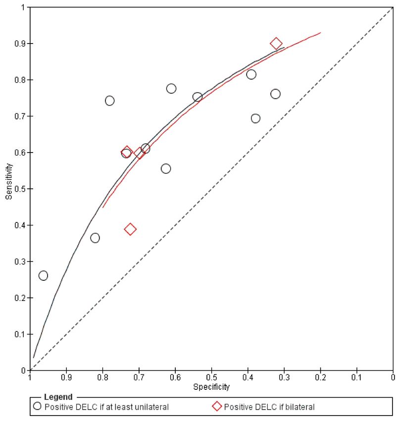
J. Clin. Med. 2021, 10, 2799                                                                                          5 of 12

                               whereas others provided detailed thresholds of the relation of the crease to the whole
                               length of the earlobe, which varied from more than one-third [39], at least half [40,41], at
                               least two-thirds [42,43], or 100% [29,44–46]. The combination of these approaches leads to
                               the finding that DELC was defined in nine different ways.
                                     All studies evaluated the diagnostic accuracy of DELC in the diagnosis of CCS. The
                               reference standard was invasive coronary angiography (ICA), except one that utilized
                               computed tomography angiography. In both, the threshold for anatomically significant
                               stenosis was 50%.
                                     Results of quality assessment using the QUADAS-2 tool are presented in Figures 3 and 4.
                               Detailed explanations of our decisions are provided in Supplementary Materials File 2.
                               Only five were judged as having a low risk of bias and concern of applicability in all do-
                               mains. The most important limitation was patient selection because of unclear indications
                               for performing ICA or inclusion of some patients that did not precisely match the review
                               question. The main issues, which resulted in reduced ratings in the index test domains,
                               were the unclear pre-specified definition of DELC and unclear blinding of the results of the
                               reference standards. Reference standard domains were rated as having the highest quality.
                               One study performed ICA and final analysis only on the subgroup of the total number of
                               patients, which resulted in a high risk of bias in the Flow and Timing domain.

                               Figure 3. Results of quality assessment of individual studies.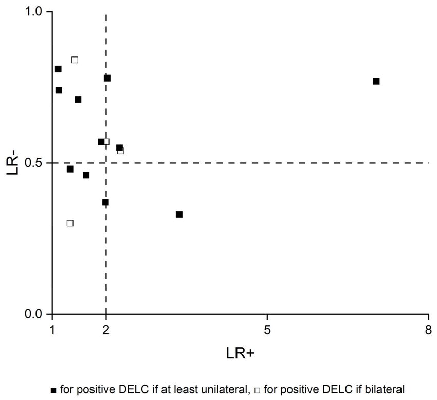
J. Clin. Med. 2021, 10, 2799                                                                                                       6 of 12

                                   Figure 4. Summarized results of quality assessment.

                                        Results of all included studies are detailed in Figures 5–8. TP, FP, FN, and TN values
                                   of each study with calculated sensitivity and specificity with 95% CIs are presented in
                                   Figure 5. Sensitivity and specificity were also summarized by the summary receiver
                                   operating characteristic plot in Figure 6. LR+ and LR− with 95% CI are presented in
                                   Figure 7 and are summarized in Figure 8.
                                        Due to the considerable heterogeneity, especially in the DELC definition, and the
                                   lack of raw data in many studies necessary to calculate diagnostic accuracy for subgroups
                                   of different sex and age, we decided not to perform additional analyses. In our opinion,
                                   such analyses of the mentioned subgroups as well as different locations or methodological
                                   quality would not change the final appraisal of the usefulness of DELC. Apart from that,
                                   we presented separately the results of studies that considered positive DELC when at least
                                   unilateral, and these where only bilateral DELC was accepted. Meta-analysis was not
                                   performed in this systematic review.

           Figure 5. Results of individual studies and forest plot of sensitivity and specificity with 95% confidence intervals.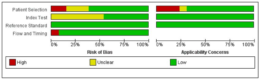
J. Clin. Med. 2021, 10, 2799                                                                                                     7 of 12

                               Figure 6. Summary receiver operating characteristic plot.

                               Figure 7. Forest plot of positive and negative likelihood ratios with 95% confidence intervals.
J. Clin. Med. 2021, 10, 2799 8 of 12
Figure 8. Scatter plot of positive and negative likelihood ratios.
4. Discussion
This systematic review aimed at summarizing the diagnostic accuracy of diagonal
earlobe crease for the diagnosis of chronic and acute coronary syndromes. Finally, only
studies evaluating CCS with anatomically significant stenosis of coronary arteries detected
mostly in invasive coronary angiography, except one which utilized computed coronary
angiography, were eligible. During the diagnostic process of CCS, the pre-test probability
of obstructive CAD is assessed basing on medical history, physical examination, and
additional tests to choose an appropriate final invasive or non-invasive test. Since ICA is
offered to patients with a high clinical likelihood of disease, the results of this systematic
review should be mainly referred to this population and carefully generalized to other
patients [3].
Sensitivity and specificity showed notable heterogeneity, whereas calculated LRs+ and
LRs− were more consistent. Generally, LR+ above 2 and LR− less than 0.5 are values that
increase or decrease pre-test probability approximately by 15 percentage points or more.
These are the most used cut-off points, which set the threshold of informativeness of the
diagnostic tests. However, values in the range from 2 to 5 and from 0.5 to 0.2, respectively,
provide only a small change in probability [47]. In most studies in this systematic review,
LR+ and LR− were even below 2 and above 0.5, respectively. Basing on the collected data,
we suppose that DELC has insufficient diagnostic value to change the clinical management
of patients. However, as its examination could slightly change CAD probability, it could be
considered as a part of the physical examination of patients with suspected CAD.
A systematic review and meta-analysis performed by Knuuti et al. investigated the
diagnostic accuracy of non-invasive tests in the detection of significant coronary steno-
sis [48]. Thirteen studies with 2442 patients focused on stress electrocardiography. Pooled
sensitivity, specificity, and LR+ and LR− with 95% CI were as follows: 58% (46–69%), 62%
(54–69%), 1.53 (1.21–1.94), and 0.68 (0.49–0.93). Thus, the authors stated that the practicalJ. Clin. Med. 2021, 10, 2799 9 of 12
utility of stress electrocardiography in this area is limited, but they also emphasized other
useful information, such as exercise tolerance or arrhythmias. Even though meta-analysis
was not performed in our systematic review, we conclude that the diagnostic accuracy of
DELC is comparable to the mentioned measures of stress electrocardiography in detecting
anatomically significant coronary stenosis.
We have found two other systematic reviews which aimed at evaluating the diagnostic
performance of DELC in this matter.
Lucenteforte et al. published a systematic review of 37 studies, of which five were
also enrolled in our review [49]. The study did not define a specific research question that
would cover all components of the patient, index test, comparison, outcome, and study
type (PICOS) framework. Among included studies there are 17 case-control studies which
mostly assigned patients to cases or controls based only on patients’ medical histories. In
only a few cases was angiographically verified. Moreover, even three autopsy research
studies were evaluated, which are detached from real-life clinical scenarios. Five records
were short letters to editors. Additionally, various thresholds for anatomically significant
stenosis—50%, 70% or 75%—were accepted. In one study, target condition was defined as
cardiovascular disease and was combined with coronary artery disease, cerebrovascular
disease, and others. Despite notable differences between studies in all areas, results were
pooled in a meta-analysis. Furthermore, methods were not established in advance in the
protocol, the extensive search strategy was not applied as authors had searched only the
MEDLINE database, and quality assessment of the included studies was not done.
At the moment, the systematic review by Curtis et al. is available at Authorea Preprint
Repository [50]. The authors aimed at including only studies where presence or absence
of DELC was compared to the diagnosis of CAD made using ICA. Of 12 included studies
there were six that were also enrolled in our systematic review. However, four others were
case-controlled, where only cases had angiographically confirmed disease and control
groups comprised asymptomatic patients without known CAD. One study was a short
letter to the editor. Similarly, various thresholds for anatomically significant stenosis—
mostly 50%, but in three studies 70% or 75%—were accepted. One research study was
assessed by us in the full-text stage but was excluded due to a not relevant population, as
it evaluated mostly invited patients who had undergone ICA in previous years. Despite
considerable heterogeneity between studies, especially in DELC definition as highlighted
in our review, diagnostic odds ratios were pooled in a meta-analysis. Additionally, the
probable reason of not including other potentially eligible studies identified by us is that it
was stated that only the MEDLINE database was searched.
Authors of both mentioned systematic reviews concluded that DELC could be con-
sidered as a marker of CAD. We generally agree with their findings, but we are far more
cautious in recommending routine examination of this sign. DELC could be a part of a
physical examination only along with thorough clinical assessment. Moreover, it must
be highlighted that available evidence comes mostly from populations of patients with
relatively high pre-test probability of CAD, and generalizability of these results is uncertain.
Additionally, the reference standard was mainly invasive coronary angiography, which has
its own limitations as well [51].
We would like to emphasize that DELC could hypothetically be used in other clinical
scenarios. For instance, it may be a part of screening for CCS in asymptomatic adults or
could be used to estimate the risk of myocardial infarction in the general population.
Limitations of this systematic review are mainly related to considerable heterogene-
ity between included studies that have been published over the last 40 years and were
conducted on four continents. Additionally, various DELC definitions were used. In a
few studies, the prevalence of DELC or CAD stood out from other studies. Second, this
systematic review finally focused only on chronic coronary syndromes.J. Clin. Med. 2021, 10, 2799 10 of 12
5. Conclusions
In conclusion, in patients with a high clinical likelihood of obstructive coronary
artery disease, diagonal earlobe crease only slightly changes pre-test disease probability,
which implies that its diagnostic accuracy in the detection of anatomically significant
coronary stenosis is insufficient. Although the mere presence or absence of diagonal earlobe
should not affect the clinical management, it could be considered as a part of physical
examination for its feasibility and easy interpretation, Nevertheless, further research is
necessary to better establish the diagnostic performance of Frank’s sign or its other potential
clinical applications.
Supplementary Materials: The following are available online at https://www.mdpi.com/article/
10.3390/jcm10132799/s1, File S1: Full search strategy, File S2: Detailed quality assessment using
QUADAS-2, File S3: List of 21 excluded studies at full-text assessment stage with reasons.
Author Contributions: Conceptualization, K.W., T.G., A.S., and B.C.; methodology, K.W., T.G., A.S.,
and B.C.; software, K.W.; validation, K.W. and T.G.; formal analysis, K.W. and T.G.; investigation,
K.W., T.G., A.S., and B.C.; resources, K.W. and T.G.; data curation, K.W.; writing—original draft
preparation, K.W. and T.G.; writing—review and editing, K.W., T.G., A.S., and B.C.; visualization,
K.W.; supervision, A.S. and B.C.; project administration, K.W. All authors have read and agreed to
the published version of the manuscript.
Funding: This research received no external funding.
Institutional Review Board Statement: Not applicable.
Informed Consent Statement: Not applicable.
Conflicts of Interest: The authors declare no conflict of interest.
References
1. Sanchis-Gomar, F.; Perez-Quilis, C.; Leischik, R.; Lucia, A. Epidemiology of coronary heart disease and acute coronary syndrome.
Ann. Transl. Med. 2016, 4, 256. [CrossRef] [PubMed]
2. Libby, P.; Theroux, P. Pathophysiology of Coronary Artery Disease. Circulation 2005, 111, 3481–3488. [CrossRef]
3. Knuuti, J.; Wijns, W.; Saraste, A.; Capodanno, D.; Barbato, E.; Funck-Brentano, C.; Prescott, E.; Storey, R.; Deaton, C.;
Cuisset, T.; et al. 2019 ESC Guidelines for the diagnosis and management of chronic coronary syndromes. Eur. Heart J. 2019, 41,
407–477. [CrossRef]
4. Boudoulas, K.D.; Triposkiadis, F.; Geleris, P.; Boudoulas, H. Coronary Atherosclerosis: Pathophysiologic Basis for Diagnosis and
Management. Prog. Cardiovasc. Dis. 2016, 58, 676–692. [CrossRef]
5. Frank, S.T. Aural Sign of Coronary-Artery Disease. N. Engl. J. Med. 1973, 289, 327–328. [CrossRef]
6. Pellen, J.-C. Frank’s Sign and Manifestations of Atherosclerosis: A Systematic Review of Literature. Master’s Thesis, Université
Paris Diderot, Paris, France, 2014.
7. Stoyanov, G.S.; Dzhenkov, D.; Petkova, L.; Sapundzhiev, N.; Georgiev, S. The histological basis of Frank’s sign. Head Neck Pathol.
2020, 15, 402–407. [CrossRef]
8. Brady, P.M.; Zive, M.A.; Goldberg, R.J.; Gore, J.M.; Dalen, J.E. A New Wrinkle to the Earlobe Crease. Arch. Intern. Med. 1987, 147,
65–66. [CrossRef]
9. Kaukola, S. The diagonal ear-lobe crease, a physical sign associated with coronary heart disease. Acta Med. Scand. Suppl. 1978,
619, 1–49. [PubMed]
10. Higuchi, Y.; Maeda, T.; Guan, J.-Z.; Oyama, J.; Sugano, M.; Makino, N. Diagonal Earlobe Crease are Associated With Shorter
Telomere in Male Japanese Patients With Metabolic Syndrome A Pilot Study. Circ. J. 2009, 73, 274–279. [CrossRef]
11. McInnes, M.D.; Moher, D.; Thombs, B.D.; McGrath, T.A.; PRISMA-DTA Group. Preferred reporting items for a systematic
review and meta-analysis of diagnostic test accuracy studies: The PRISMA-DTA statement. JAMA 2018, 319, 388–396. [CrossRef]
[PubMed]
12. Deeks, J.; Bossuyt, P.; Gatsonis, C. Cochrane Handbook for Systematic Reviews of Diagnostic Test Accuracy; The Cochrane Collaboration:
London, UK, 2010.
13. Thygesen, K.; Alpert, J.S.; Jaffe, A.S.; Chaitman, B.R.; Bax, J.J.; Morrow, A.D.; White, H.D.; Executive Group on behalf of the Joint
European Society of Cardiology (ESC); American College of Cardiology (ACC); American Heart Association (AHA); et al. Fourth
universal definition of myocardial infarction. Eur. Heart J. 2018, 40, 237–269. [CrossRef]
14. Whiting, P.F.; Rutjes, A.W.; Westwood, M.E.; Mallett, S.; Deeks, J.J.; Reitsma, J.B.; Leeflang, M.; Sterne, J.; Bossuyt, P.M. QUADAS-2:
A Revised Tool for the Quality Assessment of Diagnostic Accuracy Studies. Ann. Intern. Med. 2011, 155, 529–536. [CrossRef]J. Clin. Med. 2021, 10, 2799 11 of 12
15. Evrengül, H.; Dursunoğlu, D.; Kaftan, A.; Zoghi, M.; Tanrıverdi, H.; Zungur, M.; Kılıç, M. Bilateral diagonal earlobe crease and
coronary artery disease: A significant association. Dermatology 2004, 209, 271–275. [CrossRef]
16. Lesbre, J.P.; Castier, B.; Tribouilloy, C.; Labeille, B.; Isorni, C. Frank’s sign and coronary disease. Ann. Cardiol. d’Angéiologie 1987,
36, 37–41.
17. Kuon, E.; Pfahlbusch, K.; Lang, E. The diagonal ear lobe crease for evaluating coronary risk. Z. Kardiol. 1995, 84, 512–519.
18. Dytfeld, M.; Leśna, J.; Protasewicz, A.; Sarnowski, W.; Dyszkiewicz, W.; Paradowski, S. Ear lobe crease as a factor of potential risk
for coronary artery disease?—World news review and own research. Pol. Arch. Intern. Med. 2002, 108, 633–638.
19. Shibuya, T.; Mizuno, K.; Sugahara, H.; Arakawa, K.; Satomura, K.; Isojima, K.; Osuzu, F.; Aosaki, N.; Kurita, A.; Hosono, K.; et al.
Significance of ear-lobe crease. Shinzo 1983, 15, 557–562.
20. Elliott, W.J. Ear lobe crease and coronary artery disease. Am. J. Med. 1983, 75, 1024–1032. [CrossRef]
21. Moraes, D.; McCormack, P.; Tyrrell, J.; Feely, J. Ear lobe crease and coronary heart disease. Ir. Med. J. 1992, 85, 131–132. [PubMed]
22. Gral, T.; Thornburg, M. Earlobe Creases in a Cohort of Elderly Veterans. J. Am. Geriatr. Soc. 1983, 31, 134–136. [CrossRef]
23. Blodgett, G. The Presence of a Diagonal Ear-Lobe Crease as an Indicator of Coronary Artery Disease. Master’s Thesis, The
Univeristy of Utah, Salt Lake City, UT, USA, 1983.
24. Farrell, R.P.; Gilchrist, A.M. Diagonal ear-lobe crease: An independent risk factor in coronary heart disease? Ulst. Med. J. 1980, 49,
171–172.
25. Lichstein, E.; Chadda, K.D.; Naik, D.; Gupta, P.K. Diagonal Ear-Lobe Crease: Prevalence and Implications as a Coronary Risk
Factor. N. Engl. J. Med. 1974, 290, 615–616. [CrossRef]
26. Bawaskar, H.S.; Bawaskar, P.H.; Bawaskar, P.H. Diagonal ear lobe crease: A premonitory diagnostic sign of impeding ischemic
heart disease. J. Fam. Med. Prim. Care 2018, 7, 1361–1367. [CrossRef]
27. Lichstein, E.; Chapman, I.; Gupta, P.K.; Chadda, K.D.; Smith, H.; Schwartz, I.; Naik, D. Diagonal Ear-Lobe Crease and Coronary
Artery Sclerosis. Ann. Intern. Med. 1976, 85, 337. [CrossRef]
28. Montesinos, R.R.; Taberna, M.D.; Quilis, C.T. Frank’s sign and chest pain. Med. Clín. (Engl. Ed.) 2020, 154, 465–466. [CrossRef]
29. Shmilovich, H.; Cheng, V.Y.; Rajani, R.; Dey, D.; Tamarappoo, B.K.; Nakazato, R.; Smith, T.W.; Otaki, Y.; Nakanishi, R.;
Gransar, H.; et al. Relation of Diagonal Ear Lobe Crease to the Presence, Extent, and Severity of Coronary Artery Disease
Determined by Coronary Computed Tomography Angiography. Am. J. Cardiol. 2012, 109, 1283–1287. [CrossRef]
30. Shmilovich, H.; Cheng, V.Y.; Nakazato, R.; Smith, T.W.; Otaki, Y.; Nakanishi, R.; Paz, W.; Pimentel, R.T.; Berman, D.S.; Rajani, R.
Incremental Value of Diagonal Earlobe Crease to the Diamond-Forrester Classification in Estimating the Probability of Significant
Coronary Artery Disease Determined by Computed Tomographic Angiography. Am. J. Cardiol. 2014, 114, 1670–1675. [CrossRef]
31. Schreiber, H. Investigations of the diagonal earlobe crease in coronary heart disease. Herz Kreislauf. 1986, 18, 217–221.
32. Mirić, D.; Rumboldt, Z.; Pavić, M.; Kuzmanić, A.; Bagatin, J. The role of the diagonal ear lobe crease in the clinical evaluation of
coronary risk. Liječnički Vjesn. 1990, 112, 206–207.
33. Bernabo, J.; Rentschler, P.; Pedemonte, N. Diagonal ear-lobe crease and vascular arteriosclerotic disease. Prensa Med. Argent 1983,
70, 471–475.
34. Wermut, W.; Jaszczenko, S.; Ruszel, A. Ear lobe crease as a risk factor in coronary disease. Wiad. Lek. 1980, 33, 435–438.
35. Haft, J.I.; Gonnella, G.R.; Kirtane, J.S.; Anastasiades, A. Correlation of ear crease sign with coronary arteriographic findings.
Cardiovasc. Med. 1979, 4, 861–863.
36. Faxas, E.; Vigoa, A.; Chuckram, A.; Valdés, R.; Fariñas, H. Earlobe crease and ischemic heart disease. Rev. Cubana Med. 1995, 34,
14–21.
37. Hanna, H.; Lancaster, M.; Tolan, G.; Jackson, W., Jr. Earlobe Crease and Coronary Artery Disease; USAF School of Aerospace Medicine,
Aerospace Medical Division, Brooks Air Force Base: San Antonio, TX, USA, 1981.
38. Salamati, P.; Nazeri, I.; Alehossein, M.; Sotoudeh, K.; Rezaee, A. Earlobe crease and coronary artery disease. Pakistan J. Med. Sci.
2008, 24, 600–603.
39. Gibson, T.C.; Ashikaga, T. The ear lobe crease sign and coronary artery disease in aortic stenosis. Clin. Cardiol. 1986, 9, 388–390.
[CrossRef]
40. Kenny, D.J.; Gilligan, D. Ear Lobe Crease and Coronary Artery Disease in Patients Undergoing Coronary Arteriography. Cardiology
1989, 76, 293–298. [CrossRef]
41. Pasternac, A.; Sami, M. Predictive value of the ear-crease sign in coronary artery disease. Can. Med. Assoc. J. 1982, 126, 645–649.
42. Wang, Y.; Mao, L.-H.; Jia, E.-Z.; Li, Z.-Y.; Ding, X.-Q.; Ge, P.-C.; Liu, Z.; Zhu, T.-B.; Wang, L.-S.; Li, C.-J.; et al. Relationship between
diagonal earlobe creases and coronary artery disease as determined via angiography. BMJ Open 2016, 6, e008558. [CrossRef]
43. Wu, X.-L.; Yang, D.-Y.; Zhao, Y.-S.; Chai, W.-H.; Jin, M.-L. Diagonal earlobe crease and coronary artery disease in a Chinese
population. BMC Cardiovasc. Disord. 2014, 14, 43. [CrossRef]
44. Miot, H.A.; Medeiros, L.M.; Siqueira, C.R.; Cardoso, L.D.; Gumieiro, J.H.; Pandini Filho, M.A.; Miot, L.D. Association between
coronary artery disease and the diagonal earlobe and preauricular creases in men. An. Bras. Dermatol. 2006, 81, 29–33. [CrossRef]
45. Toyosaki, N.; Tsuchiya, M.; Hashimoto, T.; Kawasaki, K.-I.; Shiina, A.; Toyooka, T.; Noda, T.; Terao, N.; Takeda, K.;
Ishibashi, A.; et al. Earlobe crease and coronary heart disease in Japanese. Heart Vessels 1986, 2, 161–165. [CrossRef]
46. Hou, X.; Jiang, Y.; Wang, N.; Shen, Y.; Wang, X.; Zhong, Y.; Xu, P.; Zhou, L. The Combined Effect of Ear Lobe Crease and
Conventional Risk Factor in the Diagnosis of Angiographically Diagnosed Coronary Artery Disease and the Short-Term Prognosis
in Patients Who Underwent Coronary Stents. Medicine 2015, 94, e815. [CrossRef]J. Clin. Med. 2021, 10, 2799 12 of 12
47. McGee, S. Simplifying likelihood ratios. J. Gen. Intern. Med. 2002, 17, 647–650. [CrossRef]
48. Knuuti, J.; Ballo, H.; Juarez-Orozco, L.E.; Saraste, A.; Kolh, P.; Rutjes, A.W.S.; Jüni, P.; Windecker, S.; Bax, J.J.; Wijns, W. The
performance of non-invasive tests to rule-in and rule-out significant coronary artery stenosis in patients with stable angina: A
meta-analysis focused on post-test disease probability. Eur. Heart J. 2018, 39, 3322–3330. [CrossRef]
49. Lucenteforte, E.; Romoli, M.; Zagli, G.; Gensini, G.F.; Mugelli, A.; Vannacci, A. Ear lobe crease as a marker of coronary artery
disease: A meta-analysis. Int. J. Cardiol. 2014, 175, 171–175. [CrossRef] [PubMed]
50. Curtis, J.; Walford, S. Why we should be looking for ear lobe creases. A systematic review and meta-analysis of diagonal ear lobe
crease and coronary artery disease. Authorea 2020. [CrossRef]
51. Tonino, P.A.L.; Fearon, W.F.; De Bruyne, B.; Oldroyd, K.G.; Leesar, M.A.; Ver Lee, P.N.; Maccarthy, P.A.; Van’t Veer, M.; Pijls,
N.H. Angiographic versus functional severity of coronary artery stenoses in the FAME study fractional flow reserve versus
angiography in multivessel evaluation. J. Am. Coll. Cardiol. 2010, 55, 2816–2821. [CrossRef]You can also read