Discovery of prosimian and afrotherian foamy viruses and potential cross species transmissions amidst stable and ancient mammalian co-evolution
←
→
Page content transcription
If your browser does not render page correctly, please read the page content below
Katzourakis et al. Retrovirology 2014, 11:61
http://www.retrovirology.com/content/11/1/61

 RESEARCH                                                                                                                                      Open Access

Discovery of prosimian and afrotherian foamy
viruses and potential cross species transmissions
amidst stable and ancient mammalian
co-evolution
Aris Katzourakis1*†, Pakorn Aiewsakun1†, Hongwei Jia2, Nathan D Wolfe3,4,5, Matthew LeBreton6,
Anne D Yoder7 and William M Switzer2*

  Abstract
  Background: Foamy viruses (FVs) are a unique subfamily of retroviruses that are widely distributed in mammals.
  Owing to the availability of sequences from diverse mammals coupled with their pattern of codivergence with their
  hosts, FVs have one of the best-understood viral evolutionary histories ever documented, estimated to have an
  ancient origin. Nonetheless, our knowledge of some parts of FV evolution, notably that of prosimian and afrotherian
  FVs, is far from complete due to the lack of sequence data.
  Results: Here, we report the complete genome of the first extant prosimian FV (PSFV) isolated from a lorisiforme
  galago (PSFVgal), and a novel partial endogenous viral element with high sequence similarity to FVs, present in the
  afrotherian Cape golden mole genome (ChrEFV). We also further characterize a previously discovered endogenous
  PSFV present in the aye-aye genome (PSFVaye). Using phylogenetic methods and available FV sequence data, we
  show a deep divergence and stable co-evolution of FVs in eutherian mammals over 100 million years. Nonetheless,
  we found that the evolutionary histories of bat, aye-aye, and New World monkey FVs conflict with the evolutionary
  histories of their hosts. By combining sequence analysis and biogeographical knowledge, we propose explanations
  for these mismatches in FV-host evolutionary history.
  Conclusion: Our discovery of ChrEFV has expanded the FV host range to cover the whole eutherian clade, and our
  evolutionary analyses suggest a stable mammalian FV-host co-speciation pattern which extends as deep as the
  exafroplacentalian basal diversification. Nonetheless, two possible cases of host switching were observed. One was
  among New World monkey FVs, and the other involves PSFVaye and a bat FV which may involve cross-species
  transmission at the level of mammalian orders. Our results highlight the value of integrating multiple sources of
  information to elucidate the evolutionary history of viruses, including continental and geographical histories,
  ancestral host locations, in addition to the natural history of host and virus.
  Keywords: Paleovirology, Endogenous retrovirus, Foamy virus, Co-evolution, PSFVgal, PSFVaye, ChrEFV

* Correspondence: aris.katzourakis@zoo.ox.ac.uk; bis3@cdc.gov
†
 Equal contributors
1
 Department of Zoology, University of Oxford, Oxford, South Parks Road,
Oxford OX1 3PS, UK
2
 Laboratory Branch, Division of HIV/AIDS, National Center for HIV, Hepatitis,
STD, and TB Prevention, Centers for Disease Control and Prevention, Atlanta,
GA 30333, USA
Full list of author information is available at the end of the article

                                         © 2014 Katzourakis et al.; licensee BioMed Central Ltd. This is an Open Access article distributed under the terms of the
                                         Creative Commons Attribution License (http://creativecommons.org/licenses/by/4.0), which permits unrestricted use,
                                         distribution, and reproduction in any medium, provided the original work is properly credited. The Creative Commons Public
                                         Domain Dedication waiver (http://creativecommons.org/publicdomain/zero/1.0/) applies to the data made available in this
                                         article, unless otherwise stated.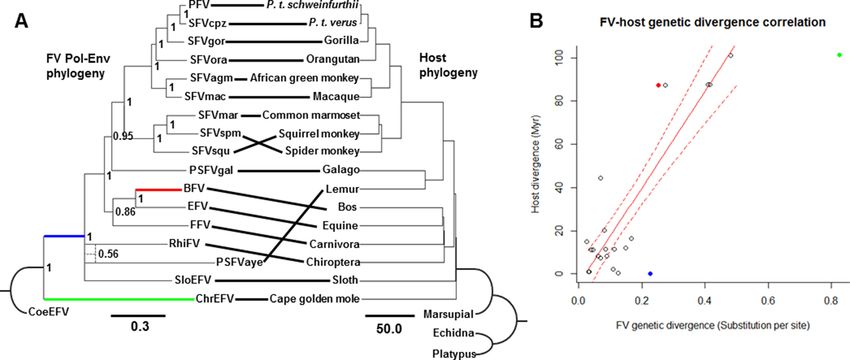
Katzourakis et al. Retrovirology 2014, 11:61                                                                   Page 2 of 17
http://www.retrovirology.com/content/11/1/61

Background                                                    and phylogenetic analysis of SloEFV [37] supported and
Foamy viruses (FVs) are complex retroviruses in the           extended the FV-host co-speciation hypothesis further to
Spumaretrovirinae subfamily [1]. All known contempor-         the basal radiation of eutherians which occurred more
ary FVs cause persistent but non-virulent, asymptomatic       than 100 Myr ago (Ma) [43] and simultaneously expanded
infections exclusively among boreoeutherian mammals           FV mammalian host range to cover xenarthrans, which
(Additional file 1: Table S1) [1]. The first FV was discov-   are one of the four superorders of the Eutheria clade
ered in a macaque in 1954 [2] and was isolated in 1955        (Laurasiatheria, Euarchontoglires and Afrotheria are the
[3]. Shortly thereafter, numerous FVs were isolated from      other superorders). This is in contrast to lentiviruses, for
other boreoeutherian mammals, including non-human             which sequence availability is comparable to that of FVs,
primates (NHPs) [4-13], cats [14], cattle [15], and horses    but their evolutionary history is not as well understood
[16], for most of which complete genomes are available        and much more difficult to investigate, as they do not al-
[16-25]. Recently, a novel FV was discovered by metage-       ways co-speciate with their hosts [44]. Nevertheless, at the
nomics in a bat (Rhinolophus affinis) from China (RhiFV)      present day, evidence of FV infection in, and sequence
and was partially sequenced [26]; however, whether RhiFV      data from prosimians and afrotherian mammals is still
is bat-specific will require confirmation with population     lacking, and this is required to better understand the FV
and epidemiological studies. Although virtually every         radiation and evolutionary history in eutherians. Here, we
NHP sampled has a species-specific FV, a human-specific       report the discovery and describe the first extant exogen-
FV has not been identified. Moreover, all FVs isolated        ous PSFV isolated from a lorisiforme galago (Otolemur
from humans have been demonstrated to originate from          crassicaudatus panganiensis) (PSFVgal) and a novel ERV,
zoonotic infections with simian FVs (SFVs) [27-36].           that is potentially an endogenous FV, present in the gen-
   Similar to other retroviruses, FVs occasionally leave      ome of an afrotherian Cape golden mole (Chrysochloris
‘fossils’ in host genomes, which are the relics of past in-   asiatica) (ChrEFV). We also further describe PSFVaye
fections. These genomic fossils are called endogenous         that was previously only partially characterized [38].
retroviruses (ERVs) and result from germ line infections      Together with the currently available FV sequences, we
that are followed by vertical inheritance and thus are        use detailed molecular analyses to re-examine the co-
present in every cell of an infected organism. In contrast,   evolutionary history of mammalian hosts and their FVs.
exogenous retroviruses and hence exogenous FVs are
transmitted horizontally from organism-to-organism via        Results
infectious body fluids and may be confined to certain         Characterization of the complete genome of the extant
cell types defined by their host tropism. Only two en-        PSFVgal from a galago
dogenous FVs have been discovered in mammalian ge-            The complete genome of PSFVgal (GenBank accession
nomes to date. One is present in the two-toed sloth           number KM233624) was obtained by long-PCR amplifica-
(Choloepus hoffmanni) genome (SloEFV) which was               tion of overlapping 5′ and 3′ genomic halves using infected
found to have a genome organization characteristic of all     HeLa cells and sequence analysis. Genomic inspection
contemporary FVs [37]. The second endogenous FV was           shows that PSFVgal exhibits all the typical structural
found in the aye-aye (Daubentonia madagascariensis)           features of mammalian FV genomes, including the pres-
genome (PSFVaye) which to date has only been partially        ence of gag, pol, env, bel1, and bel2 genes flanked by
characterized [38]. Given a broad range of FV mamma-          LTRs (Figure 1), supporting that it is a FV. There are no
lian hosts and the wealth of available whole mammalian        in-frame stop codons identified in the PSFVgal coding
genomes, the finding of only two endogenous FVs so far        regions. PCR and serological analyses also showed that
suggests that FV endogenization is a rare event. While a      PSFVgal-like sequences are not present in every tested
number of defective ERVs found in fish genomes have           Galago (7 sera and 10 DNA specimens) or Otolemur
been reported to exhibit some similarity to known ex-         species (14 sera and 6 DNA specimens) (Additional file 1:
ogenous FVs [39-41], more evidence is required to de-         Table S2) using a combination of PSFVgal-specific
finitively determine if these ERVs are truly endogenous       PCR (0/13), generic PSFVgal-PSFVaye PCR (6/16,
FVs, or alternatively, are distinct lineages that branched    37.5%) and PSFVgal Western blot (WB) testing (16/21,
early on in FV or retroviral evolution.                       76%). Combined, these findings strongly support the
   Owing to the availability of sequences from diverse        notion that PSFVgal is an exogenous FV. The length of
mammals (Additional file 1: Table S1), coupled with the       the complete genome is 12,118 nucleotides (nt). A de-
stable pattern of codivergence with their hosts, the evo-     tailed annotation of the PSFVgal genome is provided
lutionary history of mammalian FVs can be studied in          in Additional file 2: Figure S1.
great depth. For example, phylogenetic analysis of extant        Sequence similarity analysis revealed that the LTR
SFVs revealed strong evidence of SFV-host co-speciation       length is 1,267 nt. An asparagine-1,2 tRNA-utilizing pri-
over more than 30 million years (Myr) [42]. The discovery     mer binding site (PBS) was identified downstream of the
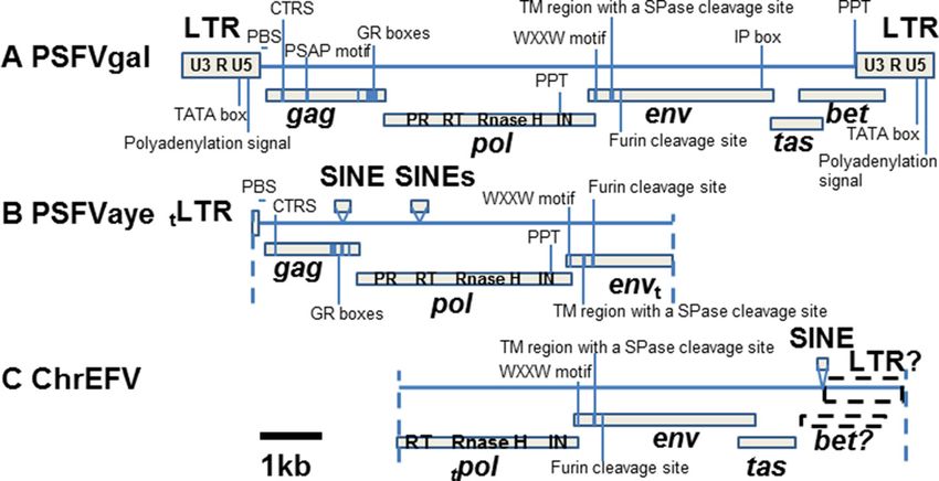
Katzourakis et al. Retrovirology 2014, 11:61                                                                                                 Page 3 of 17
http://www.retrovirology.com/content/11/1/61

 Figure 1 Complete and partial genomic organizations of PSFVgal, PSFVaye and ChrEFV. PSFVgal (A), PSFVaye (B), and ChrEFV (C) all
 exhibit at least some characteristic FV genomic features as indicated (see main text). PSFVaye and ChrEFV are interrupted by a few short
 interspersed nuclear elements (SINEs). Dashed lines indicate where genomes are truncated. Dotted boxes represent hypothetical domains which
 may be present but could not be identified. ‘t’-subscription indicates that the domain is truncated (5′-truncated: preceding the domain name;
 3′-truncated: following the domain name). The scale bar (black solid line) represents a nucleotide length of 1 kb. LTR, long-terminal repeat; PBS,
 primer binding site; CTRS, cytoplasmic retention signal; GR boxes, glycine-arginine rich boxes; PR, protease; RT, reverse transcriptase; IN, integrase;
 PPT, polypurine tract; WXXW, conserved WXXW site; TM, transmembrane; SPase, signal-peptide peptidase; IP, internal promoter.

5′-LTR before gag (TGGCGTCCCTGGGTGGGC, nt                                      position within Gag is variable [48]. The three FV-
1,270-1,287), but which is tRNALys (TGGCGCCCAA                                 specific glycine-arginine rich (GR I-III) motifs or boxes
CGTGGGGG/C) in all other mammalian FVs. We con-                                within the C-terminal region of Gag [49] were also de-
firmed the tRNAAsp PBS sequences by population-based                           termined (aa 479–493, 541–563, 567–597, respectively).
sequencing of multiple PCR products amplified on differ-                          The Pol protein is 1,156 aa long (nt 3,248-6,718) and
ent dates using primers surrounding the PBS with two dif-                      protease (PR), reverse transcriptase (RT), RNase H, and
ferent primer pairs and two different concentrations (0.1                      integrase (IN) were predicted to be located at aa posi-
and 1.0 ug) of day 52 tissue PSFVgal-infected HeLa cell                        tions 10–168, 154–355, 581–737, and 742–1,140, re-
DNA. By comparison with the SFVmac LTR [17], the cap                           spectively. The catalytic centers of PR (DTG) and RT
site defining the 5′ boundary of the R region was deter-                       (YVDD) were located at aa 20–22 and 304–307, respect-
mined to be 20 nt downstream from the TATA box                                 ively. RNase H active sites were found to be at D589 and
(TATATAA, nt 928–934) at nt 955 within the 5′-LTR, and                         D659 and IN catalytic motif (DD35E) within the IN core
the polyadenylation site within the 3′-LTR, which defines                      domain (aa 862–972) was at D926-D938-E962.
the 3′ boundary of the R region, was located at the CA                            The Env protein is 999 aa long (nt 6,603-9,602). We
dinucleotide (nt 11,955) 20 nt downstream from the                             found a highly conserved WXXW motif (WLRW, aa
polyadenylation signal (AATAAA, nt 11,930-11,935).                             10–13) in the N-terminus which is essential for Gag-Env
Thus, the PSFVgal LTR U3, R, and U5 regions are 954, 150,                      protein interaction during the budding process and is
and 163 nt long, respectively. Two polypurine tracts (PPT:                     found in all known extant FVs [50,51]. The internal pro-
AAGGAGAGGG) for the dual initiation of plus-stand DNA                          moter (TATAAAA) within the env gene, which serves
synthesis [45] were located at the 5′ boundary of the 3′-LTR                   for the initiation of bel1 transcription, is present at nt
(nt 10,842-10,853) and at the center of the genome toward                      9,299-9,305. We also identified a hydrophobic transmem-
the 3′ terminus of pol (nt 6,068-6,077) as anticipated.                        brane region, which contains a putative signal-peptide pep-
   The gag gene was predicted to be 1,932 nt long (nt                          tidase cleavage site (TMGWCIGLFCLLLILLFS↓LVIVIL), at
1,363-3,294) to generate a 644 amino acid (aa) protein.                        aa 81–104. The cleavage occurs within the endoplasmic
The cytoplasmic retention signal (CTRS), important for                         reticulum to remove the signal peptide [52]. A potential
particle assembly and viral budding [46], was located at                       furin-cleavage site was also found (TRPNYTAARSRR↓SVE)
aa 73–90 (GNWGPGDRFARIEVLLRD). The position of                                 at aa 131–145. It is this site at which the FV Env protein is
a late-assembly domain proline rich PSAP motif, which                          cleaved, either by furin or furin-like proteases, to produce
is essential for primate FV particle assembly and bud-                         an Env leader protein – a component that interacts with
ding [47,48], was located at aa positions 217–220. The                         the N-terminus of Gag via its WXXW motif and is abso-
PSAP motif is highly conserved in all SFVs but its                             lutely required for FV particle budding [52].
Katzourakis et al. Retrovirology 2014, 11:61                                                                                                 Page 4 of 17
http://www.retrovirology.com/content/11/1/61

   The bel1 and bel2 genes were predicted to be present                         PCR (Additional file 1: Table S2); all of the aye-ayes in-
at nt 9,569-10,402 and nt 10,026-11,414, respectively.                          cluding three infants were strongly PCR-positive which
The putative Bel1 protein (277 aa) exhibits only a weak                         is typical of ERVs. Serums available from 17 of these
similarity (
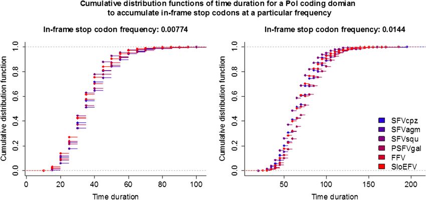
Katzourakis et al. Retrovirology 2014, 11:61                                                                     Page 5 of 17
http://www.retrovirology.com/content/11/1/61

iv) a 3′-truncated putative env gene (nt 5,612-7,867).         demonstrated by using the HMM searching technique
We also found ~113 nt on the 5′ end of the PBS to be           (HMM profile GyDB (http://www.gydb.org/): Bel1 spu-
uniquely-mapping within the aye-aye genome. This               maretroviridae; E = 0.24; note that this weak support is ra-
might represent some portion of the 5′ end of the 5′           ther typical and expected given that this is a part of an
PSFVaye LTR (nt 1–111). We confirmed the truncation            endogenous FV and that FV Bel1 protein is not conserved
of the putative 5′-LTR and env gene and the absence of         among FVs [59]). Although a number of FV-specific gen-
a 3′-LTR in the PSFVaye genome by using a genome               omic features as outlined in the PSFVgal characterization
walking of genomic DNA from two different aye-ayes.            section could not be located, possibly due to the lack of
We found that PSFVaye exhibits several characteristic          sequence data and/or divergence, this genomic informa-
FV features. For example, its Gag protein contains a de-       tion supports a FV progenitor of ChrEFV.
generate, but still recognizable, putative CTRS (GPR?            Aside from the aye-aye genome, currently available
VGD*WQRICLAFQY, aa 37–54) and three GR I-III                   partial and complete genomes of other lemurs (Lemur
boxes (box I: nt 1,248-1,358; box II: nt 1,422-1,781           catta and Microcebus murinus, respectively) do not seem
interrupted by a SINE (nt 1,449-1,748); box III nt 1,851-      to contain PSFVaye-like orthologs, confirmed by PCR
1,871); the putative pol gene harbors a PPT (AAGGA-            testing of eight lemuriform species and one tarsiiforme
CAAAG, nt 5,092-5,101); and the N-terminus of the pu-          species using generic PSFVaye and PSFVgal primers
tative Env contains a highly conserved WXXW motif              (Additional file 1: Table S2). These results support the
(WLAW, aa 10–13). A hydrophobic transmembrane re-              hypothesis that endogenization of PSFVaye occurred
gion containing a signal-peptide peptidase cleavage site       after the basal diversification of the lemurs ~55-66 Ma
(ILIWIMLFLILFSAILVS↓TLIAVF) was also located in                [60-62], in the ancestral aye-aye lineage after it diverged
the N-terminus of the putative Env at aa 63–86. We             from the ancestral lineage of all other living lemurs.
could not identify a furin-cleavage site in the Env of         Similarly, ChrEFV orthologs could not be found in ele-
PSFVaye, most likely due to its defective nature. How-         phant, hyrax, elephant shrew, and tenrec genomes, imply-
ever, by comparing the Env sequence of PSFVaye to              ing a maximum age of ~60-85 Myr based on the proposed
those of other exogenous non-defective FVs, we specu-          Afrosoricida basal split date [43,63]. To further estimate the
late that it should be located between aa 113 and 127          age of these elements, we analyzed the frequencies of
(YFSQAHV*KSRAIHF). Moreover, its putative Gag and              in-frame stop codons in their putative Pol proteins with
Env proteins only exhibit similarities to FV proteins          a Monte Carlo simulation approach, using pols of
(Table 1) and share no detectible similarities with other      PSFVgal, SFVcpz, SFVagm, SFVsqu, FFV, and SloEFV
(non-FV) retroviral proteins. All these findings strongly      (Figure 2) as model sequences. Consistently across all
suggest that the PSFVaye sequence is an endogenous             simulations, the mean age estimates of PSFVaye and
FV, and not merely a distantly related FV-like ERV.            ChrEFV are ~35.2-39.6 and ~65-78 Myr, respectively,
   ChrEFV contains i) a 5′-truncated putative Pol coding       congruent with (i.e. less than) their upper-bound age limit.
gene (nt
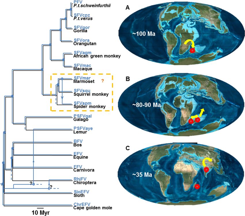
Katzourakis et al. Retrovirology 2014, 11:61                                                                                        Page 6 of 17
http://www.retrovirology.com/content/11/1/61

 Figure 2 PSFVaye and ChrEFV integration dates estimated from in-frame stop codon frequencies. Cumulative distribution functions
 (CDFs) of time duration for Pols of SFVcpz, SFVagm, SFVsqu, PSFVgal, FFV, and SloEFV to accumulate in-frame stop codons at the frequencies of
 0.00774 (right, stop codon frequency of PSFVaye Pol) and 0.0144 (left, stop codon frequency of ChrEFV Pol). Based on these CDFs, the mean age
 of PSFVaye is estimated to be ~35.2-39.6 millions of years (Myr) old (95% confidence interval: 15–75 Myr old, mode: 30–35 Myr old, median: 35
 Myr old). The mean age estimate of ChrEFV is 65.6-78.2 Myr old (95% confidence interval: 35–125 Myr old, mode: 60–65 Myr old, median: 65–75
 Myr old).

FVs, but more evidence is required to definitively de-                    we performed two analyses: (i) FV-host phylogenetic
termine that these ERVs truly have a FV origin. Since                     reconciliation analysis, and (ii) FV-host divergence cor-
these fish ERVs are phylogenetically basal to all known                   relation analysis. A Bayesian phylogeny of FVs was con-
extant mammalian FVs, it is difficult to say with cer-                    structed based on an alignment of concatenated Pol-
tainty that the progenitors of these ERVs are “true”                      Env protein sequences and compared to the previously
FVs, and not distinct lineages that branched earlier in                   published host phylogeny [43] (Figure 3A). Gag protein
retroviral evolution but which are “FV-like” enough to                    sequences were not included in the alignment since (i)
be detected by sequence similarity using the true FVs                     the Gag sequence of RhiFV is not available and (ii) the
as probes. Moreover, in terms of genomic organization,                    alignable region of Gag for the rest of the FVs is rela-
these fish ERVs do not show all the features character-                   tively short (~180 aa). Potential recombination among
istic of mammalian FV genomes. For example, the two                       the sequences within the alignment was assessed using a
identified accessory genes of CoeEFV show no signifi-                     quartet-based recombination detection program VisRD3
cant similarity to those of extant mammalian FVs [39],                    [64], and the results showed no significant evidence for re-
only two out of five determined DrFV-1 proteins, Gag                      combination, both at nucleotide and protein levels (nu-
and Pol, are FV-like [40], FV-like accessory genes in                     cleotide: p = 0.621; protein: p = 0.495). Furthermore, we
platyfishEFV have not been located [41], and only FV-                     also found that neither of the separate Pol nor Env align-
like RT could be found for CodEFV [41]. Although it is                    ments reject the topology of the best phylogeny inferred
known that the accessory genes are the least conserved                    from the concatenated Pol-Env alignment (Figure 3A),
FV genes [59], and that this partial characterization of                  congruent with the results from the recombination test
their genomes could be due to an ancient divergence of                    (approximately unbiased test: p-AUPol = 0.987; p-AUEnv =
fish and mammalian FVs or simply the lack of se-                          0.700; Additional file 4: Figure S3). The two phylogenies
quence data, it raises a possibility that the progenitors                 show remarkably similar topologies, reflecting the well-
of these ERVs may not be FVs but merely class III FV-                     established evolutionary history of stable FV co-speciation
like viruses. Resolution of this debate will require identifi-            with their hosts [37,42]. In total, 14/17 (82.4%) potential
cation and analysis of FV genomes of other vertebrates                    FV-host co-speciation events were inferred among the
like amphibians, reptiles, and birds, if they indeed exist                17 FV and host sequences using the co-phylogeny re-
[39]. Phylogenetic analysis of extant fish FVs and ERVs                   construction software Jane v4.0 [65]. The deepest co-
would also help resolve this debate.                                      speciation event occurred early on in eutherian diversifica-
   To investigate FV phylogenetic relationships in more                   tion (Figure 3A) corresponding to the Exafroplacentalia-
detail and evaluate the FV-host co-evolutionary history,                  Afrotheria split. The reconstruction that maximizes the
Katzourakis et al. Retrovirology 2014, 11:61                                                                                               Page 7 of 17
http://www.retrovirology.com/content/11/1/61

 Figure 3 Co-evolution of foamy viruses (FVs) and their mammalian hosts. (A) FV Bayesian phylogeny (left) is compared to that of their
 hosts (right, previously published in [43]). Curved branches indicate outgroups. Numbers on branch nodes are Bayesian posterior probabilities.
 Solid lines between the two trees indicate FV-host associations. FV and host phylogeny scale bars are in units of amino-acid substitutions per site
 and million years, respectively. Weak support for the sister-taxon relationship of RhiFV and PSFVaye (dotted branches, posterior probability = 0.56)
 was inferred. Among 17 FV and host sequences, 3 apparent mismatches have been inferred using co-phylogenetic reconstruction. Branches
 corresponding to FV-host co-evolution were identified and used in FV-host divergence correlation analysis (B). Branch lengths of FV tree and
 host divergence time were determined to have a linear correlation with a statistically strong support for its coefficient (linear regression: N = 23,
 R2 = 0.823, p = 3.48E-8), represented by the solid red line. Dotted lines represent the 95% confidence interval of the estimated linear relationship.
 Outliers determined by Cook’s distances (solid dots), including the bovine foamy virus (BFV) branch (red), the ChrEFV branch (green), and the
 ancestral branch leading to the exafroplacentalian FVs (blue), were not included in the linear regression. The colors of the outliers correspond to
 the colors of the branches in A.

number of co-speciation events suggests that the most re-                     divergence occurred in proportion to host divergence.
cent common ancestor (MRCA) of PSFVaye and RhiFV                              Combined, these findings strongly support stable FV co-
was present in the MRCA of their hosts and requires a du-                     divergence with their mammalian hosts for more than
plication of the virus lineage at the base of the exafropla-                  100 Myr throughout the Cretaceous and Cenozoic eras
centalian mammal clade (Additional file 5: Figure S4).                        as has been previously proposed [37].
Based on what we know about FV biology, it is extremely                         Despite ChrEFV having an inferred co-speciation event
unlikely that a FV will diversify into two lineages within a                  (posterior probability = 1), the ChrEFV branch lengths, in-
single natural host, i.e. in the absence of host diversification.             cluding both the terminal branch and the internal branch
We therefore postulate that this viral lineage duplication                    leading to other FVs, are longer than would be expected
represents an ancient host speciation event, where one of                     based on the FV-host divergence linear relationship
the resulting species is not sampled in our dataset. We                       (Figure 3B; identified as outliers, green and blue dots, re-
therefore adopt a conservative approach and do not count                      spectively, with Cook’s distance investigation). Given that
the PSFVaye and RhiFV co-speciation event, constraining                       ChrEFV is basal to all known extant FVs, this finding chal-
the number of co-speciation events in our reconstruction                      lenges the notion that ChrEFV is an ERV that has a FV ori-
to 13 (76.5%). This estimate relies on the assumption of the                  gin. Nevertheless, ChrEFV is a long terminal branch in the
existence of this un-sampled host lineage. Nevertheless,                      inferred FV phylogeny and has accumulated numerous
13 co-speciation events are still greater than expected                       neutral genetic changes since its ancient endogenisation.
by chance (random tip mapping: sample size = 1000, p <                        Coupled with long-branch attraction, this ‘pseudogene ef-
0.001), indicating that the FV-host co-speciation history                     fect’ [66] could cause artificial inflation of the branch length
is very stable. Moreover, there is a strong linear correl-                    with a concomitant deep placement of ChrEFV within the
ation between FV and host divergence which extends                            FV phylogeny. Thus, given the congruent ChrEFV/host
from the present to the exafroplacentalian/exafropla-                         phylogenies and the FV-like features of ChrEFV outlined
centalian FV radiation (linear regression: N = 23, R2 =                       above, it is still likely that ChrEFV is a genuine endogenous
0.823, p = 3.48E-8; Figure 3B), confirming previous esti-                     FV and not merely a class III FV-like ERV. Resolution of
mates [37]. This result suggests that across this pro-                        ChrEFV evolutionary history requires further analysis with
tracted time period the accumulation of FV sequence                           other extant afrotherian, marsupial or monotreme FVs
Katzourakis et al. Retrovirology 2014, 11:61                                                                      Page 8 of 17
http://www.retrovirology.com/content/11/1/61

when and if they are identified. The results from these ana-      Discussion
lyses could also elucidate the earlier events of mammalian        Consistent with previous findings [37,42], our analyses
FV diversification. To the best of our knowledge, this is the     suggest an extremely stable FV-host co-speciation his-
first (endogenous) afrotherian FV to be discovered, extend-       tory across a timescale spanning millions of years. Inter-
ing the FV host range to cover the whole eutherian clade.         estingly, this is in contrast to what is suggested by
   PSFVgal was found to be a sister taxon of SFVs with ro-        various in vitro experiments. For example, an in vitro
bust support (posterior probability = 0.95), consistent with a    study has shown prototype FV (PFV) to be capable of
FV-host co-speciation history. Additionally, we also identi-      infecting not only primate, but also rodent, laura-
fied a number of extant FVs from other lorises, including         siatheria, avian and reptile cells, with an extremely broad
the silvery greater galago (Otolemur monteiri monteiri),          range of susceptible cell types [68]. Furthermore, in vitro
southern lesser bush baby (Galago senegalensis moholi),           investigations of FV cell-attachment and entry also sug-
and a potto (Perodicticus potto) (Additional file 1: Table S2),   gested that FVs of different species likely utilize the
and partially sequenced their genomes (IN core domain:            same, perhaps promiscuous, receptor molecule(s) for
~259 nt, 85 aa). When these new sequences were in-                cell-attachment and/or entry [69-71]. Although these
cluded in our phylogenetic analysis, they were all                findings might in part help to explain FV cross-species
found to be distinct FVs forming a clade with PSFVgal             transmissions (FV speciation events by means of jump-
with relatively high statistical support (posterior prob-         ing from one host species to another, without the host
ability = 0.85) within which the branching orders gener-          speciating at the time of the jump) observed in nature
ally mirror those of their lorisiforme hosts (Additional          (see below), one would expect to see more host-switches
file 6: Figure S5). These results suggest that co-speciation      happening given these findings. Analyses of several anti-
between exogenous lorisiforme FVs and their hosts is very         retroviral restriction factors have shown that they are
stable, extending to the species level. Together, our results     specific to particular FVs. For example, while the tripar-
indicate an ancient distribution and evolution of lorisi-         tite motif protein 5αs (TRIM5αs) of most New World
forme FVs in continental Africa dating to the divergence          monkeys (NWMs) have been shown to be able to re-
between strepsirrhine primates (lemurs and lorises) and           strict some NWM FVs, PFV and a SFV from macaque,
the anthropoid primates about 70–95 Ma [43,62,63]. The            TRIM5αs from apes cannot, but instead can restrict
absence of PSFV in some lorises (Additional file 1: Table         feline FV [19,72]. Inhibition of apolipoprotein B mRNA-
S2) may be due to the testing of limited numbers of in-           editing, enzyme-catalytic, polypeptide-like 3C (APO-
dividuals and/or testing of captive animals that were             BEC3C) by FVs has also been shown to be species-specific
captured as infants and thus were likely PSFV-negative            [73]. Whether this species-specific FV-host antagonistic
at that time.                                                     interaction is one of the factors underlining the stable
   In contrast, PSFVaye was not a sister-taxon of PSFVgal         co-speciation history or the result of it is still unclear.
as would be expected, showing a conflict in the co-               To date, the factors that determine this extremely stable
evolutionary history of PSFVaye (Figure 3A). Instead, the         pattern of FV-host co-speciation in nature are still very
sequence is robustly placed outside the boreoeutherian            poorly understood.
FV clade (posterior probability = 1), but still remains              Although we found the pattern of FV-host co-speciation
together with exafroplacentalian FVs (posterior proba-            to be very strong and stable, it is not absolute. Against a
bility = 1). The same evolutionary history was inferred           clear background of FV-host co-divergence are a small
for RhiFV (Figure 3A); instead of being grouped together          number of mismatches in FV-host evolutionary history.
with other fereungulata FVs (bovine, equine, and feline           One involves NWM FVs which form a clade that is sister
FVs) as would be expected if it were to co-speciate with          to catarrhine primate FVs, reflecting the branching order
its host, it is placed robustly outside the boreoeutherian        of their hosts. However, the branching orders within the
FV clade (posterior probability = 1), but still remains           NWM FV clade clearly do not parallel that of their platyr-
together with exafroplacentalian FVs (posterior prob-             rhine hosts [60,74,75] (Figure 4), with SFVmar from a
ability = 1). We compared our best estimated phylogeny            common marmoset being more closely related to SFVspm
against hypothetical alternative phylogenies where                from a spider monkey than to SFVsqu from a squirrel
PSFVaye and/or RhiFV co-speciate with their hosts                 monkey, as would be expected under a co-evolutionary
using approximately unbiased tests [67], and found that           scenario. We compared our inferred phylogeny against an
the co-speciation hypotheses of PSFVaye and RhiFV are             alternative phylogeny in which NWM FVs co-speciate
both rejected (Additional file 4: Figure S3-A). Interest-         with their hosts using approximately unbiased tests [67],
ingly, our analysis also inferred a sister group relationship     and found that the co-speciation picture is rejected
between PSFVaye and RhiFV although statistical sup-               (Additional file 4: Figure S3-B). These results are con-
port for this relationship is extremely weak (posterior           sistent with results from a previous study that used an
probabilities = 0.56, Figure 3A).                                 alignment of Gag protein sequences [19] and implied an
Katzourakis et al. Retrovirology 2014, 11:61                                                                                            Page 9 of 17
http://www.retrovirology.com/content/11/1/61

 Figure 4 Hypothesis explaining the mismatches in foamy virus (FV)-host evolutionary history. FV phylogeny (blue) was superimposed
 upon host phylogeny (black) and is scaled to host divergence times. The scale bar is in units of millions of years. Solid circles represent FV-host
 co-speciation events. Open-circles represent possible host switching events of which the inferred switching directions are indicated by arrows. A
 hypothetical scenario of host switching (as indicated by an orange ‘?’) within the New World monkey FV clade is shown in an orange box. An
 alternative scenario is shown in Additional file 5: Figure S4. For PSFVaye and RhiFV, we speculate that, in the very early history of mammalian
 evolution, eutherians came to the Madagascar-India landmass with their FVs (red circles) upon which they established stable populations (A). The
 landmass was then split into the Madagascar and India landmasses resulting in the FV population splitting into two separate groups ~80-90 Ma
 (B); one FV variant remained on Madagascar and the other was transported to Laurasia via the Indian landmass continental drift (C). The former
 gave rise to the PSFVaye progenitor while the latter gave rise to RhiFV. Directions of infected FV host population movements are indicated by
 yellow arrows. However, it is unknown which ancestral species introduced FV to the lemur and bat lineages and when the transmissions occurred
 (as indicated by a black ‘?’). Additional sequence data from other mammals, especially bats and lemurs, are required to further resolve these
 aspects of FV evolutionary history.

ancestral NWM FV host switch (Figure 4 and Additional                       during captivity. This is unsurprising given that this
file 5: Figure S4, orange boxes). Marmosets, squirrel mon-                  type of FV cross-species transmission between closely
keys, and spider monkeys are closely related, occupy large                  related primate species has already been documented
overlapping geographic ranges [19], and are commonly                        [30]. In contrast to our results, a phylogenetic analysis of
used in biomedical research. It is thus plausible to imagine                a wider range of NWM FVs using short pol nucleotide
a scenario under which heterologous NWM FV infections                       sequences (276 nt) suggested that these three NWM FVs
might have occurred in the past, either in the wild or                      co-speciate with their hosts and that host switches
Katzourakis et al. Retrovirology 2014, 11:61                                                                          Page 10 of 17
http://www.retrovirology.com/content/11/1/61

occurred elsewhere [76]. Analysis of additional NWM FV               India landmass upon which their FVs established stable
complete genomes will be necessary to distinguish these              populations (Figure 4A). Since then, these early eutherian
possibilities.                                                       FVs evolved independently from, and hence have distant
   A more extreme case of host switching across taxonomic            evolutionary relationships with, FVs circulating on the Afri-
orders may explain the phylogenetic placement of PSFVaye             can landmass, which later would give rise to all extant
and RhiFV, where both are placed robustly outside the bor-           SFVs, lorisiforme FVs and fereungulata FVs. The India
eoeutherian FV clade (posterior probability = 1). Neverthe-          landmass was then split from the Madagascar landmass
less, one possibility for the inferred placement of PSFVaye          about 80–90 Ma [78,79] (Figure 4B), separating the FV
may be that it is an artefact due to neutral genetic changes         populations into two groups: one circulating in Madagascar
that have accumulated since its endogenization [38]. To              which gave rise to the progenitor of PSFVaye and the sec-
examine whether or not neutral evolution alone could be re-          ond transferred to Laurasia via the India landmass contin-
sponsible for the observation, Bayesian phylogenies of               ental drift which independently gave rise to RhiFV
concatenated Pol-Env protein alignments containing ar-               (Figure 4C). However, the model does not suggest which
tificially and uniformly mutated SFVagm, SFVsqu, and                 ancestral species introduced FV to the lemur and bat line-
PSFVgal sequences were inferred. These sequences were mu-            ages and when these transmissions occurred. Nonetheless,
tated in silico with an overestimate of the mammalian neu-           both PSFVaye and RhiFV are placed robustly with exafro-
tral substitution rate of 10E-9 substitutions per site per year      placentalian FVs but outside the boreoeutherian FV clade
(s/n/y) over a mean estimate of PSFVaye endogenization               (posterior probability = 1 and 1, respectively). This suggests
date of 40 Myr ago (Figure 2). The substitution process was          that the original hosts were exafroplacentalians, and not
assumed to be neutral, homogenous, and independent across            boreoeutherians, and that the host switches occurred be-
sites and base types. Re-aligning the sequences after the            tween species across deep clades long diverged from each
simulation using MUSCLE implemented in MEGA6 [77]                    other. Sequence data of FVs from additional mammals, es-
does not change the relative position of mutated sequences           pecially bats and lemurs, are required to examine further
in other SFV sequences. Our results suggest that, even with          the evolutionary histories of PSFVaye and RhiFV.
this biased substitution rate setting of 10E-9 s/n/y, the neu-          Our hypothesis helps to explain the deep phylogenetic
tral genetic change accumulation alone is insufficient to ex-        placement and dispersed geographical distribution of
plain the inferred phylogenetic position of PSFVaye. Our in          PSFVaye and RhiFV. In addition, this hypothesis also
silico analysis only increased the branch length and de-             gives an explicit prediction that PSFVaye and RhiFV
creased the branch support without changing the tree top-            should share a MRCA prior to 80–90 Ma based on the
ology (Additional file 7: Figure S6). It is thus unlikely that the   estimated Madagascar-India landmass split date [78,79].
placement of PSFVaye is an artefact. Similarly, the placement        Indeed, although not well-supported (posterior probabil-
of RhiFV is also likely to be genuine since it is an extant ex-      ity = 0.56), our best phylogenetic estimate suggests that
ogenous FV, supported by the absence of in-frame stop co-            PSFVaye and RhiFV have a sister-group relationship
dons or frameshift mutations, and that it was found to be            (Figure 3A). Assuming that our inferred PSFVaye-RhiFV
present in viral particles and not host genomic material [26].       relationship is correct, we estimated the tMRCA of
In addition, the RhiFV lineage remains in the same phylo-            PSFVaye and RhiFV to be 93 (85–101) Myr based on the
genetic position even after removal of PSFVaye from the              linear relationship of FV-host diversity (Figure 3B). Al-
phylogenetic analysis.                                               though slightly high, our estimate range largely overlaps
   We then asked whether there is a reasonable FV host               with the date range derived from this hypothesis. The
switching model that can explain both (i) the deep phylo-            accumulated neutral changes in the PSFVaye sequences
genetic placement of PSFVaye and RhiFV which is basal to             may explain both the weak branch support and the
all known exafroplacentalian FVs, and (ii) the dispersed             slightly overestimated divergence date.
geographical distribution of their hosts (Madagascar and
China, respectively). We propose the following scenario to           Conclusions
explain the phylogenetic relationships of PSFVaye and                Here, we report the characterization of the complete
RhiFV within the context of the evolutionary and geo-                PSFVgal genome obtained from a galago, and describe in
graphical history of FV hosts (Figure 4). Approximately              more detail the partial genome of PSFVaye from the
180–160 Ma, Gondwana was split into two separate land-               aye-aye. The genomic organization of PSFVgal and
masses: the Madagascar-India-Antarctica (MIA) landmass               PSFaye is characteristic of FVs, and they are phylogenet-
and the Africa-South America (ASA) landmass [78,79]. The             ically placed within the FVs. The defective nature of the
MIA landmass then split into the Madagascar-India and                PSFVaye genome, coupled with its robust amplification
the Antarctica landmasses about 135 Ma [78,79]. There-               from genomic DNA and an absence of antibodies to the
after, but still in the very early period of eutherian evolu-        genetically related PSFVgal in infected animals, indicates
tionary history, eutherians arrived onto the Madagascar-             that it is endogenous. In contrast, PSFVgal has a complete
Katzourakis et al. Retrovirology 2014, 11:61                                                                     Page 11 of 17
http://www.retrovirology.com/content/11/1/61

viral transcriptome, elicits a FV-specific immune response,      QIAmp DNA mini kit. DNA concentrations were deter-
and is not present in all individuals, consistent with other     mined using a Nanodrop spectrophotometer and DNA
exogenous FVs. We also report the discovery and describe         integrity was confirmed with ß-actin PCR.
a novel ERV present in the Cape golden mole genome,
ChrEFV. Genomic inspection and analysis suggests that            Isolation of PSFVgal
ChrEFV is also an endogenous FV, and its phylogenetic            HeLa cells were infected with PSFVgal (SFV-5; ATCC#
placement is consistent with a history of co-speciation be-      VR-644), originally isolated from the throat swab of an
tween FVs and their mammalian hosts. However, ChrEFV             Otolemur crassicaudatus panganiensis, and grown in
has a high level of accumulated sequence divergence com-         complete DMEM medium supplemented with 10% FBS,
pared to other FVs, likely an artefact of relaxation of evo-     2 mM L-glutamine, 100 ug/ml streptomycin and 100 U/
lutionary constraints since becoming endogenous.                 ml penicillin (Invitrogen). PSFVgal is the only viral iso-
   We found a general, stable pattern of mammalian FV-           late analyzed in our study. Cell cultures were incubated
host co-divergence which extends as deep as the exafro-          at 37°C and 5% CO2 and were split when 90% confluent.
placentalian basal diversification, spanning more than 100       Cellular DNA was prepared using a Qiagen kit and
Myr. To date, it is still poorly understood why FVs stably       quantified with a Nanodrop spectrophotometer when
co-speciate with their natural hosts. Furthermore, we also       cytopathic effect was observed in >50% of the cells.
identified two possible cases of host switching in the evo-
lutionary history of FVs. The first was observed in NWMs,        Western blot (WB) assay
as previously shown [19], which may have happened dur-           PSFVgal-infected and uninfected cells were pelleted by
ing captivity or in the wild and which has previously been       centrifugation at 1,500 rpm for 10 min and washed 2X
reported to rarely occur in Old World monkeys and apes           with phosphate-buffered saline. Antigen for WB testing
[30]. However, others have shown congruent NWM and               was prepared by treating cell pellets with WB sample
SFV phylogenies using a larger distribution of species-          buffer (Invitrogen) at 100°C for 10 min. Protein concen-
specific sequences [76]. The second involves PSFVaye and         trations were determined using the BioRad DC Protein
RhiFV which may involve cross-species transmission at the        Assay kit and 150 ug of infected and uninfected HeLa cell
level of mammalian orders. We propose a scenario based           lysates were applied separately to 4-12% polyacrylamide
upon geographical knowledge of continental drift and             gels (Invitrogen) for WB analysis. Serum specimens were
hypothetical migration of ancient eutherians to explain          tested using a 1:50 dilution as previously described [80].
this observation. Our results highlight the value of integrat-
ing multiple sources of information to elucidate the evolu-      PCR-amplification of 5′ and 3′ PSFVgal genome halves,
tionary history of mammals and their viruses, including          plasmid cloning, and sequence analysis
continental and geographical histories, ancestral host loca-     To obtain the full-length genomic sequence of PSFVgal we
tions, in addition to the natural history of host and virus.     first PCR-amplified small regions in the LTR and polymerase
                                                                 (pol) gene by using nested PCR with degenerate SFV primers
Methods                                                          and conditions provided elsewhere [80,81]. PSFVgal-specific
Nonhuman primate (NHP) samples and nucleic acid                  primers were then designed from the LTR and pol fragments
preparation                                                      to amplify two overlapping genomic halves using PCR and a
NHP specimens (serum, frozen or fresh whole blood, tis-          Roche Expand 20 kb PCR System Kit following the manufac-
sues) were purchased from the Duke Lemur Center,                 turer’s instructions. The Expand kit contains thermostable
were collected on an opportunistic basis from seven U.S.         Taq DNA polymerase and a thermostable DNA polymerase
zoos following approved animal use protocols, or dried           with proofreading activity to minimize PCR-induced se-
blood spots (DBS) via NHP hunters in Cameroon from               quence artefacts. Briefly, the PCR reaction mixture was pre-
freshly hunted NHP bushmeat in a study approved by               pared in 50 ul reaction volumes containing 500 uM of each
Institutional Animal Care and Use Committees and the             dNTP, 400 nM of each forward and reverse primer, and 5
Cameroon government (Additional file 1: Table S2).               units of Expand 20 kb enzyme mix using 0.5 ug of PSFVgal
Hunters were educated about the risks associated with            tissue culture DNA. The LTR-pol fragment was amplified
direct contact with NHPs and about appropriate prevention        using the primers PSFVgal-LF1 5′ GGC TTG GAT AAT
measures. Prior to processing, all specimens were stored         TAA TTG TTA GAT GCT CTG 3′ and PSFVgalpolF2R 5′
at −20°C or −80°C, except DBS which were stored in nytran        GTT CCA AAC GTA TGC CCC TCT CCT T 3′. The
ziplock bags with dessicant at room temperature. DNA ly-         PSFVgal pol-LTR fragment primers are PSFVgalpolF1 5′
sates or nucleic acids were prepared from blood specimens        GTC AGC ATT CAC CTC TTC CAC CTT G 3′ and
and DBS as previously described [28]. Nucleic acids were         PSFVGAL-LR1 5′ GAC TTA TTT ATT ACT GCA AGA
extracted from archived tissue specimens from the Duke           CCC GAG AGG G 3′. Following denaturation at 92°C for
Lemur Center (liver, spleen, kidney) using a Qiagen              2 min, amplification consisted of 10 cycles at 92°C for 10
Katzourakis et al. Retrovirology 2014, 11:61 Page 12 of 17
http://www.retrovirology.com/content/11/1/61
secs, 60°C for 30 secs, and 68°C for 6 mins, followed by downstream, PSAER2FOR/DW2-ACPN; 3rd PCR
20 cycles at 92°C for 10 secs, 60°C for 30 secs, and 68°C for primers: upstream, PSAGF1REV/UniP2; downstream,
6 mins with an additional 10 sec per cycle. PSAER1FOR/UniP2). Positive bands were cloned and
PCR products were visualized with 0.8% agarose gel Sanger sequenced using an ABI7700 instrument. Fur-
electrophoresis and amplicons of the expected size were thermore, this in silico screening process also returned
collected using a QIAGEN gel purification kit. Purified one contig from the WGS assembled sequence of a Cape
amplicons were cloned into the pCR-XL-TOPO vector golden mole (Chrysochloris asiatica), designated ‘ChrEFV’
(Invitrogen) for genome sequencing. Two plasmids were here. Both were annotated via sequence homology. The
obtained; pCR-XL-TOPO- PSFVgal-LP containing the presence of simple repetitive elements was determined by
LTR-pol insert (5,148-bp) and pCR-XL-TOPO- PSFVgal-PL CENSOR (http://www.girinst.org/censor/).
containing the pol-LTR insert (6,201-bp). The PSFVgal
plasmid DNAs were purified using the Purelink Hipure PCR testing of Prosimian genomic DNA specimens for FV
Plasmid Filter Purification Kit (Invitrogen) and sequenced 1 ug of prosimian DNA was PCR tested for FV pol se-
in both directions using a 3130XL Genetic Analyzer quences using a combination of nested primer sets or a sin-
(Applied Biosystems). Complete genomes were assembled gle round of PCR to detect PSFVaye gag sequences
using the software program Vector NTI v11.1. 5′ and 3′ depending on the species (Additional file 1: Table S2). The
LTRs were determined based on overlapping sequences first PCR assay is PSFVgal-specific and is based on the
and comparison with available FV genome sequences. PSFVgal genome and uses the first and second round PCR
Homology of PBS sequences to tRNAs was inferred using primers SIF1GAL 5′ CTT GCT GTG CAG AGC AGT
the transfer RNA database (http://trnadb.bioinf.uni-leipzig. CAC AAG GT 3′ and SIR1GAL 5′ GTT TTA TTT CAC
de/). Confirmation of the tRNA PBS sequence was done by TGT TTT TCC GTT CCA C 3′ and SIF3GAL 5′ CCA
PCR amplification with primers flanking that region and AGT CTG GAT GCA GAG CTT ATC CA 3′ and SIR3-
using 0.1 and 1.0 ug of PSFVgal-infected HeLa tissue cul- GAL 5′ ACT TTG GGG GTG ATA CGG AGT ACT 3′ to
ture DNA. Primary and nested primers were used to inde- generate 712-bp and 635-bp fragments, respectively. The
pendently amplify two products of different lengths and second assay was designed using an alignment of complete
amplification was done separate from that to obtain the 5′ Old World and New World primate SFV genomes and uses
halve of the genome containing the PBS. Primary PCR pri- the primary primers SIF5N 5′ TAC ATG GTT ATA CCC
mer sequences are F982 (5′-GAC CAG TGT GAG ATT CAC KAA GGC TCC TCC 3′ and SIR5N 5′ AAT AAW
GGT GTC TC-3′) and R1547 (5′-CCA GTT GCC TCC GGA TAC CAC TTT GTA GGT CTT CC 3′ and nested
TGT GAT TCT AAC-3′) and the internal primers are primers SIP4N 5′ TGC ATT CCG ATC AAG GAT CAG
F1133 (5′-CTC CTG GTT GAG GAC AAG GGA AC-3′) CAT T 3′ and SIR1NN 5′ GTT TTA TYT CCY TGT
and R1475 (5′-CCA ATT TGT GCT CGT GGC ACT TTT TCC TYT CCA CCA T 3′ to generate to generate
GG-3′) and standard PCR conditions were used. Numbers 282-bp and 141-bp pol sequences, respectively. The third
in the primer names reference their locations in the assay was designed using an alignment of PSFVgal and
PSFVgal genome. PCR products from both primary and PSFVaye genomes and uses the primary primers 3′ FVPF05
nested and 0.1 and 1.0 ug DNA inputs were sequenced. 5′ KKM TAY TGG TGR CCT AAT ATG 3′ and FVPFR5
5′ GGT CTW CCA ACY ART AGT TTA G 3′ and nested
Discovery and characterization of ChrEFV and PSFVaye primers FVPF01 5′ CCT TTT GAT AAA ATY TAT ATG
Publically available GenBank whole genome shotgun G 3′ and FVPR01 5′ CAS CTT TCC ACT ACT TTG G 3′
(WGS) sequences were screened using tBLASTn and to generate 483-bp and 292-bp products, respectively. To
various FV protein sequences (Additional file 1: Table S3). detect PSFVaye gag sequences the primers PSAGF1 5′
As previously reported by Han and Worobey [38], several AAG ACC CTT GCT GCC TAA TGT TGG 3′ and
matches were returned from the WGS assembled se- PSAGR1 5′ TAT TTG TAA CCA GGG CTT GAC CAG
quence of the aye-aye, Daubentonia madagascariensis, 3′ were used to amplify a 475-bp sequence. 5 ul of primary
spanning four non-overlapping contigs. The adjacency of PCR product was used as template in the nested PCR reac-
these four contigs and sequences flanking the gag and env tion. Selected PCR products were visualized by agarose gel
regions, especially the LTR and host integration se- electrophoresis analysis, extracted using a QIAquick Gel
quences, were determined using a Seegene genome walk- Extraction kit, and sequenced in both directions using an
ing kit (Seegene, MD, USA). Three rounds of PCR were ABI7700 instrument.
performed using universal primers provided in the kit
(DW2-ACP, DW2-ACPN, UNIP2) with PSFVaye-specific Estimating the integration dates of PSFVaye and ChrEFV
primers (1st PCR primers: upstream PSFVAYE-1174R/ with Monte Carlo simulation
DW2-ACP; downstream, PSFVAYE-6122 F/DW2-ACP; To estimate the age of PSFVaye and ChrEFV, we analyzed
2nd PCR primers: upstream, PSAGF2REV/DW2-ACPN; the frequencies of in-frame stop-codons in their PolKatzourakis et al. Retrovirology 2014, 11:61 Page 13 of 17
http://www.retrovirology.com/content/11/1/61
protein sequences. Excluding a hypothetical last stop geometry (Hamming) weighting option was selected. In the
codon, PSFVaye (1,163 codons) and ChrEFV (975 codons) nucleotide analysis, the window size and step size was 300
Pols contain 9 and 14 in-frame stop codon mutations, and 60 nt, respectively; while in the protein analysis, the
which are equivalent to stop codon frequencies of 0.00774 window size and step size was 100 and 20 aa, respectively.
and 0.0144, respectively. Using a Monte Carlo simulation The results showed no significant evidence for recombin-
approach and pols of PSFVgal, SFVcpz, SFVagm, SFVsqu, ation. The alignment is available from the authors upon
FFV, and SloEFV as model sequences, we estimated two request. The protein phylogeny reconstruction was per-
cumulative distribution functions (CDFs) of time duration formed using best available protein-specific model parti-
for in-frame stop codons to be accumulated at these two tions of rtREV + Γ(4) + F (Pol) and WAG + I + Γ(4) + F
frequencies for each model sequence. Each pol model se- (Env) as determined by ProtTest 2.4 [84] under the AIC
quence was mutated in silico over various hypothetical criterion. The MCMC was run for 5,000,000 steps with
time durations from 5 Myr to 200 Myr with an increment the initial 25% discarded as burn-in. Trees and parameters
of 5 Myr. The mutation (substitution) process was as- were logged every 100 steps. Parameter value convergence
sumed to be neutral, homogenous, and independent and sampling independency were manually inspected and
across all sites and base types with the rate of 2.2E-9 s/n/ had effective sample sizes >1,000.
y, an average substitution rate of mammalian genomes We evaluated the FV-host co-speciation hypothesis using
[82]. 1,000 simulations were performed for each period of two analyses: (i) FV-host phylogenetic reconciliation ana-
time and the frequency of in-frame stop codons was then lysis and (ii) FV-host divergence correlation analysis. Phylo-
calculated for each simulation. Two separate CDFs of time genetic reconciliation analysis of FV and host trees was
duration were constructed for each sequence model using performed using Jane v4.0 [65] (Genetic algorithm: number
stop-codon frequencies of 0.00774 and 0.0144. In total, we of generations = 100, population size = 100). The vertex-
constructed six CDFs for each of the two stop-codon fre- based cost mode was used, and the costs were set as fol-
quencies. Means, medians, modes and 95% confidence in- lows: co-speciation = −1, duplication = 0, duplication &
tervals of the time duration for in-frame stop codons to host switch = 0, loss = 0, and failure to diverge = 0. This
accumulate at the two frequencies were calculated. setting was adopted in order to maximize the number of
co-speciation events. The probability of observing the in-
Phylogenetic analysis ferred number of potential co-speciation events by chance
To investigate the phylogenetic relationships of PSFVgal, was calculated by random tip mapping in Jane v4.0 [65]
PSFVaye, and ChrEFV with other retroviruses, a consen- (Genetic algorithm: number of generation = 100, popula-
sus unrooted phylogenetic tree was constructed using a tion size = 100, sample size = 1,000). This method of ana-
manually curated alignment of 240 RT protein sequences lyzing phylogenetic incongruence cannot formally
(162 aa) from alpha-, beta-, gamma-, delta-, epsilon-, differentially weight transmission at different taxonomic
lenti-, and spuma-like retroviruses using RAxML 7.2.8- levels however, and the derivation of p-values relies upon
HPC2 on XSEDE [83]. The alignment is available from the interchangeability of branches. Thus, it does not dis-
the authors upon request. The LG + G + F model was criminate between distant and close interspecies transmis-
determined to fit the data best by using ProtTest 2.4 sions. Once the potential FV-host co-speciation events
[84]. Bootstrap support values for the branching order were located, we then identified FV-host corresponding
were calculated using 5,000 pseudoreplicates. branches that represent FV-host co-evolutionary histories
To further investigate the phylogenetic relationships of for the FV-host divergence correlation analysis. In this
PSFVgal, PSFVaye, and ChrEFV with other FVs, a Bayesian test, we investigated whether or not the lengths of these
FV phylogeny was estimated using MrBayes 3.2.1 [85] and corresponding branches were linearly correlated, i.e.
a manually-curated alignment of concatenated Pol-Env se- whether or not the accumulation of FV sequence diver-
quences (893 and 603 amino acids, respectively). We used gence occurred in proportion to host divergence, using
CoeEFV [39] to root our mammalian FV tree since it is the linear regression implemented in MATLAB [86]. Outliers
most immediate outgroup of mammalian FVs known to were determined by using Cook’s distance inspection.
date although it is still debatable whether this ERV is a real Outliers were removed one at a time using a threshold
endogenous FV or not. Gag sequences were not included value of 3x mean of the observed Cook’s distances and the
in the alignment since (i) the Gag of RhiFV is not available model was re-fitted until no outliers were found. The
and (ii) the alignable region of Gag for the rest of the FVs is model was then used to estimate how long the parental
relatively short (~180 aa). Potential recombination among branch of RhiFV and PSFVaye branches (0.0564 amino-acid
the sequences within the alignment were checked using substitutions per site) is in units of time. Given that the ra-
VisRD3 [64] both at nucleotide and protein levels. For both diation of exafroplacentalians was 101.1 Ma [43], we then
analyses, the null distribution was built based on 1,000 sets re-calculated when PSFVaye and RhiFV shared a MRCA to
of randomly-shuffled sequences and the extended statistical be 93.342 (85.095-101.589) Ma.You can also read