Early detection of osteoarthritis in the rat with an antibody specific to type II collagen modified by reactive oxygen species
←
→
Page content transcription
If your browser does not render page correctly, please read the page content below
Gigout et al. Arthritis Research & Therapy (2021) 23:113
https://doi.org/10.1186/s13075-021-02502-1

 RESEARCH ARTICLE                                                                                                                                   Open Access

Early detection of osteoarthritis in the rat
with an antibody specific to type II collagen
modified by reactive oxygen species
Anne Gigout1, Donata Harazin1, Louise M. Topping2, Didier Merciris3, Sven Lindemann1, Christian Brenneis1 and
Ahuva Nissim2*

  Abstract
  Background: Osteoarthritis (OA) is a disease of the whole joint, with articular cartilage breakdown as a major
  characteristic. Inflammatory mediators, proteases, and oxidants produced by chondrocytes are known to be
  responsible for driving cartilage degradation. Nevertheless, the early pathogenic events are still unclear. To
  investigate this, we employed an antibody that is specific to oxidative post-translationally modified collagen type II
  (anti-oxPTM-CII) to detect early cartilage pathogenic changes in two rat models of OA.
  Methods: The animals underwent surgery for destabilization of the medial meniscus (DMM) and were sacrificed
  after 3, 5, 7, 14, and 28 days. Alternatively, anterior cruciate ligament transection with partial meniscectomy
  (ACLT+pMx) was performed and animals were sacrificed after 1, 3, 5, 7, and 14 days. Joints were stained with
  toluidine blue and saffron du Gatinais for histological scoring, anti-oxPTM-CII, and anti-collagen type X antibodies
  (anti-CX).
  Results: We observed positive oxPTM-CII staining as early as 1 or 3 days after ACLT+pMx or DMM surgeries,
  respectively, before overt cartilage lesions were visible. oxPTM-CII was located mostly in the deep zone of the
  medial tibial cartilage, in the pericellular and territorial matrix of hypertrophic chondrocytes, and co-localized with
  CX staining. Staining was weak or absent for the lateral compartment or the contralateral knees except at later time
  points.
  Conclusion: The results demonstrate that oxidant production and chondrocyte hypertrophy occur very early in the
  onset of OA, possibly initiating the pathogenic events of OA. We propose to use anti-oxPTM-CII as an early
  biomarker for OA ahead of radiographic changes.
  Keywords: Reactive oxygen species, Osteoarthritis, Hypertrophy, Collagen type II, Collagen type X

* Correspondence: a.nissim@qmul.ac.uk
2
 Barts and the London School of Medicine and Dentistry, Queen Mary
University of London, Chaterhouse Square, London EC1M 6BQ, UK
Full list of author information is available at the end of the article

                                        © The Author(s). 2021 Open Access This article is licensed under a Creative Commons Attribution 4.0 International License,
                                        which permits use, sharing, adaptation, distribution and reproduction in any medium or format, as long as you give
                                        appropriate credit to the original author(s) and the source, provide a link to the Creative Commons licence, and indicate if
                                        changes were made. The images or other third party material in this article are included in the article's Creative Commons
                                        licence, unless indicated otherwise in a credit line to the material. If material is not included in the article's Creative Commons
                                        licence and your intended use is not permitted by statutory regulation or exceeds the permitted use, you will need to obtain
                                        permission directly from the copyright holder. To view a copy of this licence, visit http://creativecommons.org/licenses/by/4.0/.
                                        The Creative Commons Public Domain Dedication waiver (http://creativecommons.org/publicdomain/zero/1.0/) applies to the
                                        data made available in this article, unless otherwise stated in a credit line to the data.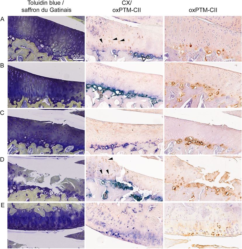
Gigout et al. Arthritis Research & Therapy   (2021) 23:113                                                    Page 2 of 11

Background                                                   validation studies as well as for detection and moni-
Osteoarthritis (OA) is one of the leading causes of re-      toring of early OA in patients.
duced quality of life worldwide, due to the associated         We previously developed a panel of human single chain
chronic pain and various degrees of disability. Although     fragment variable (scFv) that binds specifically to oxidative
OA affects all the tissues of the articular joint, degrad-   post-translationally modified collagen type II (oxPTM-
ation and loss of articular cartilage is a central feature   CII) [12]. We showed that anti-oxPTM-CII (i) binds spe-
[1, 2]. Cartilage degradation in OA results from a disrup-   cifically to arthritic cartilage from patients with RA and
tion in homeostasis due to activation of the chondro-        OA; (ii) stains cartilage in murine models of inflammatory
cytes by various factors that promote the production of      arthritis (antigen induced arthritis (AIA) and OA, namely
matrix degrading enzymes, in excess of the capacity of       DMM); (iii) localizes in the arthritic joint in vivo in a
the chondrocyte to replace damaged and degraded              mouse model of AIA and DMM following systemic ad-
matrix components. The factors that activate chondro-        ministration of labeled anti-oxPTM-CII with Alexa Fluor
cytes to promote matrix degradation include excessive        680 or Cy5.5 [12, 13]; and (iv) was able to target thera-
and abnormal mechanical loading, pro-inflammatory cy-        peutic scaffolds specifically to arthritic joint [14].
tokines, and chemokines, as well as Wnt ligands and fac-       In the current study, we evaluate longitudinally the
tors activating the innate immune system [1, 3]. Many of     presence of oxPTM-CII staining in early OA in two rat
these OA factors stimulate chondrocytes to produce re-       models: DMM and ACTL+pMx. Our goal was to pos-
active oxidants (ROS). ROS are utilized as secondary         sibly unveil some of the very early events in OA and
messengers in mediating intracellular signaling events       evaluate the possibility to detect the initiation of the dis-
that regulate expression of matrix degrading enzymes [4,     ease before the appearance of cartilage lesions. Rats were
5] and pro-death signaling pathways, thus compromise         sacrificed 3, 5, 7, 14, and 28 days after DMM and 1, 3, 5,
chondrocyte integrity and promote cartilage damage [6].      7, and 14 days after ACTL+pMx surgery and the lateral
The most abundant ROS produced by chondrocytes in-           and contralateral knees were stained with toluidine blue
clude superoxide, hydrogen peroxide, the reactive nitro-     and saffron du Gatinais, or for oxPTM-CII and type X
gen species nitric oxide, and the nitric oxide derived       collagen (CX). OxPTM-CII and CX were detectable
product peroxynitrite [2].                                   already at the earliest time points in the medial tibial
   Experimental OA models induced by joint instability       cartilage and strongly co-localized together. We con-
have been highly valuable in identifying key pathogenic      clude that (i) ROS production and increased type X col-
pathways in disease and for validating new treatments.       lagen expression are early events in OA and prefigure
They produce robust degradation of the articular cartil-     cartilage lesions and (ii) anti-oxPTM-CII detection could
age and changes in the subchondral bone and can be           be a powerful tool to detect initiation of OA.
used to investigate pain-like symptoms. Widely used
models of animal OA involve surgically induced instabil-     Method
ity of the knee [7]. These models are characterized by an    Antibody preparation
acute injury to the joint that causes mechanical instabil-   Anti-oxPTM-CII scFv was expressed in HB2151 bacteria
ity, resulting in OA. Many types of operations on various    as described [15]. ScFv was converted to full length anti-
animals have been developed, including cruciate or col-      body by cloning VH domain into pFUSEss-CHIg-hG1e3
lateral ligament transections and partial or total menisc-   and VL domain into pFUSEss-CLIg-hk (InvivoGen).
ectomies on dogs, goats, rabbits, and rodents. Most of       Plasmid DNA was isolated using a QIAFilter Plasmid
the studies detect mild changes in the articular cartilage   Maxi Kit according to the manufacturer’s instruction
at 2 to 4 weeks post-operatively. For example, in the an-    (QIAGEN). Following transient expression in Expi293F
terior cruciate ligament transection (ACLT) model, car-      Expression System according to the manufacturer’s in-
tilage destruction is seen 2–4 weeks after surgery [8].      structions (Thermo Fisher Scientific), supernatants were
Alternatively, when OA is induced by destabilization of      collected and purified using protein A Sepharose CL-4B
the medial meniscus (DMM), structural-change pro-            (GE Healthcare). The ability to retain specific binding of
gression is slower [9–11]. Currently, the size of the        anti-oxPTM-CII over native CII was assessed by ELISA
animal precludes prospective assessment of disease by        as described [12].
conventional radiographic approaches, and disease is
assessed by serial histology of the joint, which is time     Animal models
consuming, costly, and requires large number of ani-         Male Lister Hooded (Crl:LIS) outbred SPF rats (aged 8–
mals as they need to be culled at each experimental          9 weeks and within 150–175 g weight range, Charles
time point under investigation. Powerful non-invasive        River) were housed in colony cages as described [16]
small-animal imaging techniques for longitudinal             with 48 rats/cage at the start of the study. After 4 weeks
studies are therefore highly desirable for preclinical       of acclimatization, rats underwent surgery under
Gigout et al. Arthritis Research & Therapy   (2021) 23:113                                                  Page 3 of 11

anesthesia. Anterior cruciate ligament transection with      CX and oxPTM-CII a double staining was first per-
partial meniscectomy (ACLT+pMx) was performed as             formed. However, because the blue staining of CX was
described elsewhere [16] with the exception that only        very dark and both staining overlapped, it was difficult
50% of the meniscus was removed. For destabilization of      to visualize clearly the brown staining of the oxPTM-
the medial meniscus (DMM), a skin incision was made          CII. For this reason, for the DMM study, an additional
from distal the patella proximal to the tibial plateau (of   single staining for oxPTM-CII was conducted.
the right joint). The muscle layer was opened in knee          For the CX staining, the sections were first deparaffi-
flexion with a scalpel and prepared to visualize the med-    nized and rehydrated. An epitope retrieval using Protein-
ial meniscus tendon which was ligated using scissors.        ase K (Leica Biosystems) [18] was performed. Sections
Finally, the joint capsule, associated muscles and con-      were subsequently incubated with a monoclonal mouse
nective tissue were sutured in layers. For postsurgical      antibody specific for Collagen X (#1-CO097-05, Quar-
analgesia, rats received meloxicam (0.5 mg/kg s.c.; Meta-    tett) diluted 1:50, for 30 min at room temperature. For
cam injection solution, Boehringer Ingelheim). At the        the detection, the Leica Polymer Refine Red Detection
specified time points following surgery, rats (N = 9–10      System (#DS9390, Leica Biosystems) was used where the
per time point) were humanely euthanized by transtho-        RED dye was substituted with NBT/BCIP (#ab7468,
racic heart puncture under isoflurane anaesthetization.      Abcam). The type X collagen staining was developed
                                                             using a fully automated immunohistochemistry stainer
Histology and scoring                                        (Bond III, Leica Microsystems).
The ipsilateral knees of all animals and contralateral         For the staining of oxPTM-CII, epitope retrieval was
knees of two animals per time points (N = 10) were fixed     performed with pepsin. Sections were first equilibrated
for 7 days in paraformaldehyde (VWR) 4% in phosphate-        in HCl (Merck KGaA) 0.02% in ddH2O, 37 °C, 15 min
buffered saline (PBS, VWR) and decalcified for 6 weeks       and digested with 15 mg/mL pepsin (Merck KGaA) in
in formic acid (Sigma-Aldrich) 4% in PBS and embedded        HCl 0.02%, 37 °C, 45 min [19]. The sections were incu-
in paraffin. Coronal sections of 7 μm (including medial      bated with the anti-oxPTM-CII antibody 6.5 μg/mL
tibial plateau, femur condyle, and menisci) were cut with    overnight at 4 °C and detected using Polink-2 Plus HRP
a microtome within the weight-bearing area. Every 35th       human IgG with 3,3′-diaminobenzidin (DAB) (#D88,
section was collected. The sections were deparaffinized      GBI Labs).
and rehydrated, stained with toluidine blue (VWR,              For both staining protocols, negative controls were
0.05% in PBS, 6 min) and saffron du Gatinais (Mor-           performed where the primary antibody was omitted.
phisto GmbH, diluted 1:3 in absolute Ethanol, 1 min).          CX and oxPTM-CII staining were quantified in a re-
Slides were then dehydrated, coverslipped, and after         gion of interest (ROI) selected on the tibial medial plat-
drying digitized using slide scanner SCN400 (Leica           eau using the image analysis software, Calopix® v 4.1.0.3
Microsystems). For histopathological grading, we             (Tribvn, France). Positive areas for each staining were
adapted the modified Mankin score described in [17]          expressed as the percentage of the total cartilage area in
and recommended by the OsteoArthritis Research So-           the ROI. The ROI was placed where lesions develop and
ciety (OARSI) for guinea pig. The changes we made            was 1 mm long (the width corresponded to cartilage
to this score had the purpose to make it usable for          thickness). Because the growth plate was found to be
several species, to be able to compare results obtained      positive for both oxPTM-CII and CX, sections that were
with various types of animals. In the present work,          not or only weakly stained in this area were excluded
we used it to describe rat OA. Our score has a max-          from the analysis.
imum SUM score of 28. Sub-scores are described in
supplementary material Table S1. Scanned sections
were analyzed by two independent observers and the           Statistical analysis
most severe lesion within the weight-bearing area for        Data were analyzed for their normality with the D’Agos-
each rat was selected for evaluation. For each sub-          tino Person or the Shapiro-Wilk normality test (for n <
score, the values from two consecutive sections were         8). For most of the data, several groups did not follow a
averaged to determine overall values for each animal.        normal distribution and the Kruskal-Wallis test cor-
                                                             rected for multiple comparison with a Dunn’s test was
Immunostaining                                               applied. For the oxPTM-CII quantification results in the
Single or double staining for type X collagen (CX) and       DMM and ACLT+pMx model and the CX quantifica-
ROS-modified type II collagen (oxPTM-CII) were ap-           tion in the ACLT model, all groups followed a normal
plied. For the ACLT+pMx study, two single staining           distribution and in this case, a one-way ANOVA cor-
were conducted, one for CX and one for oxPTM-CII.            rected for multiple comparison with a Dunnet test was
For the DMM study, to evaluate the co-localization of        applied. GraphPad Prism v8.4.2 was used.
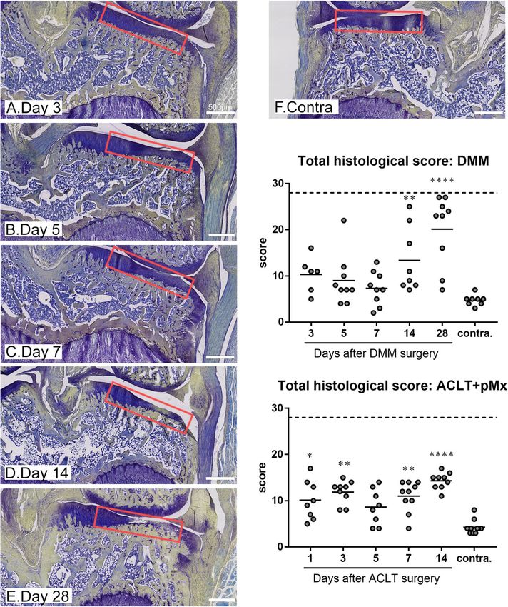
Gigout et al. Arthritis Research & Therapy      (2021) 23:113                                                                                Page 4 of 11

Results                                                                        loss of matrix staining in cartilage was apparent already
DMM surgery affects more profoundly and ACLT+pMx                               after 3 days as illustrated on Fig. 1a and on the matrix
more strongly the medial tibial compartment                                    staining sub-score (Figure S1) but cartilage defects ap-
After both DMM and the ACLT+pMx surgeries, osteo-                              peared only at day 28 in most of the animals (Fig. 1e
arthritis develops mostly in the medial tibial plateau and                     and sub-score cartilage structure in Figure S1). Most of
this area was scored according to the histochemical-                           the other sub-scores such as cellularity, alteration of the
histological scoring system modified from [17]. For the                        tidemark and thickening of the subchondral bone started
DMM model, animals were sacrificed at days 3, 5, 7, 14,                        to be evident at day 14. Small to medium-sized osteo-
and 28. Because ACLT with meniscectomy results in                              phytes were observed in a minority of animals 14 or 28
more severe OA compared to DMM [9], earlier time                               days after the surgery. As a result, the total histological
points were chosen for this model and animals were                             score was significantly elevated at day 14 and 28 in com-
sacrificed at days 1, 3, 5, 7, and 14. After DMM surgery,                      parison to the contralateral medial tibial plateau (Fig. 1)

 Fig. 1 Histological scoring of OA for the medial tibial plateau in the ACLT+pMx and DMM models. Rats underwent ACLT+pMx or DMM surgery
 and were sacrificed at various time points (N = 9–10 rats per time points). The ipsilateral or contralateral knees were taken for histological analysis.
 Slides were stained with toluidine blue and saffron du Gatinais and scored as detailed in the “Method”. Histological sections for the DMM model
 are shown with the region selected for scoring marked in red. The total histological score is shown for both models. Data on the graphs
 represent the total score for each animal (N = 9–10) and the mean for each time point and for the selected contralateral knees. Double asterisks
 and quadruple asterisks mean significantly different from contralateral with p < 0.01 or p < 0.0001, respectively
Gigout et al. Arthritis Research & Therapy (2021) 23:113 Page 5 of 11
for this model. After the ACLT+pMx surgery, the total staining was weak all along the tidemark. Similarly, most
histological score (Fig. 1) was significantly elevated from of the cartilage was negative for oxPTM-CII but a posi-
the first day compared to the contralateral knee and this tive staining was evident in the deep zone in the region
was mainly driven by the cartilage structure and the where cartilage degradation occurred. Interestingly, both
matrix staining sub-scores (Figure S2). The other sub- staining were already observable at early time points, be-
scores were barely affected, and no osteophyte was fore visible cartilage damage started to develop. Both
observed. The comparison of both models at days 3–14 staining were also particularly strong where hypertrophic
shows that the total histological scores were higher (ex- chondrocytes in the deep zone were visible. In addition,
cept at day 5) for the ACLT+pMx model, and this seems in the DMM model at day 28, more severe damage
to be mostly driven by the higher sub-scores for cartilage could be observed. In this case, oxPTM-CII and CX
structure and matrix staining. However, more sub-scores staining extended beyond the deep zone at the damage
were affected in the DMM compared to ACLT+pMx. site and the full depth of cartilage was positive for CX.
We also looked at the lateral tibial compartment of
OxPTM-CII and CX signal in the deep zone are early the operated knees (Figure S3) and the contralateral
markers of OA knees (Figure S4). Both the ACLT+pMx and DMM
ACLT+pMx and DMM knees were stained with anti- models showed weak or no visible staining in the lateral
oxPTM-CII and antibody specific to CX (staining for the tibial compartment at early time points (days 1 or 7 or
medial tibial plateau is shown in Fig. 2 and for the lateral days 3 or 14, respectively) for CX and oxPTM-CII irre-
tibial plateau in Figure S3). On the medial compartment spective of the histological score obtained on the medial
(Fig. 2), for both the ACLT+pMx and DMM knees, a side (indicated in brackets Figure S3A). However, at day
strong CX staining is observable in the deep zone lo- 14 in the ACLT+pMx model and day 28 in the DMM
cated where matrix loss or cartilage damages were also model signal for both collagens appeared. Contrary to
visible. Otherwise, in the rest of the deep zone, the the medial tibial cartilage, the staining was not restricted
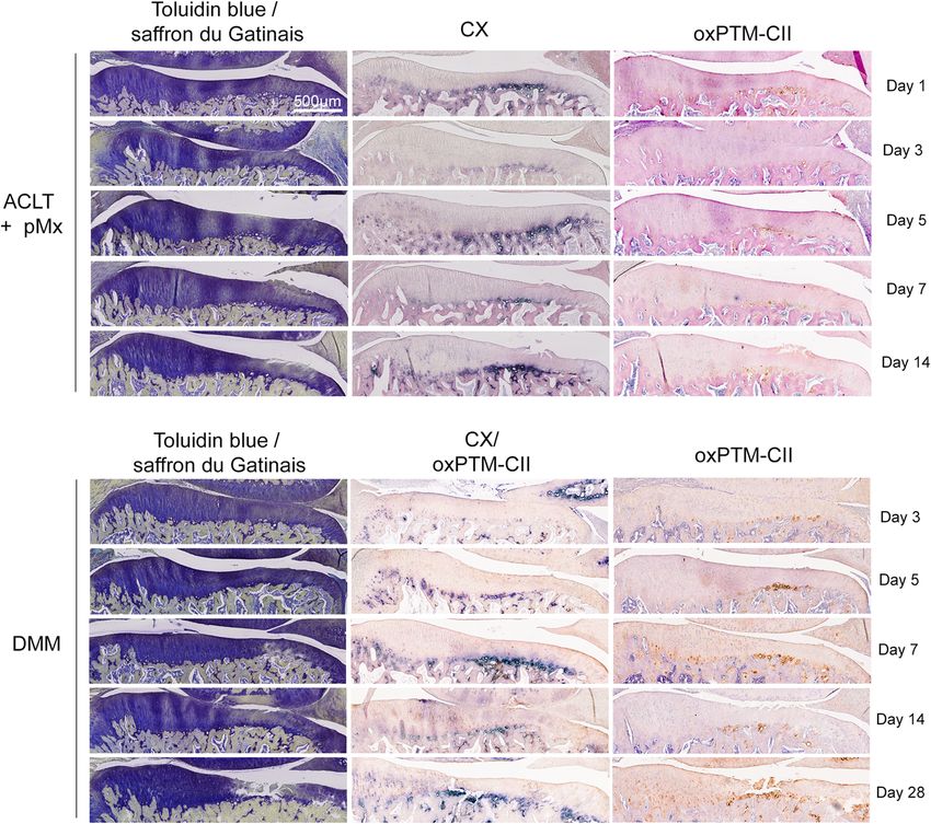
Fig. 2 CX and oxPTM-CII staining of the medial tibial plateau cartilage in the ACLT+pMx and DMM models. Representative results for toluidine
blue and saffron Gatinais as well as type X collagen (blue, CX) and oxPTM type II collagen (brown, oxPTM-CII) immunostainings are shown. For
the ACLT+pMx, study single immunostainings were realized. For the DMM study, a CX and oxPTM-CII double staining was performed as well as
an oxPTM-CII collagen single stainingGigout et al. Arthritis Research & Therapy (2021) 23:113 Page 6 of 11 to a specific location in the cartilage. These results illus- Finally, condylar cartilage was found to be mostly trate that at later time points the disease progressed to negative for oxPTM-CII (not shown) but started to be the lateral side of the operated joint. The lateral tibial positive when large damages develop on the tibial side compartment was scored as well (Figure S3B) and the (see example on Fig. 3d, e). Condylar cartilage was also histological scores were low in all groups and no differ- weakly positive for CX, which was strictly localized in ence could be observed to the contralateral lateral tibial the deep zone. A stronger CX staining appeared with compartment at all time point tested. the progression of the disease but at later time points Similarly, the contralateral knees (Figure S4) showed compared to the tibial cartilage (data not shown). only a weak CX and oxPTM-CII staining at day 1 or 3 in the ACLT+pMx or DMM models respectively but an OxPTM-CII and CX staining partially co-localize increased staining intensity was visible on the medial Higher magnifications of the medial tibial cartilage from side for both CX and oxPTM-CII after ACLT+pMx at DMM knees are shown in Fig. 3 for cartilage with matrix day 14 and for oxPTM-CII after DMM at day 28. As for staining loss but no defect (Fig. 3a, b) or for cartilage the ipsilateral knee, CX and oxPTM-CII were strictly lo- with a small (Fig. 3c) or a large defect (Fig. 3d). An ex- calized in the deep zone around the large hypertrophic ample for condylar cartilage is also shown (Fig. 3e). Only chondrocytes in the medial tibial compartment, and the the DMM knees are shown as the double staining for staining appeared more diffuse in the lateral tibial CX and oxPTM-CII enabled to better determine compartment. whether both markers co-localized. In the cartilage with Fig. 3 Partial co-localization of CX and oxPTM-CII staining in the cartilage of the medial tibial plateau and condyle in the DMM model. Toluidin blue and saffron du Gatinais, type X collagen (blue, CX) and oxPTM type II collagen (brown, oxPTM-CII) staining are shown for cartilage of the medial tibial plateau presenting different levels of degeneration: a, b loss of matrix staining but no defect (both from day 14) and c, d cartilage with a small and a large defect, respectively (from day 5 and 28, respectively). One example of staining for cartilage form the medial condyle is shown in e. Black arrows show examples of chondrocytes positive for oxPTM-CII but negative for CX
Gigout et al. Arthritis Research & Therapy      (2021) 23:113                                                                               Page 7 of 11

no apparent defect, CX and oxPTM-CII staining co-                             The area of the cartilage positive for oxPTM-CII and CX
localized in the deep zone mostly in the pericellular and                     increases with the progression of the disease
territorial matrix of the hypertrophic chondrocytes. In                       The areas positively stained for oxPTM-CII and CX
panel A, chondrocytes that were positive for oxPTM-                           were quantified in the medial tibial cartilage for both
CII but negative for CX can also be observed (see ar-                         models (Fig. 4). In the DMM model, the oxPTM-CII
rows). In cartilage presenting a small defect (panel C),                      and CX positive areas were larger in the operated knee
the pattern was similar. However, in the case of larger                       compared to the contralateral knee and their size in-
defect (panel D), CX staining was found in the inter-                         creased with disease progression. Compared to contra-
territorial matrix and extended to the middle zone                            lateral knees, this increase was not significant at early
while oxPTM-CII staining extended to the middle                               time points and became significant at day 28 for
and superficial zone. Interterritorial staining of                            oxPTM-CII and days 14 and 28 for CX. These results
oxPTM-CII was also observed in the middle and                                 strengthen the previous observations that oxPTM-CII
superficial zones in the area of the damaged fibrillar                        and CX extend progressively beyond the deep zone as
cartilage (panel D). In addition, in the middle and                           OA progresses.
superficial zones, cells positive for oxPTM-CII but no                            For the ACLT+pMx model, the stained area for
CX were observed (see arrows). These results indicate                         oxPTM-CII remained small, and similarly to the
that oxPTM-CII and CX staining mainly co-localize                             DMM model no difference to the contralateral med-
(around the larger chondrocytes in the deep zone) in                          ial cartilage could be observed at day 14 or before.
early OA but might show a slightly different pattern                          However, a trend to increased oxPTM-CII can be
at later OA stages. Finally, condylar cartilage facing a                      observed at day 14 (p = 0.0920). The CX staining was
large defect (here picture from panels D and E are                            found to be highly variable. There was a trend to an
from the same animal) were positive for both staining                         increased positive area at day 3 to 14 (only signifi-
and the staining were observed in all zones of                                cant at day 5) but no clear progression can be ob-
cartilage.                                                                    served. These results are in accordance with the

 Fig. 4 Quantification of oxPTM-CII and CX stainings in the cartilage of the medial tibial plateau in the DMM and ACLT+tMx models. A region of
 interest (ROI) was defined in the medial tibial plateau and the area stained in blue (for CX) or in brown (for oxPTM-CII) was measured and
 normalized by the total cartilage area in the ROI. Data on the graphs represent the % area of the ROI for each animal (N = 4–10) and the mean
 for each time point and for the selected contralateral knees. A single asterisk, double asterisks, and quadruple asterisks mean significantly different
 from contralateral with p < 0.05, 0.01, or 0.0001, respectively
Gigout et al. Arthritis Research & Therapy   (2021) 23:113                                                                          Page 8 of 11

histological score (Fig. 1) which did not progress for                    ACLT+pMx. Prolonging both studies for a longer time
the ACLT+pMx study between day 1 and 14.                                  could have enabled to observe bigger difference between
  Finally, it would have been interesting to compare the                  both models [20]. Interestingly, when looking at the sub-
operated knees to healthy rat knees instead of collateral                 scores, the ACLT+pMx influenced only two categories
knee of OA rats, to evaluate if a significant difference for              (cartilage structure and matrix staining) until day 14,
oxPTM-CII and CX can appear at earlier time points.                       whereas after DMM all sub-scores were affected.
Indeed, in this study, the contralateral knees were se-                     Oxidative stress is known to play a major role in OA [2,
lected from all time points (2 rats per time point) and as                21]. To investigate if oxidative stress is an early or late
we described above, the positive areas for oxPTM-CII                      event in OA and to better understand how it affects cartil-
and CX started to increase contralateral knees at day 14                  age and chondrocytes, we used an antibody against oxi-
for the ACLT+pMx and day 28 for the DMM models.                           dized type II collagen (oxPTM-CII). Anti-oxPTM-CII was
This would suggest that for this readout contralateral                    developed to recognize different forms of oxidized type II
knees are not equivalent to those of healthy controls.                    collagen and was demonstrated to bind human OA and
                                                                          RA cartilage, but not healthy cartilage [12]. OxPTM-CII
The growth plate and the meniscus are positive for                        was detected in the medial tibial cartilage of the operated
oxPTM-CII                                                                 knees as early as 1 and 3 days after ACTL+pMx and
As expected, the growth plate and the calcified part of                   DMM surgeries, respectively, and before any cartilage
the meniscus were positive for CX (Fig. 5). These tissues                 damage was visible. Until 14 days post-surgery, the stain-
were also found to be positive for oxPTM-CII. In these                    ing localized in the deep zone in the pericellular and terri-
two tissues, both staining appear to co-localize.                         torial matrix of large hypertrophic chondrocytes. When
                                                                          larger defects occurred however, oxPTM-CII extended to
Discussion                                                                the interterritorial matrix through the complete depth of
The ACLT+pMx (or tMx) and DMM models are both                             cartilage. The staining was absent from the lateral tibial
surgically induced instability OA models that are broadly                 and femoral condylar cartilage but with the progression of
used to study disease progression. In the present study,                  the disease, the staining became visible in both these com-
we investigated early disease progression for both                        partments (day 14 for ACLT+pMx, day 28 for DMM).
models and evaluated the presence of oxidized type II                     This is in accordance with the observations from others
collagen in cartilage and its co-localization with type X                 [9, 22] that in these OA models, lesions develop primarily
collagen—a marker of chondrocyte hypertrophy.                             in the medial tibial plateau. We also looked at the contra-
  In the current study, we used an ACLT+pMx and                           lateral knees and found that oxPTM-CII staining was ab-
DMM model in rats housed in colony cages enabling                         sent or weak at early time points but started to appear at
free movement of the animals [9, 16]. In accordance                       later time points. This finding suggests that the disease
with previous results from others [9], the total histo-                   also starts to develop in the contralateral knee, possibly
pathological scores were slightly higher with the ACLT+                   because the animal changes their gait thus inducing mech-
pMx model than the DMM model for the same time                            anical stress in the contralateral joint [23]. This is also ex-
points and the diseased developed earlier with the                        pected to be accelerated in the colony housing where

 Fig. 5 CX and oxPTM-CII staining in the meniscus and in the growth plate. One example of a double staining for type X collagen (blue, CX) and
 oxPTM type II collagen (brown, oxPTM-CII) and the single staining for oxPTM-CII is shown for the growth plate and the meniscus
Gigout et al. Arthritis Research & Therapy (2021) 23:113 Page 9 of 11 animal are free to perform more weight-bearing activities the middle zone and superficial layer are more resistant compared to smaller cages [16, 20]. It was already demon- to hypertrophy [31] increasing the delay between ROS strated that under mechanical stress chondrocytes pro- production and CX production in these zones. However, duce ROS [24]. It is also known that removal or the chondrocytes in this zone might have already been displacement of the meniscus increases peak stress in the pre-hypertrophic and with this in mind it would have medial compartment [25] and that cartilage normally cov- been of interest to investigate the presence of Runx2, an ered by the meniscus possesses decreased load-bearing early hypertrophy marker. Taken together, our results capacity and less resilience to damage compared to cartil- confirm the therapeutic potential of inhibiting ROS to age not covered by menisci [26]. We postulate that the treat OA. Indeed, it has already been demonstrated that DMM and the ACLT+pMx surgeries produce a strong oral administration of the anti-oxidant N-acetyl cysteine mechanical stress in the tibial cartilage that was covered (NAC) protects against OA in the rat [32] and reduces by the meniscus before the surgery resulting in ROS pro- hypertrophy in the growth plate of mice [28]. Future duction [2] and the subsequent appearance of oxidized studies could use NAC to evaluate its impact on type II collagen. The localization of the staining likely cor- oxPTM-CII generation, hypertrophy, and OA initiation responds to the zone where mechanical stress was the and progression in animal models. highest. It is also interesting to note that on the lateral Interestingly, the quantification of the medial tibial compartment where the meniscus remained, and conse- cartilage area positive for oxPTM-CII and CX revealed quently mechanical stress was lower, the staining was that in the DMM model, the CX staining spread across a more diffuse and not restricted to the deep zone. A simi- larger cartilage area at day 14 already when compared lar pattern was observed in the medial condyles. Pos- with day 28 for oxPTM-CII. This may indicate that be- sibly, in medial tibial cartilage, the production of ROS side ROS production other mechanisms might induce and the resulting oxPTM-CII arises from excessive cartilage hypertrophy in OA. loading while on the lateral side and the condyles the In addition, our results are in accordance with previ- ROS production and oxPTM-CII staining might be ous observations that the localization of oxPTM-CII is rather due to the diffusion of ROS and other inflam- predominantly in the ipsilateral joint and that it is de- matory components from the medial tibial cartilage to tected ahead of cartilage structural changes [12, 13]. In other joint compartments. previous work, we used Cy5.5-anti-ROS CII antibody or Because oxPTM-CII was predominantly found in the Cy5.5-anti-ROS CII scFv injected i.v. or i.a. to detect OA deep zone and given that ROS are known to stimulate changes in vivo with non-invasive imaging in DMM chondrocyte hypertrophy [27, 28], we also evaluated if mice after 4 or 8 weeks. The present study demonstrates oxPTM-CII co-localizes with type X collagen (CX). In- that labeled oxPTM-CII antibody or scFv could diagnose deed, we observed a strong co-localization and both OA even earlier in the DMM model and could be used staining became more intense with disease progression. to monitor disease progression. Future longitudinal stud- We also observed that 28 days after DMM surgery, CX ies will need to assess the utility of anti-oxPTM-CII as a staining extended to the complete depth of cartilage at a novel molecular imaging tool to both detect early onset time when large defects were also observed, resulting in and to longitudinally monitor OA in small animal a larger area positively stained for CX. Previous studies models. In addition, because at early time points the also described an increased CX expression during OA oxPTM-CII staining was restricted to few cells in the [29, 30] and in line with our observation, a pericellular deep zone, the anti-oxPTM-CII might need to be opti- staining in the deep zone and in advanced OA also in mized to diffuse efficiently in cartilage and provide a the middle zone was reported [30]. However, to our high signal-to-noise ratio. If proven to be successful, knowledge, the present study is the first that shows an anti-oxPTM-CII may be exploited for molecular imaging increase of CX expression early in the disease. Similarly in parallel with future developments of MRI capabilities to oxPTM-CII, CX staining was detected before any ap- in human. Imaging with anti-oxPTM-CII may be inter- parent cartilage damage develops. At later time points preted in association with MRI/radiography for en- (14 days for ACLT+pMx and 28 days for DMM), staining hanced overall clinical management of patients, as well no longer fully co-localized. The co-localization was still as improvement of outcome readouts in clinical trials. found in the deep zone as observed at earlier time points In conclusion, oxPTM-CII and CX staining of ACLT+ but staining only partially co-localized in the middle and pMx and DMM rat knees showed that the disease starts the superficial zone. This finding demonstrates that the extremely early (day 1 and 3, respectively) in the deep formation of oxPTM-CII was not restricted to hyper- zone of tibial medial cartilage and that the load-bearing trophic cells. We hypothesize that chondrocytes first zone that was covered by the meniscus before surgery produce ROS, which induces hypertrophy [27, 28] and was affected first. Our results confirm that chondrocyte subsequently lead to CX deposition. Possibly cells from hypertrophy is an integral part of the OA pathobiology,
Gigout et al. Arthritis Research & Therapy         (2021) 23:113                                                                                   Page 10 of 11

and we propose that it might be an initiating event of the                       organized the in vivo studies. Donata Harazin optimized and performed the
disease. In addition, because oxPTM-CII and CX staining                          staining and Didier Merciris performed the quantification of the oxPTM-CII
                                                                                 and CX immunohistochemistry stainings. Sven Lindemann supervised the
were strictly localized in the pericellular matrix at early                      histology work and helaped at critically intrepretig the data. Anne Gigout,
time points, this study also supports the hypothesis that                        Ahuva Nissim, and Louise Topping drafted the manuscript which was critic-
OA is a disease of the pericellular matrix [33, 34].                             ally reviewed by all authors. All authors have read and approved the final
                                                                                 submitted manuscript.

Conclusions                                                                      Funding
We propose that oxPTM-CII antibodies or oxPTM-CII                                No funding was received for this article.

scFv labeled with a fluorescent probe is a promising bio-                        Availability of data and materials
marker to detect OA initiation ahead of radiographic                             All data generated during this study are included in this published article
changes and monitor its progression.                                             and in its supplementary information files or are available from the
                                                                                 corresponding author on reasonable request.
Abbreviations
OA: Osteoarthritis; CII: Collagen type II; oxPTM-CII: Oxidative post-            Declarations
translationally modified collagen type II; CX: Collagen type X;
ACLT+pMx: Anterior cruciate ligament transection with partial                    Ethics approval and consent to participate
meniscectomy; DMM: Destabilization of the medial meniscus; ROS: Reactive         All procedures were approved by the animal protection authorities of the
oxidants                                                                         local district government (Regional Authorities of Hessen, Germany, approval
                                                                                 number DA 4/1019, approved on 29 March 2017).

Supplementary Information                                                        Consent for publication
Supplementary information accompanies this paper at https://doi.org/10.          Not applicable.
1186/s13075-021-02502-1.
                                                                                 Competing interests
 Additional file 1: Table S1. Sub-scores for the histological scoring of         Anne Gigout, Sven Lindemann, Christian Brenneis, and Donata Harazin were
 OA. Figure S1. Histological sub-scores for the medial tibial plateau in the     employees of Merck KGaA at the time of the study. Anne Gigout and Didier
 DMM model. Rats underwent DMM surgery and were sacrificed at days 3,            Merciris are currently employees of Galapagos SASU. The other authors
 5, 7,14 and 28. The ipsilateral or contralateral knees were taken for histo-    declare that they have no competing interests.
 logical analysis. Slides were stained with toluidine blue and saffron du
 Gatinais and evaluated according to the sub-scores detailed in the Table        Author details
                                                                                 1
 S1. Individual data for each animal (N = 9–10) and the mean is shown for         Osteoarthritis Research, Merck KGaA, Darmstadt, Germany. 2Barts and the
 each timepoint as well as for the selected contralateral knees. *, ** and       London School of Medicine and Dentistry, Queen Mary University of London,
 *** means significantly different from contralateral with p < 0.05, p < 0.01    Chaterhouse Square, London EC1M 6BQ, UK. 3Galapagos SASU, Romainville,
 or p < 0.001 respectively. Figure S2. Histological sub-scores for the med-      France.
 ial tibial plateau in the ACLT+pMx model. Rats underwent ACLT+pMx sur-
 gery and were sacrificed at days 1, 3, 5, 7 and 14. The ipsilateral or          Received: 22 July 2020 Accepted: 3 April 2021
 contralateral knees were taken for histological analysis. Slides were
 stained with toluidine blue and saffron du Gatinais and evaluated accord-
 ing to the sub-scores detailed in the Table S1. Individual data for each        References
 animal (N = 9–10) and the mean is shown for each timepoint as well as           1. Loeser RF, Goldring SR, Scanzello CR, Goldring MB. Osteoarthritis: a disease
 for the selected contralateral knees. *, **, *** and **** means significantly       of the joint as an organ. Arthritis Rheum. 2012;64(6):1697–707. https://doi.
 different from contralateral with p < 0.05, 0.01, 0.001, and 0.0001 respect-        org/10.1002/art.34453.
 ively. The sub-score chondroosteophyte is not shown because it was              2. Bolduc JA, Collins JA, Loeser RF. Reactive oxygen species, aging and
 equal to 0 in all groups. Figure S3. Staining and total histological score          articular cartilage homeostasis. Free Radic Biol Med. 2019;132:73–82. https://
 for the lateral tibial plateaus in the ACLT+pMx and DMM models. A. At               doi.org/10.1016/j.freeradbiomed.2018.08.038.
 various timepoints, the knees were taken for histological analysis and          3. Liu-Bryan R, Terkeltaub R. Emerging regulators of the inflammatory process
 stained with toluidine blue and saffron du Gatinais or for type X collagen          in osteoarthritis. Nature Rev Rheum. 2015;11(1):35–44. https://doi.org/10.103
 (blue, CX) and oxPTM type II collagen (brown, oxPTM-CII). Staining for the          8/nrrheum.2014.162.
 lateral tibial plateau for different timepoints and scores obtained for the     4. Collins JA, Diekman BO, Loeser RF. Targeting aging for disease modification
 medial plateau (in brackets) are shown. B. The total histological score for         in osteoarthritis. Curr Opin Rheumatol. 2018;30(1):101–7. https://doi.org/10.1
 the lateral tibial plateau was determined according to the sub-scores de-           097/BOR.0000000000000456.
 tailed in the Table S1. Individual data for each animal (N = 9–10) and the      5. Henrotin YE, Bruckner P, Pujol JP. The role of reactive oxygen species in
 mean is shown for each timepoint and selected contralateral knees. *                homeostasis and degradation of cartilage. Osteoarthr Cartil. 2003;11(10):
 means significantly different from contralateral with p < 0.05. Figure S4.          747–55. https://doi.org/10.1016/S1063-4584(03)00150-X.
 Staining of the contralateral tibial plateaus in the ACLT+pMx and DMM           6. Carlo MD Jr, Loeser RF. Increased oxidative stress with aging reduces
 models. Representative pictures obtained with the toluidine blue and saf-           chondrocyte survival: correlation with intracellular glutathione levels.
 fron du Gatinais staining, type X collagen (in blue, CX) and oxPTM-type II          Arthritis Rheum. 2003;48(12):3419–30. https://doi.org/10.1002/art.11338.
 collagen (in brown, oxPTM-CII) single or double immunostainings are             7. Cope PJ, Ourradi K, Li Y, Sharif M. Models of osteoarthritis: the good, the
 shown.                                                                              bad and the promising. Osteoarthr Cartil. 2019;27(2):230–9. https://doi.org/1
                                                                                     0.1016/j.joca.2018.09.016.
                                                                                 8. Aizah N, Chong PP, Kamarul T. Early alterations of subchondral bone in the
Acknowledgements                                                                     rat anterior cruciate ligament transection model of osteoarthritis. Cartilage.
The authors thank Andreas Westhof, Herbert Ziegler, Jennifer Freiwald,               2019:1947603519878479. https://doi.org/10.1177/1947603519878479.
Julianne Dalchow, and Nicole Ellinghaus who contributed to this work.            9. Glasson SS, Blanchet TJ, Morris EA. The surgical destabilization of the medial
                                                                                     meniscus (DMM) model of osteoarthritis in the 129/SvEv mouse. Osteoarthr
Authors’ contributions                                                               Cartil. 2007;15(9):1061–9. https://doi.org/10.1016/j.joca.2007.03.006.
All authors contributed to the study conception and design. Ahuva Nissim         10. Inglis JJ, McNamee KE, Chia SL, Essex D, Feldmann M, Williams RO, et al.
and Louise Topping provided the oxPTM-CII antibody, Christian Brenneis               Regulation of pain sensitivity in experimental osteoarthritis by the
Gigout et al. Arthritis Research & Therapy (2021) 23:113 Page 11 of 11
endogenous peripheral opioid system. Arthritis Rheum. 2008;58(10):3110–9. endochondral ossification. J Exp Med. 2007;204(7):1613–23. https://doi.
https://doi.org/10.1002/art.23870. org/10.1084/jem.20062525.
11. Little CB, Barai A, Burkhardt D, Smith SM, Fosang AJ, Werb Z, Shah M, 29. Aigner T, Reichenberger E, Bertling W, Kirsch T, Stoss H, von der Mark K.
Thompson EW. Matrix metalloproteinase 13-deficient mice are resistant to Type X collagen expression in osteoarthritic and rheumatoid articular
osteoarthritic cartilage erosion but not chondrocyte hypertrophy or cartilage. Virchows Arch B Cell Pathol Incl Mol Pathol. 1993;63(4):205–11.
osteophyte development. Arthritis Rheum. 2009;60(12):3723–33. https://doi. https://doi.org/10.1007/BF02899263.
org/10.1002/art.25002. 30. Boos N, Nerlich AG, Wiest I, von der Mark K, Ganz R, Aebi M.
12. Hughes C, Faurholm B, Dell'Accio F, Manzo A, Seed M, Eltawil N, Marrelli A, Immunohistochemical analysis of type-X-collagen expression in
Gould D, Subang C, al-Kashi A, de Bari C, Winyard P, Chernajovsky Y, Nissim A. osteoarthritis of the hip joint. J Orthop Res. 1999;17(4):495–502. https://doi.
Human single-chain variable fragment that specifically targets arthritic org/10.1002/jor.1100170406.
cartilage. Arthritis Rheum. 2010;62(4):1007–16. https://doi.org/10.1002/art.27346. 31. Jiang J, Leong NL, Mung JC, Hidaka C, Lu HH. Interaction between zonal
13. Lim NH, Vincent TL, Nissim A. In vivo optical imaging of early osteoarthritis populations of articular chondrocytes suppresses chondrocyte
using an antibody specific to damaged arthritic cartilage. Arthritis Res Ther. mineralization and this process is mediated by PTHrP. Osteoarthr Cartil.
2015;17(1):376. https://doi.org/10.1186/s13075-015-0898-5. 2008;16(1):70–82. https://doi.org/10.1016/j.joca.2007.05.014.
14. Topping LM, Thomas BL, Rhys HI, Tremoleda JL, Foster M, Seed M, 32. Kaneko Y, Tanigawa N, Sato Y, Kobayashi T, Nakamura S, Ito E, Soma T,
Voisin MB, Vinci C, Law HL, Perretti M, Norling LV, Azevedo HS, Nissim Miyamoto K, Kobayashi S, Harato K, Matsumoto M, Nakamura M, Niki Y,
A. Targeting extracellular vesicles to the arthritic joint using a damaged Miyamoto T. Oral administration of N-acetyl cysteine prevents osteoarthritis
cartilage-specific antibody. Front Immunol. 2020;11:10. https://doi.org/1 development and progression in a rat model. Sci Rep. 2019;9(1):18741.
0.3389/fimmu.2020.00010. https://doi.org/10.1038/s41598-019-55297-2.
15. Harrison JL, Williams SC, Winter G, Nissim A. Screening of phage antibody 33. Chery DR, Han B, Li Q, Zhou Y, Heo SJ, Kwok B, et al. Early changes in
libraries. Methods Enzymol. 1996;267:83–109. https://doi.org/10.1016/S0076- cartilage pericellular matrix micromechanobiology portend the onset of
6879(96)67007-4. post-traumatic osteoarthritis. Acta Biomater. 2020;
16. Brenneis C, Westhof A, Holschbach J, Michaelis M, Guehring H, 34. Guilak F, Nims RJ, Dicks A, Wu CL, Meulenbelt I. Osteoarthritis as a disease
Kleinschmidt-Doerr K. Automated tracking of motion and body weight for of the cartilage pericellular matrix. Matrix Biol. 2018;71-72:40–50.
objective monitoring of rats in colony housing. J Am Assoc Lab Anim Sci.
2017;56(1):18–31.
17. Kraus VB, Huebner JL, DeGroot J, Bendele A. The OARSI histopathology
Publisher’s Note
Springer Nature remains neutral with regard to jurisdictional claims in
initiative - recommendations for histological assessments of osteoarthritis in
published maps and institutional affiliations.
the guinea pig. Osteoarthr Cartil. 2010;18(Suppl 3):S35–52. https://doi.org/1
0.1016/j.joca.2010.04.015.
18. Ramos-Vara JA, Beissenherz ME. Optimization of immunohistochemical
methods using two different antigen retrieval methods on formalin-fixed
paraffin-embedded tissues: experience with 63 markers. J Vet Diagn
Investig. 2000;12(4):307–11. https://doi.org/10.1177/104063870001200402.
19. Dell'accio F, De Bari C, Eltawil NM, Vanhummelen P, Pitzalis C. Identification
of the molecular response of articular cartilage to injury, by microarray
screening: Wnt-16 expression and signaling after injury and in osteoarthritis.
Arthritis Rheum. 2008;58(5):1410–21. https://doi.org/10.1002/art.23444.
20. Brenneis C, Menges S, Westhof A, Lindemann S, Thudium CS, Kleinschmidt-
Doerr K. Colony housing promotes structural and functional changes during
surgically induced osteoarthritis in rats. Osteoarthritis Cartilage Open. 2020;
2. https://doi.org/10.1016/j.ocarto.2020.100100.
21. Abramson SB. Osteoarthritis and nitric oxide. Osteoarthr Cartil. 2008;
16(Suppl 2):S15–20. https://doi.org/10.1016/S1063-4584(08)60008-4.
22. Iijima H, Aoyama T, Ito A, Tajino J, Nagai M, Zhang X, Yamaguchi S, Akiyama
H, Kuroki H. Destabilization of the medial meniscus leads to subchondral
bone defects and site-specific cartilage degeneration in an experimental rat
model. Osteoarthr Cartil. 2014;22(7):1036–43. https://doi.org/10.1016/j.joca.2
014.05.009.
23. Zhu J, Zhu Y, Xiao W, Hu Y, Li Y. Instability and excessive mechanical
loading mediate subchondral bone changes to induce osteoarthritis. Ann
Transl Med. 2020;8(6):350. https://doi.org/10.21037/atm.2020.02.103.
24. Goodwin W, McCabe D, Sauter E, Reese E, Walter M, Buckwalter JA, Martin
JA. Rotenone prevents impact-induced chondrocyte death. J Orthop Res.
2010;28(8):1057–63. https://doi.org/10.1002/jor.21091.
25. Arunakul M, Tochigi Y, Goetz JE, Diestelmeier BW, Heiner AD, Rudert J,
Fredericks DC, Brown TD, McKinley TO. Replication of chronic abnormal
cartilage loading by medial meniscus destabilization for modeling
osteoarthritis in the rabbit knee in vivo. J Orthop Res. 2013;31(10):1555–60.
https://doi.org/10.1002/jor.22393.
26. Yeow CH, Lau ST, Lee PV, Goh JC. Damage and degenerative changes in
menisci-covered and exposed tibial osteochondral regions after simulated
landing impact compression-a porcine study. J Orthop Res. 2009;27(8):1100–
8. https://doi.org/10.1002/jor.20861.
27. Kim KS, Choi HW, Yoon HE, Kim IY. Reactive oxygen species generated by
NADPH oxidase 2 and 4 are required for chondrogenic differentiation. J Biol
Chem. 2010;285(51):40294–302. https://doi.org/10.1074/jbc.M110.126821.
28. Morita K, Miyamoto T, Fujita N, Kubota Y, Ito K, Takubo K, Miyamoto K,
Ninomiya K, Suzuki T, Iwasaki R, Yagi M, Takaishi H, Toyama Y, Suda T.
Reactive oxygen species induce chondrocyte hypertrophy inYou can also read