ESTROGEN DEFICIENCY - A CENTRAL PARADIGM IN AGE-RELATED IMPAIRED HEALING? - EXCLI Journal
←
→
Page content transcription
If your browser does not render page correctly, please read the page content below
EXCLI Journal 2021;20:99-116 – ISSN 1611-2156
Received: November 26, 2020, accepted: January 04, 2021, published: January 11, 2021
Review article:
ESTROGEN DEFICIENCY –
A CENTRAL PARADIGM IN AGE-RELATED IMPAIRED HEALING?
Mohamed El Mohtadi1, Kathryn Whitehead2, Nina Dempsey-Hibbert2, Amina Belboul2,
Jason Ashworth2,*
1
Department of Biology, Edge Hill University, Ormskirk, Lancashire, L39 4QP, UK
2
Centre for Bioscience, Manchester Metropolitan University, Chester Street, Manchester,
M1 5GD, UK
* Corresponding author: Dr Jason Ashworth, Centre for Bioscience, Manchester
Metropolitan University, Chester Street, Manchester, M1 5GD, UK,
E-mail: J.Ashworth@mmu.ac.uk
http://dx.doi.org/10.17179/excli2020-3210
This is an Open Access article distributed under the terms of the Creative Commons Attribution License
(http://creativecommons.org/licenses/by/4.0/).
ABSTRACT
Wound healing is a dynamic biological process achieved through four sequential, overlapping phases; hemostasis,
inflammation, tissue proliferation and remodeling. For effective wound healing, all four phases must occur in the
appropriate order and time frame. It is well accepted that the wound healing process becomes disrupted in the
elderly, increasing the propensity of non-healing wound states that can lead to substantial patient morbidity and
an enormous financial burden on healthcare systems. Estrogen deprivation in the elderly has been identified as the
key driver of age-related delayed wound healing in both genders, with topical and systemic estrogen replacement
reversing the detrimental effects of aging on wound repair. Evidence suggests estrogen deprivation may contribute
to the development of chronic wound healing states in the elderly but research in this area is somewhat limited,
warranting further investigations. Moreover, although the beneficial effects of estrogen on cutaneous healing have
been widely explored, the development of estrogen-based treatments to enhance wound repair in the elderly have
yet to be widely exploited. This review explores the critical role of estrogen in reversing age-related impaired
healing and evaluates the prospect of developing more focused novel therapeutic strategies that enhance wound
repair in the elderly via activation of specific estrogen signaling pathways in regenerating tissues, whilst leaving
non-target tissues largely unaffected.
Keywords: Wound healing, aging, age-related impaired healing, estrogen, estrogen receptors
BACKGROUND of estrogen-based treatments to reverse de-
layed healing in the elderly has not been
Declining levels of estrogen in both gen-
widely adopted outside research settings.
ders with increasing age suggests that age-re-
Moreover, the potential role of the sex steroid
lated impaired wound healing may result in
hormones in chronic wounds remains unclear
part from the loss of protection that was once
but evidence suggests that being male is a risk
afforded by estrogen during youth. Indeed, es-
factor for venous ulceration, whilst the use of
trogen treatments appear to reverse the detri-
hormone replacement therapy (HRT) by post-
mental effects of age-related impaired heal-
menopausal women appears to reduce the risk
ing, resulting in accelerated wound repair in
both genders. Despite these findings, the use of venous ulceration (Bérard et al., 2001;
Margolis et al., 2002). Furthermore, polymor-
phisms in the estrogen receptor-beta (ER-)
99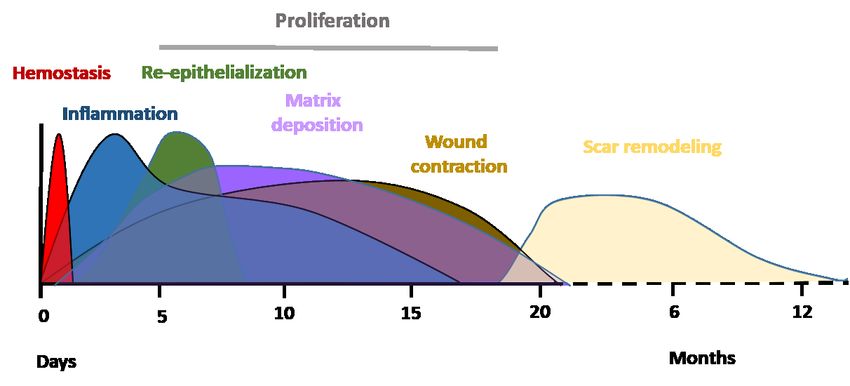
EXCLI Journal 2021;20:99-116 – ISSN 1611-2156
                         Received: November 26, 2020, accepted: January 04, 2021, published: January 11, 2021

gene are associated with venous ulceration            in numbers at 24 to 36 hours post-injury (Dovi
(Ashworth et al., 2005, 2008). Thus, it is fea-       et al., 2004). Neutrophils remove foreign ma-
sible that estrogen deprivation may contribute        terials and invading microorganisms, such as
to the development of chronic wound healing           bacteria, via the release of reactive oxygen
states in the elderly. The lack of extensive re-      species (ROS) and lysosomal enzymes, and
search in this area highlights the need for fur-      degrade damaged matrix tissues by colla-
ther investigations to explore the precise            genases and proteinases (Mosser and Ed-
mechanisms by which estrogen deficiency               wards, 2010). The majority of neutrophils are
may contribute to the development or pro-             enclosed in the wound clot and are either
gression of chronic wounds in the elderly.            eliminated with the eschar or by macrophages
This review explores current knowledge in             via phagocytosis (Newman et al., 1982;
this field, highlighting the critical role of es-     Kondo and Ishida, 2010).
trogen in reversing age-related impaired heal-             In response to chemoattractants such as
ing and prospects for developing more fo-             TGF-β, macrophage chemoattractant protein
cused therapies in the form of local dressings        1 (MCP-1), and macrophage inflammatory
that promote healing in the elderly via activa-       protein (MIP), monocytes from the blood-
tion of specific estrogen signaling pathways          stream subsequently arrive at the wound area
in regenerating tissues, whilst leaving other         and differentiate into tissue macrophages,
non-target tissues in the body largely unaf-          peaking in numbers around day 5 to day 7
fected.                                               post-injury (Lorenz and Longaker, 2008; Sen
                                                      and Roy, 2008). Macrophages replace neutro-
                                                      phils as the predominant inflammatory cells
       ACUTE WOUND HEALING
                                                      at the wound site and carry out the process of
     Acute wound healing is a complex and             phagocytosis of invading microorganisms, re-
dynamic biological process divided into four          moval of damaged tissues and dead neutro-
sequential, overlapping phases; hemostasis,           phils, and the release of growth factors such
inflammation, tissue proliferation and remod-         as PDGF and TGF-β (Beanes et al., 2003; El
eling of the tissue scar (Figure 1). Immedi-          Mohtadi et al., 2020). Damaged extracellular
ately after trauma, degranulating platelets ad-       matrix is degraded by the action of macro-
here to damaged blood vessels and start a he-         phage-derived proteolytic enzymes such as
mostatic reaction, increasing the coagulation         metalloproteases. Macrophages also release
cascade and producing a fibrin clot to prevent        growth factors that induce the proliferative
extreme blood loss and provide a temporary            phase including insulin-like growth factor-1
protection for the wound against foreign bod-         (IGF-1), keratinocyte growth factor (KGF),
ies (Vaughan et al., 2000; Weyrich and Zim-           epidermal growth factor (EGF) and vascular
merman, 2004; Gilliver et al., 2007). Platelets       endothelial growth factor (VEGF) (Shaw et
in the clot release a variety of pro-inflamma-        al., 1990). Three to ten days after injury, tis-
tory cytokines and growth factors including           sue proliferation starts. It is characterized by
platelet-derived growth factor (PDGF), trans-         the creation of new extracellular matrix
forming growth factor-beta (TGF-β), fibro-            (ECM) by fibroblasts, re-epithelialization (the
blast growth factor-2 (FGF-2), vascular endo-         restoration of an intact epidermis) by
thelial growth factor (VEGF) and epidermal            keratinocytes and angiogenesis (revasculari-
growth factor (EGF) (Bauer et al., 1985; Guo          zation) by endothelial cells. The final phase is
and DiPietro, 2010). These cytokines, chem-           remodeling of the tissue scar, which can take
okines and growth factors attract inflamma-           several months or, in some cases, up to a year
tory cells from circulation to the wound site,        post-injury. It is characterized by the remod-
initiating the inflammatory phase. Neutro-            eling of collagen and the vascular maturation
phils are the first inflammatory cells recruited      of newly formed capillaries, allowing vascu-
from circulation (Ley et al., 2007). They peak        lar density to return to normal within the

                                                  100
EXCLI Journal 2021;20:99-116 – ISSN 1611-2156
Received: November 26, 2020, accepted: January 04, 2021, published: January 11, 2021
Figure 1: Typical timescale and phases of acute wound healing stages in young adult humans.
Immediately after injury, healing initiates with hemostasis. This results in the formation of a fibrin clot
within minutes following injury. The inflammatory phase overlaps with hemostasis and occurs within
minutes after injury, with neutrophils being recruited from circulation, followed by monocytes. Monocytes
undergo a series of changes to differentiate into tissue macrophages, which carry out phagocytosis and
release cytokines that encourage the recruitment and activation of further leukocytes to the injury site
and initiation of the proliferation phase. Three to ten days after injury, the proliferation phase starts
enabling granulation tissue formation, re-epithelialization and angiogenesis. The final phase is the re-
modeling of a mature tissue scar, which can take several months or, in some cases, up to a year post-
injury. 0 = day of wounding/injury (El Mohtadi, 2019).
wound (Guo and DiPietro, 2010). For suc- and reduced matrix deposition (Figure 2)
cessful healing, wound repair requires pro- (Ashcroft et al., 1997b, 2002). Although the
gression through all four phases in the correct inflammatory response becomes more pro-
order and time frame (Singer and Clark, 1999; nounced with increasing age, the propensity
Guo and DiPietro, 2010). for wounds to become infected increases in
the elderly (Ashcroft et al., 2002; Cooper et
AGING AND WOUND HEALING al., 2015), due to the delay in wound repair
With increasing age, acute wound healing and the impaired ability of inflammatory cells
proceeds but becomes delayed. This detri- to eliminate bacteria from the wound site
mental change in acute wound healing in the (Emori et al., 1991; Thomas, 2001).
elderly is called age-related impaired healing In contrast, chronic wounds are character-
and is linked with intrinsic cellular aging pro- ized by failure of tissue repair processes to
cesses, including an elevated but delayed in- proceed through an orderly set of wound heal-
flammatory response, reduced cell prolifera- ing phases within an expected time frame.
tion and migration, decreased extracellular Wounds are deemed chronic if they do not
matrix (ECM) production and increased enzy- heal within three months and in many cases
matic degradation of tissues leading to skin they can take several months or even years to
fragility (Thomas, 2001). Delayed wound heal (if they heal at all) (Mustoe, 2005; Adeyi
healing in the elderly is associated with de- et al., 2009). Chronic wounds typically affect
layed hemostasis (Ashcroft et al., 1999), pro- the elderly (over 65 years of age) and arise
longed and excessive inflammation, delayed from one or more underlying pathologies,
re-epithelialization, impaired angiogenesis with more than 90 % of chronic wounds being
venous, diabetic or pressure ulcers (Boulton
101EXCLI Journal 2021;20:99-116 – ISSN 1611-2156
Received: November 26, 2020, accepted: January 04, 2021, published: January 11, 2021
et al., 2005). Chronic wounds have major leads to tissue breakdown (Snyder, 2005;
clinical implications and cause an enormous Taylor et al., 2005; Fazli et al., 2009). A shift
burden on healthcare services, in terms of in the balance between the formation and deg-
medical effort and cost (Harding et al., 2002; radation of ECM occurs, leading to ECM
Boulton et al., 2005). Chronic wound treat- breakdown by destructive inflammatory me-
ment costs the UK National Health Service diators such as proteases (Edwards et al.,
(NHS) about £5 billion per annum (Guest et 2004; Schönfelder et al., 2005). Chronic
al., 2015). wounds also have defective macrophage
At present, effective therapies/treatments function that leads to increased propensity of
for chronic wounds are somewhat limited, bacterial infection, decreased growth factor
making this an area of research that needs ur- secretion, impaired angiogenesis and delayed
gent attention. Chronic wounds become re-epithelialization (Hohn et al., 1976; Har-
trapped within the inflammatory phase of ding et al., 2002; Frykberg and Banks, 2015).
wound repair and are characterized by an ex-
cessive, unabated inflammatory response that
Figure 2: Schematic representation of the effect of age on acute wound healing. Age-related im-
paired healing is linked to delayed but excessive inflammation, delayed re-epithelialization, reduced
angiogenesis and decreased fibroblast proliferation and matrix deposition (El Mohtadi, 2019).
102EXCLI Journal 2021;20:99-116 – ISSN 1611-2156
Received: November 26, 2020, accepted: January 04, 2021, published: January 11, 2021
ESTROGEN AND AGING sults in a dramatic fall in the synthesis of ac-
tive androgens and estrogens in peripheral tis-
Endogenous estrogens are produced from
sues, a phenomenon which could be associ-
cholesterol, initially by several enzymes to
ated with several age-related diseases (Labrie
create androgens, such as testosterone and an-
et al., 1998). Estrogen synthesized locally in
drostenedione, which are then converted to
peripheral tissues becomes progressively
estrogens through the action of the P450 en-
more important after the menopause in
zyme aromatase, in the endoplasmic reticu-
women, when systemic levels are lost (Picard
lum of estrogen-producing cells (Payne and
et al., 2000). However, the rapid decline in lo-
Hales, 2004). In adipose tissues, andros-
cal production of active estrogens with in-
tenedione is converted to estrone whilst in
creasing age means peripheral estrogen pro-
ovarian granulosa cells testosterone is con-
duction is insufficient to compensate for the
verted into estradiol. Aromatase is found in
loss in systemic estrogen levels in elderly
many peripheral tissues such as skin, bone,
women.
adipose tissue, brain and vascular smooth
muscle (Nawata et al., 1995; Simpson, 2000;
Azcoitia et al., 2001; Ling et al., 2004). In fe- ESTROGEN RECEPTORS
males at the age of reproduction, systemic es- Over the past decades, the existence of
trogen is produced mainly by the ovary. It is two nuclear estrogen receptor (ER) proteins
predominantly biosynthesized in granulose have been identified, ER-alpha (ER-α) and
cells of the ovarian follicles and the corpora ER-beta (ER-β), that are part of the nuclear
lutea. In males, the gonad is the principle pro- receptor (NR) family. ER-α was first discov-
ducer of systemic estrogen. However, a sub- ered in 1958 (Jensen and Jacobson, 1960) and
stantial amount of estrogen is also produced is known to be predominant in reproductive
locally in peripheral tissues in both genders, tissues (Kuiper et al., 1997; Ali and Coombes,
acting in an autocrine and paracrine manner 2000; Campbell et al., 2010) whereas ER-β
(Labrie et al., 1998). A significant amount of was first identified in rat prostate and ovary in
inactive steroid precursors including dehy- 1996 (Mosselman et al., 1996) and predomi-
droepiandrosterone (DHEA), its sulphate nates in peripheral, non-reproductive tissues
(DHEA-S), and androstenedione (4-dione) (Kuiper et al., 1997; Ali and Coombes, 2000;
are produced by the adrenals and converted Campbell et al., 2010). The biological effects
into active steroid hormones in peripheral tis- of estrogens are largely mediated by the bind-
sues (Labrie et al., 1998). Several peripheral ing of estrogen to nuclear ER homodimers or
human tissues, such as adipose tissue, bone heterodimers (Matthews and Gustafsson,
and skin can produce active estrogens and an- 2003), and subsequent activation or repres-
drogens locally from conversion of adrenal- sion of gene transcription (Paige et al., 1999).
derived inactive precursors (Nelson and However, rapid, non-genomic estrogen sig-
Bulun, 2001). Plasma DHEA-S is the major naling involving membrane-bound ER pro-
adrenal-derived steroid precursor and levels teins has also been described (Gruber et al.,
in adult men and women are around 100 to 2002; Ascenzi et al., 2006). Recent research
500 times higher than those of testosterone suggests estrogen can have direct effects on
and as much as 1000 to 10 000 times higher inflammatory cells, such as monocytes and
than those of estradiol (Labrie et al., 2000). macrophages, and skin-associated cells such
Thus, inactive adrenal-derived steroid precur- as keratinocytes, due to the presence of nu-
sors provide a large circulating reservoir for clear and membrane-bound ER proteins
conversion into potent sex steroid hormones (Weusten et al., 1986; Stimson, 1988; Cocchi-
in peripheral tissues. However, the sharp de- ara et al., 1990). The response of particular in-
cline in DHEA and DHEA-S production by flammatory cells depends on the local levels
the adrenals during aging in both genders re-
103EXCLI Journal 2021;20:99-116 – ISSN 1611-2156
Received: November 26, 2020, accepted: January 04, 2021, published: January 11, 2021
of estrogen and the maturity (stage of differ- Both ER-α and ER-β enhance aspects of
entiation) of the cells (Ashcroft and Ash- acute wound repair but their roles are some-
worth, 2003). what different; although ER-α regulates in-
Estrogen signals predominantly by bind- flammatory cell activity, ER-β appears to
ing to inactive ER proteins in the nucleus of modulate the overall wound healing response
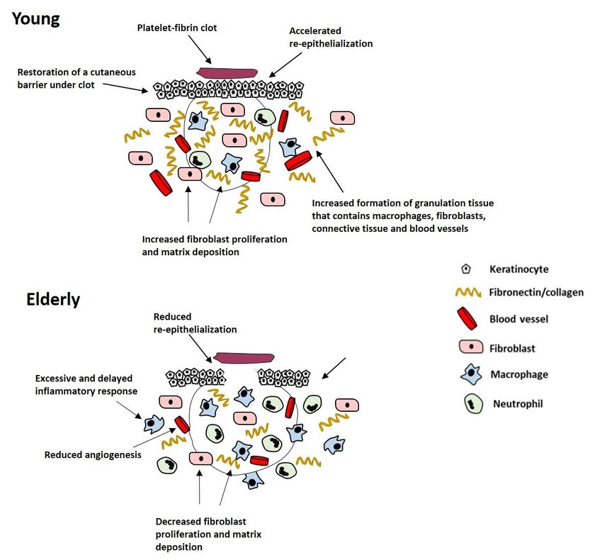
the cell (Klinge, 2000). ER proteins share a (Emmerson and Hardman, 2012). The de-
structure (Figure 3) that is typical of the NR layed wound repair observed in ovariecto-
family, consisting of six domains (A-F) (Kui- mized mice can be reversed by stimulation of
per et al., 1998; Klinge, 2000; Begam et al., ER-β alone, whilst ER-α activation alone fails
2017). ER proteins are expressed in skin, sug- to enhance murine wound repair (Campbell et
gesting estrogen regulates skin function, al., 2010). Moreover, estrogen replacement
maintenance and/or turnover (Ashworth, therapy in ovariectomized mice lacking func-
2005). While ER-α and ER-β have 97 % ho- tional ER-β retards wound healing, suggest-
mology in the C domain that acts as a DNA- ing ER-β may be critical to establishing
binding domain (DBD), they only have 55 % prompt tissue formation during wound repair
homology in the E domain which forms the (Campbell et al., 2010). In addition, a human
ligand-binding domain (LBD) (Barkhem et study conducted by Ashworth (2005) indi-
al., 1998; Webb et al., 1999; Klinge, 2000), cates that polymorphisms in the 0N promoter
enabling targeted ER activation using artifi- region of the human ER-β gene are signifi-
cial ligands with ER-specific binding affinity. cantly associated with chronic venous ulcera-
When estrogen binds to ER proteins, they tion in the British Caucasian population.
become activated and dimerize (Klinge,
2000). The DBD of each activated ER then EFFECT OF ESTROGEN ON SKIN
binds to an estrogen response element (ERE) MAINTENANCE
in the DNA of target genes and induces gene It is commonly accepted that the age-re-
transcription (Kuiper et al., 1998; Klinge, lated reduction in estrogen levels is linked
2000). In cells expressing a single ER sub- with skin degeneration. However, most evi-
type, homodimers of ER-α or ER-β are dence in humans comes from studies per-
formed (Kuiper et al., 1998). In cells that ex- formed in pre- and/or post-menopausal
press both ER subtypes, a heterodimer con- women. During pregnancy, skin syndromes
taining one ER-α and one ER-β may form such as psoriasis have been shown to im-
(Kuiper et al., 1998). ER heterodimers and prove, an effect that is directly linked to in-
ER-α homodimers bind to DNA with a similar creased estrogen levels in the circulation
affinity. However, ER-β homodimers bind to (Boyd et al., 1996). Moreover, oral contracep-
DNA with a lower affinity (Kuiper et al., tive pills are frequently used to treat severe
1998). acne. During the menopause, estrogen defi-
Figure 3: The structure of human ER-α and ER-β. Homology between domains (A-F) is represented
as percentage (%) similarity. NHD = N-terminal homology domain, DBD = DNA-binding domain, LBD =
ligand-binding domain (Webb et al., 1999; Klinge, 2000; Begam et al., 2017; El Mohtadi, 2019)
104EXCLI Journal 2021;20:99-116 – ISSN 1611-2156
Received: November 26, 2020, accepted: January 04, 2021, published: January 11, 2021
ciency results in detrimental changes in skin the vascularization of dermis and in terms of
appearance including sagging, wrinkling, skin appendages, estrogen extends the life cy-
dryness and fragility (Ashcroft et al., 1999; cle of human hair follicles but retards hair
Shah and Maibach, 2001). These changes can growth and sebum secretion by sebaceous
often be reversed during the first 6 months of glands (Stumpf et al., 1974).
topical or systemic estrogen replacement ther- In summary, the age-related fall in the lev-
apy (Brincat et al., 1987). els of estrogen detrimentally affects the
There is a reduction in mainly collagen maintenance and turnover of intact skin,
type III, but also type I to some degree, in the whilst estrogen supplementation reverses
skin of post-menopausal women compared to these effects in the elderly by stimulating
pre-menopausal women, resulting in a de- keratinocyte proliferation, increasing ECM
crease in the ratio of type III/type I collagen deposition and quality, and enhancing skin
within the dermis that is associated with es- turgor and moisture retention.
trogen deficiency (Affinito et al., 1999; Horng
et al., 2017). When applied locally to the skin ESTROGEN AND WOUND HEALING
of post-menopausal women, estradiol signifi- The influence of estrogen on wound heal-
cantly increases the production of hydroxy- ing was first studied in animals in 1947
proline, reflecting elevated collagen synthesis (Sjövall, 1947; Horng et al., 2017) and then in
in the dermis (Albright et al., 1941; Affinito humans in 1953 (Sjöstedt, 1953; Horng et al.,
et al., 1999; Sator et al., 2001; Horng et al., 2017). Subsequently, there has been an accu-
2017). Indeed, topical estrogen improves the mulating body of evidence supporting estro-
external facial appearance of post-menopau- gen as a global regulator of wound healing
sal women by reducing skin sagging and (Brincat et al., 1987; Varila et al., 1995; Af-
wrinkling (Schmidt et al., 1994). Not only finito et al., 1999; Sauerbronn et al., 2000;
topical but also systemic estrogen supplemen- Mills et al., 2005; Hardman and Ashcroft,
tation conserves skin thickness by promoting 2008; Brufani et al., 2009; Lee et al., 2013;
dermal collagen deposition in post-menopau- Midgley et al., 2016; Mukai et al., 2016;
sal women (Savvas et al., 1993; Sauerbronn et Chenu et al., 2017; Leblanc et al., 2017;
al., 2000). Horng et al., 2017; Pepe et al., 2017; Wil-
It has also been reported that estrogen re- kinson and Hardman, 2017).
placement therapy can improve skin elasticity Research has demonstrated the key role of
by 5 % per year (Brincat et al., 1987). In line sex-steroid hormones in inflammation and the
with this finding, topical estrogen supplemen- wound healing process (Guo and DiPietro,
tation improves the elasticity of ECM fibres 2010; Gilliver et al., 2007). Estrogen has pro-
in the dermis (Albright et al., 1941; Sator et tective, anti-inflammatory properties in sev-
al., 2001). Topical estrogen ointments notably eral tissues (Straub, 2007). Estrogen has also
increase the number and thickness of elastin been reported to stimulate wound repair pro-
fibres in the ECM, with histological examina- cesses such as re-epithelialization and ECM
tion demonstrating improved orientation and
production independently from its anti-in-
reduced fibre fragmentation in the dermis flammatory effects in elderly subjects of both
(Punnonen et al., 1987). Estrogen also pro- genders (Ashcroft et al., 1997b). HRT-treated
motes the synthesis of glycosaminoglycans in post-menopausal women heal acute wounds
the ECM, restoring skin turgor and moisture faster than their age-matched control counter-
levels (Brincat, 2000). parts, who have taken no estrogen supplemen-
Topical estrogen application enhances tation (Ashcroft et al., 1997b). Other reports
stratum corneum barrier function of skin in indicate that topical estrogen supplementation
post-menopausal women and increases the enhances wound healing in elderly male and
rate of mitosis and turnover of epidermal cells
(Stumpf et al., 1974). Estrogen also enhances
105EXCLI Journal 2021;20:99-116 – ISSN 1611-2156
Received: November 26, 2020, accepted: January 04, 2021, published: January 11, 2021
female patients, connected with a reduced in- (Ashcroft et al., 1997b; Ashcroft and Ash-
flammatory response (Ashcroft et al., 1997b, worth, 2003; Thornton, 2013; Archer, 2012;
1999). Stevenson and Thornton, 2007).
Variances in the human immune system
between male and female subjects have been Effect of estrogen on the inflammatory
identified in several epidemiological and phase of wound healing
medical studies (McGowan et al., 1975; It is commonly known that age-related
Bone, 1992), with evidence indicating that impaired healing is associated with an exces-
women have a superior immune system com- sive and prolonged inflammatory response,
pared to men (Gulshan et al., 1990; Wich- linked with increased but delayed inflamma-
mann et al., 1996). Other experiments have tory cell recruitment, and increased secretion
indicated that estrogen has immune-enhanc- of pro-inflammatory cytokines such as TNF-
ing properties during stress, including in- α (Ralston et al., 1990; Pottratz et al., 1994).
creased resistance to several pathogenic in- Moreover, TNF-α is elevated in elderly pa-
fections (Yamamoto, 1999). tients with venous ulcers compared to age-
Since systemic and peripheral estrogens matched healthy controls, with the highest
decline with age, it is suggested that estrogen levels of TNF-α typically found in patients
deprivation in the elderly could increase the carrying polymorphisms of the promoter re-
propensity for chronic wounds. Margolis et al. gion of the ER- gene that predispose to ve-
(2002) performed a case-cohort study to in- nous ulceration (Ashworth et al., 2008).
vestigate the protective effects of estrogen Recent research has indicated that chronic
against chronic wounds. Patients aged oved wounds are associated with elevated levels of
65 years receiving HRT treatment were elastase and MMPs, which are released by
shown to be 30-40 % less likely to develop a macrophages, keratinocytes and fibroblasts,
venous leg ulcer than age-matched patients and linked with excessive tissue destruction
lacking HRT supplementation (Margolis et (Wysocki et al., 1993). Estrogen has been de-
al., 2002). scribed to control and dampen the early in-
Chronic wounds are characterized by an flammatory response during acute wound
excessive and chronic prolonged inflamma- healing by inhibiting neutrophil infiltration to
tion. High levels of inflammatory mediators, the wound via a reduction in the expression of
including tumor necrosis factor alpha (TNF- cell adhesion molecules (Ashcroft et al.,
α), interleukin-1 beta (IL-1β), IL-6, IGF-1 and 1999; Sproston et al., 2018). Furthermore, es-
matrix metalloproteinases (MMPs), that are trogen increases the oxidative metabolism of
present in chronic wound exudate (Ashcroft neutrophils, suggesting estrogen deprivation
et al., 1997b, 1999) are downregulated via the could lead to diminished phagocytic capabil-
action of estrogen (Vural et al., 2006; Straub, ity of neutrophils, an increased risk of infec-
2007; Wira et al., 2015). In particular, TNF- tion and a postponement in healing (Magnus-
α is elevated in humans that are predisposed son and Einarsson, 1990). Estrogen has been
to chronic wounds and has been identified as shown to have a direct influence on mono-
a therapeutic target for impaired wound heal- cytes and macrophages, due to their posses-
ing in the elderly (Ashcroft et al., 2012). Both sion of both nuclear and membrane-bound es-
systemic and topical estrogen treatments en- trogen receptor (ER) proteins (Weusten et al.,
hance wound healing in elderly men and 1986; Suenaga et al., 1996, 1998). In addition,
women by stimulating re-epithelialization, 17β-estradiol has been reported to reverse the
angiogenesis, matrix deposition and wound substantial delay in cutaneous murine wound
contraction whilst dampening the inflamma- healing induced by bacterial lipopolysaccha-
tory response and expression of pro-inflam- ride (Crompton et al., 2016).
matory cytokines and proteolytic mediators Increased levels of epidermal pro-matrix
metalloproteinase-2 (pro-MMP-2) have been
106EXCLI Journal 2021;20:99-116 – ISSN 1611-2156
Received: November 26, 2020, accepted: January 04, 2021, published: January 11, 2021
observed in intact aging skin and is immedi- menopausal women (Ashcroft et al., 1997b).
ately activated following cutaneous injury, It has been reported that the rate of wound re-
explaining the reported rise in MMP-2 and epithelialization of post-menopausal women
ECM degeneration observed in the wounds of treated with HRT for more than 3 months was
the elderly (Ashcroft et al., 1997a). In addi- similar to levels of re-epithelialization in pre-
tion, research suggests that estrogen defi- menopausal females, whereas a non-HRT
ciency inhibits the differentiation of mono- post-menopausal group showed diminished
cytes into tissue macrophages during the in- re-epithelialization. This improved re-epithe-
flammatory phase of wound healing, leading lialization following estrogen supplementa-
to an increase in protease expression (Calvin tion is due to increased proliferation of epi-
et al., 1998a). Estrogen decreases tissue-dam- dermal keratinocytes (Raja et al., 2007).
aging protease levels, including elastase and In addition to its effect on epithelial mi-
MMP secretion, leading to an overall increase gration and proliferation, estrogen indirectly
in the content of collagen and fibronectin in effects matrix deposition by mesenchymal
the dermis (Ashcroft et al., 1999). cells. Various in vivo animal studies report
In skin, the anti-inflammatory effect of es- that estrogen increases fibroblast infiltration
trogen is predominantly mediated through in- and collagen deposition. In contrast, a small
hibition of the pro-inflammatory cytokine, number of studies report a decrease in fibro-
macrophage migration inhibitory factor blast infiltration and collagen deposition fol-
(MIF) (Hardman et al., 2005). Macrophage lowing treatment with estrogen in mice
migration inhibitory factor (MIF) has been (Lundgren, 1973; Pallin et al., 1975). A pos-
identified as a global regulator of wound heal- sible explanation for these contradictions in-
ing mediated by estrogen and released by clude differences in the wound models, hor-
monocytes, macrophages, neutrophils, endo- mone concentrations and intervals of admin-
thelial cells and keratinocytes (Hardman et istration used. Furthermore, the duration of
al., 2005; Emmerson et al., 2009). Ashcroft et estrogen insufficiency results in distinct ef-
al. (2003) reported that mice with estrogen de- fects on several healing parameters; for in-
ficiency have higher MIF levels, resulting in stance, wound contraction becomes reduced
an elevated inflammatory response and de- after 4 months of estrogen deprivation
layed wound healing, whereas MIF null-mice whereas matrix deposition becomes reduced
displayed enhanced wound healing, with after only 1 month (Calvin et al., 1998b). In
lower inflammation and greater matrix for- humans, topical estrogen supplementation in
mation. Estrogen downregulates MIF expres- elderly men and women results in reduced
sion leading to a decline in inflammation, en- wound size via stimulation of wound contrac-
hanced matrix deposition, increased re-epi- tion (Ashcroft et al., 1999). Estrogen pro-
thelialization and an overall accelerated motes PDGF expression by monocytes and
wound repair (Hardman et al., 2005). macrophages (Mendelsohn and Karas, 1999),
leading to mitogenesis and chemotaxis of fi-
Effect of estrogen on the proliferative phase broblasts and a subsequent increase in wound
of wound healing contraction and ECM deposition (Seppä et al.,
Age-related impaired healing is linked 1982). Estrogen also enhances the secretion
with reduced growth factor expression, re- of TGF-β1 by dermal fibroblasts in vivo (Ash-
duced keratinocyte proliferation and in- croft et al., 1997b, 1999), resulting in en-
creased response to inhibitory cytokines, hanced formation of ECM, particularly colla-
causing a delayed re-epithelialization in vivo gen deposition (Ashcroft and Ashworth,
(Butcher and Klingsberg, 1963; Rattan and 2003).
Derventzi, 1991; Holt et al., 1992). Estrogen Estrogen promotes angiogenesis, leading
enhances the mitogenesis of keratinocytes to increased granulation tissue (Iyer et al.,
and increases re-epithelialization in post-
107EXCLI Journal 2021;20:99-116 – ISSN 1611-2156
Received: November 26, 2020, accepted: January 04, 2021, published: January 11, 2021
2012) through a direct stimulation of endothe- 1991). Another in vivo study reports that top-
lial cells (Rubanyi et al., 2002). Estrogen ical estrogen treatment increases collagen
modulates the synthesis of IL-1 by tissue deposition in elderly males and females after
macrophages, a key protein implicated in the 7 and 80 days post-injury (Ashcroft et al.,
creation of a new granulation tissue (Hu et al., 1999a). It was also noticed in other in vivo
1988). Estrogen increases endothelial cell at- studies that matrix collagen deposition at 7
tachment to laminin, fibronectin and colla- and 84 days post-wounding decreased in post-
gens I and IV in vitro. In addition, estrogen menopausal women lacking HRT treatment.
enhances the creation of capillary-like struc- In contrast, post-menopausal females who
tures by endothelial cells, when positioned on took HRT for more than 3 months had similar
a reconstructed basement membrane (Mo- levels of matrix collagen deposition and
rales et al., 1995). Paradoxically, other in wound remodeling as younger pre-menopau-
vitro studies report a reduction in vascularity sal females (Ashcroft et al., 1997b, 1999).
following stimulation with estrogen (Nyman, Estrogen stimulates the expression of
1971; Lundgren, 1973). The precise effect of TGF-β1 in vivo. This results in improving col-
estrogen on angiogenesis remains unknown, lagen deposition in the dermis (Ashcroft et al.,
and additional investigations are needed to 1997b). Reports suggest a decreased wound
define the impact of estrogen on vascularzsa- collagen deposition associated with MMP-
tion in acute and impaired wound healing. mediated collagenolysis in ovarectomized
In summary, despite some contradictions rats (Pirila et al., 2001). These effects were re-
in the literature, estrogen appears on balance versed by estrogen replacement, implicating
to enhance most tissue formation occurring in estrogen as a pivotal mediator involved in
the proliferative phase of wound healing, par- shifting the balance from matrix degradation
ticularly re-epitheliazation and ECM for- to matrix synthesis (Pirila et al., 2001). Inter-
mation. estingly, an in vivo study indicated that the
quality of mature tissue scars was greater in
Effect of estrogen on the remodeling phase post-menopausal women in comparison with
of wound healing pre-menopausal women. This suggests that
The age-related decline in estrogen levels estrogen enhances wound repair at the ex-
causes a decrease in wound collagen and fi- pense of scar quality (Ashcroft et al., 1997b).
bronectin in vivo. This has been associated
with elevated levels of inflammatory cell-de- FUTURE PERSPECTIVES FOR
rived elastase, MMP-2 and MMP-9 (Herrick ESTROGEN THERAPIES
et al., 1997; Ashcroft et al., 1997a). Estrogen Although many known effects of estrogen
supplementation reverses the degradation of on wound healing have been established in
ECM by inhibiting the synthesis of wound the past two decades, fewer recent develop-
proteases such as MMPs during wound re- ments have been made and there remain sub-
modeling (Ashcroft and Ashworth, 2003; stantial areas for further investigation. It has
Brincat, 2000).
been established that estrogen plays a funda-
Topical estrogen supplementation in- mental beneficial role in skin maintenance
creases the deposition of collagen during the and acute wound healing processes. Moreo-
remodeling phase of wound repair in elderly ver, the systemic and peripheral decline in es-
patients (Ashcroft et al., 1997b, 1999). Previ- trogen with increasing age suggests estrogen
ous animal studies report that 17β-estradiol deprivation could be linked with chronic
increases the production of tissue inhibitor of wounds in the elderly. However, systemic es-
metalloproteinases (TIMPs) by rabbit uterine trogen replacement therapy is an unfocused,
fibroblasts, but reduces the production of pro- biological sledgehammer rather than a tar-
collagenase and pro-stromelysin (Sato et al., geted treatment strategy. Although estrogen is
108EXCLI Journal 2021;20:99-116 – ISSN 1611-2156
Received: November 26, 2020, accepted: January 04, 2021, published: January 11, 2021
protective against photoaging, an extrinsic ag- merous peripheral tissues, but are anti-estro-
ing process that correlates with higher mortal- genic in the breast tissue and are therefore
ity rates from skin cancers in men than used extensively in breast cancer research
women (Weinstock, 1994; Miller and Neil, (Furr and Jordan, 1984; Morris and Wakeling,
1997), unopposed systemic estrogen replace- 2002; Park and Jordan, 2002; Mirkin and
ment therapy is a risk factor for breast and en- Pickar, 2015). Tamoxifen was discovered and
dometrial cancer development (Nuttall et al., reported by the Food and Drug Administra-
2001), thereby restricting its exploitation in tion (FDA) in 1977 (Park and Jordan, 2002;
clinical practice. The widespread distribution Jordan, 2006; Mirkin and Pickar, 2015;
of estrogen-responsive tissues exposes non- Quirke, 2017). Tamoxifen binds to both ER
target cells to the potential hyper-proliferative proteins and its effect depends on the cell and
and neoplastic effects of systemic estrogen tissue type, being anti-estrogenic in breast tis-
therapies, suggesting either local estrogen or sue and therefore commonly used to prevent
targeted therapies are needed. Interestingly, and/or treat breast cancer in both post- and
studies performed in vitro have shown that pre-menopausal females (Zidan et al., 2004;
ER- is the dominant partner in heterodimers, Quirke, 2017). Tamoxifen has also been re-
resulting in an ER-predominant effect with ported to maintain the density of bone in rats
repressed ER- transcriptional activity (Pet- and humans (Jordan et al., 1987; Zidan et al.,
tersson and Gustafsson, 2001). Thus, by mod- 2004). However, it has multiple side effects
ulating ER--mediated gene transcription, and is frequently linked with endometrial can-
ER- may decrease the overall cellular sensi- cer due to its estrogenic effects in the uterus
tivity to estrogen and provide protection (Kedar et al., 1994).
against the hyper-proliferative and neoplastic There have been some investigations on
effects of ER- (Rollerova and Urbancikova, the effect of SERMs on skin and wound heal-
2000). Thus, a clear understanding of tissue- ing processes. Tamoxifen and raloxifene have
specific regulation of ER expression and been shown to stimulate fibroblast prolifera-
downstream cellular and molecular mecha- tion in vitro (Stevenson et al., 2009). While
nisms of estrogen action might enable con- raloxifene improves skin elasticity and colla-
trolled manipulation of estrogen signaling gen deposition in post-menopausal females
pathways during wound repair, potentially (Sumino et al., 2009), genistein has been re-
leading to the development of more targeted ported to improve the vascularization of the
therapies with fewer side effects on non-target dermis and augment the loss of epidermal
tissues. thickness typically observed in post-meno-
Selective estrogen receptor modulators pausal females (Moraes et al., 2009). Another
(SERMs) are ER-interacting molecules that study on mice indicated genistein stimulates
have the ability to bind ER proteins and act as wound healing via synthesis of TGF-β1 (Ma-
agonists in some tissues whilst acting as an- rini et al., 2010). Moreover, tamoxifen, ralox-
tagonists in different tissues (Brzozowski et ifene and genistein all significantly enhance
al., 1997; Cho and Nuttall, 2001). SERMs wound healing in ovariectomized mice by
have been used clinically to promote the ben- stimulating re-epithelialization and dampen-
eficial effects of estrogen in target tissues ing inflammation via activation of ER-β
whilst reducing the detrimental effects of es- (Hardman et al., 2008; Emmerson et al.,
trogen (e.g. increased risk of breast cancer) in 2010). However, the use of existing SERMs
non-target tissues (Mirkin and Pickar, 2015). have not yet been exploited in the treatment
Tamoxifen, raloxifene and the dietary phy- of chronic wound states.
toestrogen genistein are the most frequently The repurposing of existing pharmaceuti-
documented SERMs in the literature. They cal drugs or the development of novel thera-
are known to have estrogenic effects in nu- pies that act as ER-specific ligands or exhibit
tissue-specific estrogenic effects, delivered
109EXCLI Journal 2021;20:99-116 – ISSN 1611-2156
Received: November 26, 2020, accepted: January 04, 2021, published: January 11, 2021
locally within specialized wound dressings REFERENCES
may have potential clinical applications in the Adeyi A, Muzerengi S, Adeyi IGA, Gupta I. Leg ulcers
treatment of chronic wound states in the el- in older people: A review of management. Br J Med
derly. Understanding the differential effects Practit. 2009;2(3):21-8.
on downstream gene transcription or repres-
Affinito P, Palomba S, Sorrentino C, Di Carlo C, Bi-
sion in various tissue/cell types may help de- fulco G, Arienzo MP, et al. Effects of postmenopausal
velop more focused treatments for impaired hypoestrogenism on skin collagen. Maturitas. 1999;
wounds that can mediate specific estrogen-re- 33:239-47.
sponsive signaling pathways in injured tissues
Albright F, Smith PH, Richardson AM. Postmenopau-
whilst reducing unwanted side effects in non- sal osteoporosis: Its clinical features. J Am Med Assoc.
target tissues. 1941;116:2465-74.
CONCLUSION Ali S, Coombes RC. Estrogen receptor alpha in human
breast cancer: Occurrence and significance. J Mam-
The literature indicates estrogen defi- mary Gland Biol Neoplasia. 2000;5:271-81.
ciency is a central paradigm of age-related
Archer DF. Postmenopausal skin and estrogen. Gyne-
impaired wound healing in both genders, with
col Endocrinol. 2012;28(Suppl 2):2-6.
topical and systemic estrogen replacement re-
versing the detrimental effects of aging on Ascenzi P, Bocedi A, Marino M. Structure–function
both wound repair and skin maintenance. relationship of estrogen receptor α and β: Impact on hu-
There is growing evidence indicating estro- man health. Mol Aspects Med. 2006;27:299-402.
gen deprivation may also contribute to the de- Ashcroft GS, Ashworth JJ. Potential role of estrogens
velopment of chronic wounds in the elderly in wound healing. Am J Clin Dermatol. 2003;4:737-43.
but further research is needed in this area. In-
Ashcroft, Horan MA, Herrick SE, Tarnuzzer RW,
terestingly, although the beneficial effects of
Schultz GS, Ferguson MWJ. Age-related differences in
estrogen on wound repair have been widely the temporal and spatial regulation of matrix metallo-
explored, the development of estrogen-based proteinases (MMPs) in normal skin and acute cutane-
treatments to promote healing has failed to ous wounds of healthy humans. Cell Tissue Res.
gain traction to date, most likely due to unde- 1997a;290:581-91.
sired cellular activity (including hyper-prolif- Ashcroft GS, Dodsworth J, Van Boxtel E, Tarnuzzer
erative/neoplastic effects) in non-target tis- R, Horan MA, Schultz GS, et al. Estrogen accelerates
sues. However, a rekindled interest may be cutaneous wound healing associated with an increase
stimulated by prospects of developing tar- in TGF-b1 levels. Nat Med. 1997b;3(11):1209-15.
geted therapeutic strategies that might pro- Ashcroft GS, Greenwell-Wild T, Horan MA, Wahl
mote healing through selective activation of SM, Ferguson MW. Topical estrogen accelerates cuta-
estrogen-responsive signaling pathways in re- neous wound healing in aged humans associated with
generating peripheral tissues, whilst leaving an altered inflammatory response. Am J Pathol. 1999;
non-target tissues largely unaffected. 155:1137-46.
Ashcroft GS, Mills SJ, Ashworth JJ. Ageing and
Conflict of interest wound healing. Biogerontology. 2002;3:337-45.
The authors declare no conflict of interest.
Ashcroft GS, Mills SJ, Lei K, Gibbons L, Jeong M-J,
Taniguchi M, et al. Estrogen modulates cutaneous
Acknowledgment wound healing by downregulating macrophage migra-
The authors wish to acknowledge that this tion inhibitory factor. J Clin Invest. 2003;111:1309-18.
review article includes some aspects of the in-
troductory chapters of the PhD thesis submit- Ashcroft, Jeong MJ, Ashworth JJ, Hardman M, Jin W,
Moutsopoulos N, et al. Tumor necrosis factor‐alpha
ted by the lead author (Mohamed El Moh- (TNF‐α) is a therapeutic target for impaired cutaneous
tadi). wound healing. Wound Repair Regen. 2012;20:38-49.
110EXCLI Journal 2021;20:99-116 – ISSN 1611-2156
Received: November 26, 2020, accepted: January 04, 2021, published: January 11, 2021
Ashworth JJ. Estrogen receptor polymorphisms and Brincat MP, Versi E, Moniz CF, Magos A, de Trafford
wound healing. Manchester, UK: The University of J, Studd JW. Skin collagen changes in postmenopausal
Manchester, Faculty of Life Sciences, 2005. women receiving different regimens of estrogen ther-
apy. Obstet Gynecol. 1987;70:123-7.
Ashworth JJ, Smyth JV, Pendleton N, Horan M, Pay-
ton A, Worthington J, et al. The dinucleotide (CA) re- Brufani M, Ceccacci F, Filocamo L, Garofalo B,
peat polymorphism of estrogen receptor beta but not Joudioux R, La Bella A, et al. Novel locally active es-
the dinucleotide (TA) repeat polymorphism of estrogen trogens accelerate cutaneous wound healing. A prelim-
receptor alpha is associated with venous ulceration. J inary study. Mol Pharm. 2009;6:543-56.
Steroid Biochem Mol Biol. 2005;97:266-70.
Brzozowski AM, Pike AC, Dauter Z, Hubbard RE,
Ashworth JJ, Smyth JV, Pendleton N, Horan M, Pay- Bonn T, Engström O, et al. Molecular basis of agonism
ton A, Worthington J, et al. Polymorphisms spanning and antagonism in the oestrogen receptor. Nature.
the 0N exon and promoter of the estrogen receptor‐beta 1997;389(6652):753.
(ERβ) gene ESR2 are associated with venous ulcera-
tion. Clin Genet. 2008;73:55-61. Butcher EO, Klingsberg J. Age, gonadectomy, and
wound healing in the palatal mucosa of the rat. Oral
Azcoitia I, Sierra A, Veiga S, Honda Si, Harada N, Surg Oral Med Oral Pathol. 1963;16:484-93.
Garcia‐Segura LM. Brain aromatase is neuroprotec-
tive. J Neurobiol. 2001;47:318-29. Calvin M, Dyson M, Rymer J, Young SR. The effects
of ovarian hormone deficiency on wound contraction
Barkhem T, Carlsson B, Nilsson Y, Enmark E, Gus- in a rat model. Br J Obstet Gynaecol. 1998a;105:223-7
tafsson J-Å, Nilsson S. Differential response of estro-
gen receptor α and estrogen receptor β to partial estro- Calvin M, Dyson M, Rymer J, Young SR. The effect
gen agonists/antagonists. Mol Pharmacol. 1998;54: of ovarian hormone deficiency on macrophage infiltra-
105-12. tion during the inflammatory phase of wound healing
in a rat model. Wounds: A compendium of clinical re-
Bauer E, Cooper T, Huang J, Altman J, Deuel T. Stim- search and practice. 1998b;10(5):158-63.
ulation of in vitro human skin collagenase expression
by platelet-derived growth factor. Proc Natl Acad Sci Campbell L, Emmerson E, Davies F, Gilliver SC,
U S A. 1985;82:4132-6. Krust A, Chambon P, et al. Estrogen promotes cutane-
ous wound healing via estrogen receptor β independent
Beanes SR, Dang C, Soo C, Ting K. Skin repair and of its antiinflammatory activities. J Exp Med. 2010;
scar formation: The central role of TGF-[beta]. Expert 207:1825-33.
Rev Mol Med. 2003;5(08):1-22.
Chenu C, Adlanmerini M, Boudou F, Chantalat E, Gui-
Begam AJ, Jubie S, Nanjan M. Estrogen receptor ago- hot A-L, Toutain C, et al. Testosterone prevents cuta-
nists/antagonists in breast cancer therapy: A critical re- neous ischemia and necrosis in males through comple-
view. Bioorg Chem. 2017;71:257-74. mentary estrogenic and androgenic actions. Arterio-
scler Thromb Vasc Biol. 2017;37:909-19.
Bérard A, Kahn SR, Abenhaim L. Is hormone replace-
ment therapy protective for venous ulcer of the lower Cho CH, Nuttall ME. Therapeutic potential of oestro-
limbs? Pharmacoepidemiol Drug Saf. 2001;10:245-51. gen receptor ligands in development for osteoporosis.
Emerg Drugs. 2001;6(1):137-54.
Bone RC. Toward an epidemiology and natural history
of SIRS (systemic inflammatory response syndrome). Cocchiara R, Albeggiani G, Di Trapani G, Azzolina A,
JAMA. 1992;268:3452-5. Lampiasi N, Rizzo F, et al. Modulation of rat peritoneal
mast cell and human basophil histamine release by es-
Boulton AJM, Vileikyte L, Ragnarson-Tennvall G, trogens. Int Arch Allergy Immunol. 1990;93:192-7.
Apelqvist J. The global burden of diabetic foot disease.
Lancet. 2005;366(9498):1719-24. Cooper RL, Segal RA, Diegelmann RF, Reynolds AM.
Modeling the effects of systemic mediators on the in-
Boyd AS, Morris LF, Phillips CM, Menter MA. Psori- flammatory phase of wound healing. J Theor Biol.
asis and pregnancy: Hormone and immune system in- 2015;367:86-99.
teraction. Int J Dermatol. 1996;35:169-72.
Crompton R, Williams H, Ansell D, Campbell L,
Brincat MP. Hormone replacement therapy and the Holden K, Cruickshank S, et al. Oestrogen promotes
skin. Maturitas. 2000;35:107-17. healing in a bacterial LPS model of delayed cutaneous
wound repair. Lab Invest. 2016;96:439-49.
111EXCLI Journal 2021;20:99-116 – ISSN 1611-2156
Received: November 26, 2020, accepted: January 04, 2021, published: January 11, 2021
Dovi JV, Szpaderska AM, DiPietro LA. Neutrophil Guest JF, Ayoub N, McIlwraith T, Uchegbu I, Gerrish
function in the healing wound: Adding insult to injury? A, Weidlich D, et al. Health economic burden that
Thromb Haemost. 2004;92:275-80. wounds impose on the National Health Service in the
UK. BMJ Open. 2015;5(12):e009283.
Edwards JV, Howley P, Cohen IK. In vitro inhibition
of human neutrophil elastase by oleic acid albumin for- Gulshan S, McCruden A, Stimson W. Oestrogen recep-
mulations from derivatized cotton wound dressings. Int tors in macrophages. Scand J Immunol. 1990;31:691-
J Pharm. 2004;284(1-2):1-12. 7.
El Mohtadi M. Effect of estrogen on host-pathogen in- Guo Sa, DiPietro LA. Factors affecting wound healing.
teractions in ex vivo and in vitro models of the inflam- J Dental Res. 2010;89:219-29.
matory phase of age-related impaired healing. Man-
chester, UK: Manchester Metropolitan University, De- Harding KG, Morris HL, Patel GK. Science, medicine
partment of Life Sciences, Ph.D. Thesis, 2019. and the future: Healing chronic wounds. BMJ.
2002;324(7330):160-3.
El Mohtadi M, Pilkington L, Liauw CM, Ashworth JJ,
Dempsey-Hibbert N, Belboul A, et al. Differential en- Hardman M, Ashcroft GS. Estrogen, not intrinsic ag-
gulfment of Staphylococcus aureus and Pseudomonas ing, is the major regulator of delayed human wound
aeruginosa by monocyte-derived macrophages is asso- healing in the elderly. Genome Biol. 2008;9(5):R80.
ciated with altered phagocyte biochemistry and mor-
phology. EXCLI J. 2020;19:1372-84. Hardman M, Waite A, Zeef L, Burow M, Nakayama T,
Ashcroft GS. Macrophage migration inhibitory factor:
Emmerson E, Hardman MJ. The role of estrogen defi- A central regulator of wound healing. Am J Pathol.
ciency in skin ageing and wound healing. Biogerontol- 2005;167:1561-74.
ogy. 2012;13(1):3-20.
Hardman MJ, Emmerson E, Campbell L, Ashcroft GS.
Emmerson E, Campbell L, Ashcroft GS, Hardman MJ. Selective estrogen receptor modulators accelerate cu-
Unique and synergistic roles for 17β-estradiol and taneous wound healing in ovariectomized female mice.
macrophage migration inhibitory factor during cutane- Endocrinology. 2008;149:551-7.
ous wound closure are cell type specific. Endocrinol-
ogy. 2009;150:2749-57. Herrick S, Ashcroft G, Ireland G, Horan M, McCollum
C, Ferguson M. Up-regulation of elastase in acute
Emmerson E, Campbell L, Ashcroft GS, Hardman MJ. wounds of healthy aged humans and chronic venous
The phytoestrogen genistein promotes wound healing leg ulcers are associated with matrix degradation. Lab
by multiple independent mechanisms. Mol Cell Endo- Invest. 1997;77:281-8.
crinol. 2010;321:184-93.
Hohn DC, MacKay RD, Halliday B, Hunt TK. Effect
Emori TG, Banerjee SN, Culver DH, Gaynes RP, of O2 tension on microbicidal function of leukocytes in
Horan TC, Edwards JR, et al. Nosocomial infections in wounds and in vitro. Surg Forum. 1976;27(62):18-20
elderly patients in the United States, 1986–1990. Am J
Med. 1991;91:S289-S93. Holt DR, Kirk SJ, Regan MC, Hurson M, Lindblad WJ,
Barbul A. Effect of age on wound healing in healthy
Fazli M, Bjarnsholt T, Kirketerp-Møller K, Jørgensen human beings. Surgery. 1992;112:293-8.
B, Andersen AS, Krogfelt KA, et al. Nonrandom dis-
tribution of Pseudomonas aeruginosa and Staphylococ- Horng HC, Chang WH, Yeh CC, Huang BS, Chang
cus aureus in chronic wounds. J Clin Microbiol. 2009; CP, Chen YJ, et al. Estrogen effects on wound healing.
47:4084-9. Int J Mol Sci. 2017;18(11):2325.
Frykberg RG, Banks J. Challenges in the treatment of Hu S-K, Mitcho YL, Rath NC. Effect of estradiol on
chronic wounds. Adv Wound Care. 2015;4:560-82. interleukin 1 synthesis by macrophages. Int J Im-
munopharmacol. 1988;10:247-52.
Furr B, Jordan V. The pharmacology and clinical uses
of tamoxifen. Pharmacol Ther. 1984;25:127-205. Iyer V, Klebba I, McCready J, Arendt LM, Betancur-
Boissel M, Wu M-F, et al. Estrogen promotes ER-Neg-
Gilliver SC, Ashworth JJ, Ashcroft GS. The hormonal ative tumor growth and angiogenesis through mobili-
regulation of cutaneous wound healing. Clin Dermatol. zation of bone marrow–derived monocytes. Cancer
2007;25(1):56-62. Res. 2012;72:2705-13.
Gruber CJ, Tschugguel W, Schneeberger C, Huber JC.
Production and actions of estrogens. N Engl J Med.
2002;346:340-52.
112EXCLI Journal 2021;20:99-116 – ISSN 1611-2156
Received: November 26, 2020, accepted: January 04, 2021, published: January 11, 2021
Jensen EV, Jacobson HI. Fate of steroid estrogens in Ling S, Dai A, Dilley RJ, Jones M, Simpson E,
target tissues. In: Pincus G, Vollmer EP (eds). Biolog- Komesaroff PA, et al. Endogenous estrogen deficiency
ical activities of steroids in relation to cancer (pp 161- reduces proliferation and enhances apoptosis-related
78). New York: Academic Press, 1960. death in vascular smooth muscle cells: insights from
the aromatase-knockout mouse. Circulation. 2004;109:
Jordan VC. Tamoxifen (ICI46, 474) as a targeted ther- 537-43.
apy to treat and prevent breast cancer. Br J Pharmacol.
2006;147(S1):S269-S76. Lorenz HP, Longaker MT. Wounds: Biology, pathol-
ogy, and management. In: Norton JA et al. (eds). Sur-
Jordan VC, Phelps E, Lindgren JU. Effects of anti-es- gery (pp 77-88). NewYork: Springer, 2008.
trogens on bone in castrated and intact female rats.
Breast Cancer Res Treat. 1987;10(1):31-5. Lundgren D. Influence of estrogen and progesterone on
exudation, inflammatory cell migration and granula-
Kedar R, Bourne TH, Collins W, Campbell S, Powles tion tissue formation in preformed cavities. Scand J
T, Ashley S, et al. Effects of tamoxifen on uterus and Plastic Reconstruct Surg. 1973;7(1):10-4.
ovaries of postmenopausal women in a randomised
breast cancer prevention trial. Lancet. 1994;343 Magnusson U, Einarsson S. Effects of exogenous oes-
(8909):1318-21. tradiol on the number and functional capacity of circu-
lating mononuclear and polymorphonuclear leukocytes
Klinge CM. Estrogen receptor interaction with co-acti- in the sow. Vet Immunol Immunopathol. 1990;25:235-
vators and co-repressors. Steroids. 2000;65:227-51. 47.
Kondo T, Ishida Y. Molecular pathology of wound Margolis DJ, Bilker W, Knauss J, Baumgarten M,
healing. Forensic Sci Int. 2010;203:93-8. Strom BL. The incidence and prevalence of pressure
ulcers among elderly patients in general medical prac-
Kuiper GG, Carlsson B, Grandien K, Enmark E, tice. Ann Epidemiol. 2002;12:321-5.
Häggblad J, Nilsson S, et al. Comparison of the ligand
binding specificity and transcript tissue distribution of Marini H, Polito F, Altavilla D, Irrera N, Minutoli L,
estrogen receptors α and β. Endocrinology. 1997;138: Calo M, et al. Genistein aglycone improves skin repair
863-70. in an incisional model of wound healing: a comparison
with raloxifene and oestradiol in ovariectomized rats.
Kuiper GG, Shughrue PJ, Merchenthaler I, Gustafsson Br J Pharmacol. 2010;160:1185-94.
J-Å. The estrogen receptor β subtype: A novel media-
tor of estrogen action in neuroendocrine systems. Front Matthews J, Gustafsson J-Å. Estrogen signaling: A
Neuroendocrinol. 1998;19:253-86. subtle balance between ERα and ERβ. Mol Intervent.
2003;3(5):281.
Labrie F, Bélanger A, Luu-The V, Labrie C, Simard J,
Cusan L, et al. DHEA and the intracrine formation of McGowan JE, Barnes MW, Finland M. Bacteremia at
androgens and estrogens in peripheral target tissues: Its Boston City Hospital: Occurrence and mortality during
role during aging. Steroids. 1998;63:322-8. 12 selected years (1935-1972), with special reference
to hospital-acquired cases. J Infect Dis. 1975;132:316-
Labrie F, Luu-The V, Lin S-X, Simard J, Labrie C. 35.
Role of 17β-hydroxysteroid dehydrogenases in sex
steroid formation in peripheral intracrine tissues. Mendelsohn ME, Karas RH. The protective effects of
Trends Endocrinol Metab. 2000;11:421-7. estrogen on the cardiovascular system. N Engl J Med.
1999;340:1801-11.
Leblanc D, Schneider M, Angele P, Vollmer G, Do-
cheva D. The effect of estrogen on tendon and ligament Midgley AC, Morris G, Phillips AO, Steadman R. 17β‐
metabolism and function. J Steroid Biochem Mol Biol. estradiol ameliorates age‐associated loss of fibroblast
2017;172:106-16. function by attenuating IFN‐γ/STAT 1‐dependent
miR‐7 upregulation. Aging Cell. 2016;15:531-41.
Lee W-L, Cheng M-H, Tarng D-C, Yang W-C, Lee F-
K, Wang P-H. The benefits of estrogen or selective es- Miller JG, Neil SM. Gender and cutaneous melanoma.
trogen receptor modulator on kidney and its related dis- Br J Dermatol. 1997;136:657-65.
ease - chronic kidney disease - mineral and bone disor-
der: Osteoporosis. J Chin Med Assoc. 2013;76:365-71. Mills SJ, Ashworth JJ, Gilliver SC, Hardman MJ, Ash-
croft GS. The sex steroid precursor DHEA accelerates
Ley K, Laudanna C, Cybulsky MI, Nourshargh S. Get- cutaneous wound healing via the estrogen receptors. J
ting to the site of inflammation: the leukocyte adhesion Invest Dermatol. 2005;125:1053-62.
cascade updated. Nat Rev Immunol. 2007;7:678.
113You can also read