Ex Vivo Imaging and Genetic Manipulation of Mouse Hair Follicle Bulge Stem Cells
←
→
Page content transcription
If your browser does not render page correctly, please read the page content below
Methods in Molecular Biology
DOI 10.1007/7651_2018_136
© Springer Science+Business Media New York 2018

Ex Vivo Imaging and Genetic Manipulation of Mouse Hair
Follicle Bulge Stem Cells
Daniel Haensel, Melissa A. McNeil, and Xing Dai

Abstract
Stem cells that reside in the bulge of adult mouse hair follicles are a leading model of tissue stem cell
research. Ex vivo culturing, molecular and cell biological characterizations, as well as genetic manipulation
of fluorescence-activated cell sorting-isolated bulge stem cells offer a useful experimental pipeline to
complement in vivo studies. Here we describe detailed methods for culturing, immunostaining, live cell
imaging, and adenoviral infection of bulge stem cells for downstream applications such as in vitro clonal and
in vivo patch assays.

     Keywords Adenovirus, Bulge stem cells, Clonal assay, Hair follicle, Immunofluorescence, Live cell
     imaging, Patch assay

1    Introduction

                              The hair follicle is a skin appendage that undergoes dramatic remo-
                              deling during the postnatal hair cycle, which includes a growth
                              phase (anagen), a destruction phase (catagen), and a resting phase
                              (telogen) [1]. Fueling the hair cycle are the hair follicle stem cells
                              (HFSCs) that reside in the bulge, a part of the outer root sheath
                              that bulges out from the base of the non-cycling portion of the hair
                              follicle, and their immediate progeny in the secondary hair germ
                              (HG) [2, 3]. Transition from telogen into anagen occurs when the
                              HG cells receive signals from the adjacent dermal papilla and begin
                              to proliferate and differentiate into multiple cell types of the regen-
                              erative portion of the hair follicle, including the inner root sheath
                              and the hair matrix that later produces the actual hair shaft [2]. The
                              bulge HFSCs are generally quiescent but become proliferative
                              during early anagen and differentiate into the outer root sheath
                              that encases the inner parts of the growing hair follicle [2]. In
                              catagen, most of the hair follicle cells that are located beneath the
                              bulge undergo apoptosis, leaving a structure called strand which
                              retracts up to the base of the bulge [1]. Some of the outer root
                              sheath cells escape apoptosis and are incorporated into the base of
                              the non-cycling portion of the hair follicle where they form a new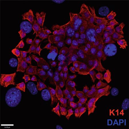
Daniel Haensel et al.

                           bulge and a new HG, which are HFSCs for the next hair cycle
                           [2]. Understanding the function of HFSCs and the molecular
                           regulation of their quiescence, activation, and differentiation repre-
                           sents an important direction in adult stem cell biology and may
                           have implications on how we prevent and treat hair loss.
                                Various tools and techniques have been generated and/or
                           applied to study HFSCs. With the knowledge of unique markers
                           of the various hair follicle cell types, immunofluorescence can be
                           used to examine the presence, absence, or fate alterations of cells in
                           the context of various genetic perturbations. HFSCs can be identi-
                           fied by immunofluorescence using classical HFSC markers, such as
                           CD34, or with pulse chase methods to identify HFSCs as label-
                           retaining cells [2]. These analyses provide static snapshots of
                           HFSCs, as skin must be frozen or fixed before sectioning and
                           further analysis. More sophisticated imaging techniques now have
                           abilities to track the migration, proliferation, and overall behavior
                           of individual cells within the hair follicle in real time during various
                           stages of regeneration [4, 5]. Coupled with fluorescent genetic
                           labeling, these pioneering live imaging techniques uncover unprec-
                           edented information about stem cell dynamics in vivo. With flow
                           cytometry and fluorescence-activated cell sorting (FACS), HFSCs
                           can be quantified and isolated for downstream purposes such as
                           gene expression analysis and in vitro assays [6, 7]. FACS-sorted
                           HFSCs can be cultured and expanded on mitotically inactivated
                           fibroblast feeders [6]. Cultured HFSCs can be functionally assessed
                           for clonal growth potential and, with serial passaging, long-term
                           self-renewal capability, as well as for regenerative capacity in host
                           animals [6, 8, 9].
                                Here we describe methods for culturing and downstream ana-
                           lyses of sorted bulge HFSCs. Specifically, we expand on the existing
                           clonal growth assay by incorporating live cell imaging to monitor
                           the division events and movement tracks of individual cells within
                           the growing bulge HFSC colonies. Furthermore, we provide pro-
                           tocols for growing bulge HFSCs on glass for immunofluorescence
                           and for efficient adenovirus infection that minimally impacts their
                           regenerative capacity in vivo.

2   Materials

2.1 J2-3T3                  1. J2-3T3 fibroblasts.
Fibroblast Culture,         2. F media (see [6] for detailed instructions).
Mitotic Inactivation,
                            3. Mitomycin C (Fisher Scientific, Cat. No. BP2531-2).
and Feeder Layer
Preparation                 4. 0.22-μm filter (Millex, Cat. No. SLGV033RS).
                            5. 10-mL syringe (BD, Cat. No. 309604).
                            6. 1 PBS.
                            7. 15-mL conical vials.
Experimentation with Hair Follicle Stem Cells
2.2 Plating and 1. Tissue culture dishes with mitotically inactivated J2-3T3 fibro-
Culture of Primary blast layer (requires reagents from Sect. 2.1).
HFSCs 2. E media (see [6] for detailed instructions).
3. 1 PBS.
4. Versene:
(a) 200 mL 10 PBS.
(b) 0.4 g EDTA disodium salt (Sigma, Cat. No. E-6511).
(c) Bring up to 2 L with deionized H2O.
(d) Autoclave.
(e) 8 mL sterile 25% glucose solution in deionized H2O.
5. 0.1% trypsin (Sigma, Cat. No. T4799).
6. 15-mL conical vials.
2.3 Clonal Assay 1. 6-well tissue culture plate (Falcon, Cat. No. 353046) or
60-mm gridded plates (Corning, Cat. No. 430166).
2. Tissue culture dishes with mitotically inactivated J2-3T3 fibro-
blast layer in F media.
3. E media.
4. 0.5% crystal violet (Sigma, Cat. No. HT90132-1L) in a 1:1
methanol/H2O solution.
2.4 Live Cell Imaging 1. Keyence BZ-X700 microscope (or equivalent microscope capa-
ble of live cell imaging).
2. 6-well tissue culture plate (Falcon, Cat. No. 353046).
3. Mitotically inactive 3T3 fibroblast feeders.
4. F media.
5. E media.
6. Freshly sorted or passaged HFSCs.
2.5 Immuno- 1. Collagen I solution at a concentration of 25 μg/mL in 0.02 M
fluorescence acetic acid (Sigma, Cat. No. C9791).
2. Glass coverslips (Fisher Scientific, Cat. No. 12-546).
3. Optional: 0.22-μm low protein-binding filter and 10-mL
syringe.
4. F media.
5. E media.
6. 1 PBS.
7. 4% paraformaldehyde, made from powder (MP Biomedicals,
Cat. No. 150146) in 1 PBS.
8. 1 PBS with 0.1% Triton X-100 (Sigma, Cat. No. T9284).Daniel Haensel et al.
9. 20% normal goat serum (NGS)-gelatin solution (20 mL):
(a) 4 mL NGS (Invitrogen, Cat. No. 16210-064).
(b) 200 μL 10% Triton X-100 (Sigma, Cat. No. T9284).
(c) 2 mL 10 PBS.
(d) 200 μL 2% NaN3 (Alfa Products, Cat. No. 50101).
(e) 200 μL 5% Tween 20 (Fisher Scientific, Cat. No. EC500-
018-3).
(f) 200 μL 1% gelatin (Sigma, Cat. No. G-1890).
(g) 13.2 mL distilled H2O.
l Mix and heat inactivate in 55 C water bath for 30 min.
Store at 4 C for about 2 weeks.
10. 10% NGS-gelatin solution (10 mL):
(a) 5 mL 20% NGS-gelatin solution (from step 9).
(b) 50 μL 10% Triton X-100 (Sigma, Cat. No. T9284).
(c) 500 μL 10 PBS.
(d) 50 μL 2% NaN3 (Alfa Products, Cat. No. 50101).
(e) 50 μL 5% Tween 20 (Fisher Scientific, Cat. No. EC500-
018-3).
(f) 50 μL 1% gelatin (Sigma, Cat. No. G-1890).
(g) 4.3 mL distilled H2O.
11. 2% NGS-gelatin solution:
(a) 1 mL 20% NGS-gelatin (from step 9).
(b) 200 μL 10% Triton X-100 (Sigma, Cat. No. T9284).
(c) 900 μL 10 PBS.
(d) 200 μL 2% NaN3 (Alfa Products, Cat. No. 50101).
(e) 200 μL 5% Tween 20 (Fisher Scientific, Cat. No. EC500-
018-3).
(f) 200 μL 1% gelatin (Sigma, Cat. No. G-1890).
(g) 7.75 mL distilled H2O.
12. Primary antibodies of interest.
13. Appropriate secondary antibodies.
14. DAPI (Life, Cat. No. D1306).
15. Vectashield (Vector, Cat. No. H1000).
2.6 Adenoviral 1. 6-well tissue culture dishes (Falcon, Cat. No. 353046) with
Infection of HFSCs mitotically inactivated J2-3T3 fibroblast layer.
2. 1 PBS.
3. E media.
4. Versene.Experimentation with Hair Follicle Stem Cells
5. 0.1% trypsin (Sigma, Cat. No. T4799).
6. 15-mL conical vial.
7. IRES-GFP adenovirus (Vector Biolabs, Cat. No. 1761).
2.7 Patch 1. Detailed lists of required reagents for the (1) isolation of neo-
Reconstitution Assay natal dermal cells and (2) patch reconstitution assay can be
with Virally Infected found in [7].
HFSCs 2. Reagents from Sect. 2.6 are required for generating virally
infected HFSCs.
3 Methods
3.1 J2-3T3 Culturing healthy fibroblasts and generation of a mitotically inac-
Fibroblast Culture, tive feeder layer are a critical component of HFSC culture.
Mitotic Inactivation,
1. For J2-3T3 growth and propagation, see the detailed protocol
and Feeder Layer in [6].
Preparation
2. Allow cells to reach 100% confluency.
3. Prepare the mitomycin C solution.
(a) Add 5 mL 1 PBS to 2 mg vial of mitomycin C and mix
(0.4 mg/mL).
(b) Filter sterilize with 0.22-μm filter.
4. Combine 12 mL F media with 240 μL of filter sterilized
0.4 mg/mL mitomycin C, and add to a confluent 100-mm
plate of J2-3T3 fibroblasts (see Note 1).
5. Swirl gently to mix and incubate at 37 C in incubator with 5%
CO2 for 2 h.
6. Aspirate media and rinse cells five times with 2 mL 1 PBS (see
Note 2).
7. Add 2 mL 0.1% trypsin and incubate until cells start to lift off
the plate (about 10 min).
8. Gently remove cells with P1000.
9. Add 8 mL F media to inactivate trypsin and transfer cell sus-
pension to a 15-mL conical vial and centrifuge to pellet at 1000
RPM for 5 min.
10. Aspirate the supernatant and resuspend the cell pellet in 5 mL F
media and count cells.
11. Seed appropriate number of mitomycin C-treated J2-3T3
fibroblasts in F media based on plate size. Use Table 1 for
guide.
12. Swirl plate gently to evenly distribute feeders (see Note 3).Daniel Haensel et al.
Table 1
Desired number of mitotically inactive J2-3T3 fibroblasts to plate
depending on plate size
Plate type J2-3T3 Fibroblast number
100 mm 1,000,000
60 mm ~360,000
35 mm ~170,000
6 Well ~170,000
13. Culture at 37 C in incubator with 5% CO2 for 2 days to allow
fibroblasts to attach fully and spread to cover the entire dish
before adding HFSCs (see Note 4).
14. Fibroblast feeder plates can be cultured for up to 1 week before
adding HFSCs. F media should be changed every 3 days until
usage.
3.2 Plating Freshly To plate HFSCs, a media switch from F media to E media must first
Isolated HFSCs occur.
1. Epidermal single cell suspension and sorting of HFSCs can be
done using the detailed protocol in [6].
2. Prepare mitomycin C-treated J2-3T3 feeder layer by removing
F media and then rinsing twice with 2 mL 1 PBS.
3. Add appropriate amount of E media to plate or well.
4. After HFSCs are obtained by FACS, count cells and add appro-
priate number of cells to each plate or well.
5. Swirl plate to mix.
6. Culture HFSCs at 35 C in incubator with 5% CO2 (see Note 5).
7. Replace E media every 3 days.
8. Colonies should become visible after ~7 days and then rapidly
expand (see Note 6).
3.3 Passaging HFSCs After ~2 weeks of culture and significant colony growth, HFSCs
can be passaged onto a new mitomycin C-treated J2-3T3 fibroblast
feeder layer.
1. Aspirate media. Add 2 mL versene and let sit for 2 min.
2. Vigorously pipette the versene with a P1000 to spray off all
fibroblasts (see Note 7).
3. Aspirate versene, wash plate with 5 mL E media to remove
residual fibroblasts, and then aspirate.Experimentation with Hair Follicle Stem Cells
4. Add 2 mL 0.1% trypsin (no EDTA) and let incubate until cells
start to lift up (about 10 min).
5. Add 8 mL E media to inactivate trypsin and spray to remove
residual cells.
6. Put cells in a 15-mL conical vial and centrifuge to pellet at 1000
RPM for 5 min.
7. Aspirate media, resuspend pellet in 5 mL E media, and count
cells.
8. Prepare mitomycin C-treated J2-3T3 feeder layer by removing
F media and then rinsing twice with 2 mL 1 PBS.
9. Add appropriate amount of E media to plate or well.
10. Add appropriate number of HFSCs from step 7 onto mitomy-
cin C-treated J2-3T3 feeder layer.
11. Culture HFSCs at 35 C in incubator with 5% CO2 (see Note 5).
12. Replace E media every 3 days.
13. Colonies should become visible after ~4 days and then rapidly
expand (see Note 8).
3.4 Clonal Assay Clonal assays involve plating a low number of cells (1000 cells/
cm2) such that each colony is generated by a single cell. There is
flexibility in terms of what plate size to use. Generally use of a 6-well
plate is recommended for ease of generating technical replicates.
Gridded plates are useful for tracking the same colony over time
and are also ideal for live cell imaging (Sect. 3.5).
3.4.1 Clonal Growth 1. Generate mitomycin C-treated J2-3T3 fibroblast feeder layer as
Assay described in Sect. 3.1 using 6-well plates.
2. Prepare mitomycin C-treated J2-3T3 fibroblast feeder layer for
HFSC as described in Sect. 3.2.
3. Add 1000 cells/cm2 of HFSCs (~9,500 cells for 6-well plate)
to appropriate volume of E media (2–3 mL for 6-well plate)
in 15-mL conical vial, gently mix, and then add to well
(see Note 9).
4. Swirl plate to mix.
5. Culture HFSCs at 35 C in incubator with 5% CO2 (see Note 5).
6. Replace E media every 3 days.
7. Culture HFSCs for 2 weeks.
8. After 2 weeks of culture, aspirate media. Add 2 mL versene and
let sit for 2 min.
9. Vigorously pipette the versene with a P1000 to spray off all
fibroblasts (see Note 7).
10. Aspirate versene, wash plate with 2 mL 1 PBS to remove
residual fibroblasts, and then aspirate.Daniel Haensel et al.
11. Add 2 mL 0.5% crystal violet staining solution in a 1:1 solution
of water:methanol and incubate for 30 min.
12. Remove staining solution and then rinse with deionized water
until water goes clear.
13. Allow plates to dry and then image and count/measure
colonies.
3.4.2 Tracking Individual 1. Prepare mitomycin C-treated J2-3T3 fibroblast feeder layer
Colonies Using Gridded using 60-mm gridded plates.
Plates 2. Add 1000 cells/cm2 of HFSCs (~21,000 cells per 60-mm
gridded plate) to appropriate volume of E media (3 mL per
60-mm gridded plate) in 15-mL conical vial, gently mix, and
then add to pre-prepared feeder plate (see Note 9).
3. Swirl plate to mix.
4. Culture HFSCs at 35 C in incubator with 5% CO2 (see Note
5).
5. Replace E media every 3 days.
6. After 7 days of culture, go through grids and look for colonies
to track, and note colony locations.
7. Image individual colonies every 24 h (Fig. 1).
8. End-point clonal analysis can also be done by completing steps
8–13 in Sect. 3.4.1.
Fig. 1 Growth of a single HFSC colony over time. Note rapid expansion of colony size during the indicated time
frame. Arrow indicates location of a grid on plateExperimentation with Hair Follicle Stem Cells
3.5 Live Cell Imaging Live cell imaging of HFSC colonies enables analysis of the migra-
tory and proliferative behaviors of cells within the expanding colo-
nies. We use the Keyence BZ-X700 live imaging system, which can
utilize multiple tissue culture plate types.
1. Prepare cells for imaging by completing steps 1–6 in Sect.
3.4.2.
2. Consider starting live imaging at 7 days after plating of HFSCs
as colonies should be visible at this time (see Note 10).
3. Adjust microscope setup to maintain temperature and CO2
levels during live imaging.
4. Image each colony at 10 magnification every 15 min or less
(see Note 11) for an 18-h duration.
5. Analysis of individual cells within colonies can be done using
the “Manual Tracking” plugin in the ImageJ software from
FIJI.
(a) Carefully track individual cells through each image frame
during the 18-h period to generate cell movement tracks
with coordinates.
(b) Set the original (X,Y) coordinate to the origin (0, 0),
adjust to be in microns based on microscope’s field of
view, and then graph.
(c) Consider calculating directionality and velocity of
migration.
6. If desired, individual cell divisions can be counted by manually
going through each frame.
3.6 Immuno- Immunofluorescence of HFSCs presents a challenge due to the
fluorescence technical complications of growing HFSCs on glass to allow for
optimal colony growth, morphology, and imaging. We have
assessed several commonly used glass-coating procedures, such as
using poly-L-lysine, collagen I or IV, laminin, or fibronectin to coat
glass coverslips before culturing, and found them all to yield less
than ideal growth and morphology. Ultimately, co-culture using
coverslips pre-seeded with a fibroblast feeder layer is the best at
preserving HFSC viability and morphology for immunofluorescent
imaging.
1. Generate working collagen solution (25 μg/mL in 0.02 M
acetic acid) as per manufacturer’s instructions. Filter through
a low protein-binding filter to sterilize.
2. Cover surface of glass coverslips or glass chamber slides with
working collagen solution for 1 h at room temperature in tissue
culture hood.
3. Remove collagen and rinse three times with 1 PBS.
4. Coated coverslips/chamber slides can be store at 4 C for up to
a month if not immediately used.Daniel Haensel et al.
5. Prepare mitomycin C-treated J2-3T3 fibroblast feeder layer as
described in Sect. 3.2 using either chamber slides or glass
coverslips. If using glass coverslips, place coverslips in 6-well
plates, and add appropriate number of mitomycin C-treated
J2-3T3 fibroblasts.
6. Add the needed volume of E media along with the desired
number of HFSCs in 15-mL conical vial, gently mix, and
then add to plate (see Note 9 regarding media volume and
cell density).
7. Culture HFSCs at 35 C in incubator with 5% CO2 (see Note 5).
8. Replace E media every 3 days.
9. Colonies become visible in approximately 7 days and fixation
for immunofluorescence can be done when colonies reach the
desired size.
10. Gently wash cells with 1 PBS, and then fix cells for 15 min in
ice cold 4% paraformaldehyde.
11. Wash for 10 min with 1 PBS, and then twice in 1 PBS
containing 0.1% Triton X-100 for 10 min each.
12. Block with 20% NGS-gelatin solution for 30 min at room
temperature to overnight at 4 C.
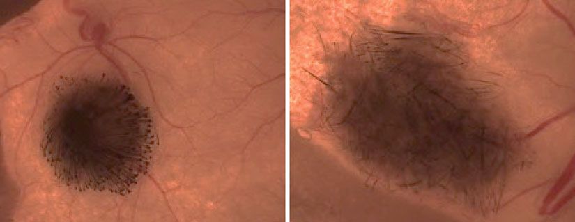
13. Incubate with primary antibody diluted in 10% (or lower,
depending on the specific antibody used) NGS-gelatin solution
overnight at 4 C. Use of an anti-keratin 14 (K14) antibody
helps to distinguish HFSC colonies from the fibroblast feeders
(Fig. 2).
Fig. 2 Immunofluorescent staining for K14 expression in cultured HFSCs. DAPI
stains the nucleiExperimentation with Hair Follicle Stem Cells
14. Wash three times with 1 PBS for 10 min at room tempera-
ture, and then wash with 2% NGS-gelatin solution for 15 min
at room temperature.
15. Incubate with secondary antibody diluted in 2% NGS-gelatin
solution for 1 h at room temperature in the dark.
16. Wash three times with 1 PBS containing 0.1% Triton X-100
for 10 min at room temperature in the dark.
17. Stain with DAPI at a final concentration of 2 μg/mL in 1 PBS
for 10–20 min in the dark.
18. Wash three times with 1 PBS for 5 min each.
19. Mount slides with Vectashield and seal edges with nail polish.
20. Image colonies using epifluorescence or confocal microscopy.
Note that the HFSCs often push the fibroblast feeders away as
the colonies expand, resulting in a single layer of cells. How-
ever, the HFSC colonies may begin to stratify if cultured for
too long.
3.7 Adenoviral Viral infection of HFSCs enables manipulation of gene expression
Infection of HFSCs and functional characterization. We found it necessary to tempo-
rarily remove the mitomycin C-treated J2-3T3 fibroblast feeders in
order to maximize the viral infection efficiency of HFSCs. In our
experiments, we utilized commercially available adenoviruses that
express GFP so infection efficiency can be estimated based on GFP
expression (Fig. 3).
1. Prepare mitomycin C-treated J2-3T3 fibroblast feeder layer as
described in Sect. 3.2.
2. Add 1000 cells/cm2 of HFSCs (~9500 cells for 6-well plate) to
appropriate volume of E media (2–3 mL for 6-well plate) in
15-mL conical vial, gently mix, and then add to well (see Note 9).
3. Swirl plate to mix.
4. Culture HFSCs at 35 C in incubator with 5% CO2 (see Note 5).
5. Replace E media every 3 days.
6. Culture HFSCs for 2 weeks so there are enough cells to infect.
7. Harvest HFSCs in a single well by following steps 1–7 in Sect.
3.3, and use for counting the number of cells (see Note 12).
8. To the remaining wells, add 2 mL versene and let sit for 2 min.
9. Vigorously pipette the versene solution with a P1000 to spray
off all fibroblasts (see Note 7).
10. Aspirate versene, wash plate with 2 mL E media to remove
residual fibroblasts, and then aspirate.
11. Mix appropriate amount of viruses based on cell count in step
7 to achieve multiplicity of infection (MOI) of 50 to 1 mL E
media in a 1.5-mL Eppendorf tube and then carefully add to
a well.Daniel Haensel et al.
A
24 h 48 h 72 h
B
100
90
80
70
% GFP+
60
50
40
30
20
10
0
1 2 3
Days After Infection
Fig. 3 GFP expression as a measure for efficiency of adenovirus infection in HFSCs. GFP protein was visualized
by (a) fluorescence microscopy and (b) flow cytometry
12. Incubate cells with viruses overnight at 35 C in incubator with
5% CO2.
13. The next day, check cells under the microscope for GFP expres-
sion (Fig. 3).
14. Prepare mitomycin C-treated J2-3T3 fibroblast feeder layer as
described in Sect. 3.2.
15. Remove the virus-containing media from HFSCs and then
briefly rinse with 2 mL 1 PBS.
16. Add 1 mL 0.1% trypsin (no EDTA) and let incubate until cells
start to lift up (about 10 min).
17. Add 4 mL E media to inactivate trypsin and spray to remove
residual cells.
18. Put cells in a 15-mL conical vial and centrifuge at 1000 RPM
for 5 min to pellet cells.
19. Aspirate media, resuspend pellet in 2 mL E media, and count
cells.Experimentation with Hair Follicle Stem Cells
20. Add 1000 cells/cm2 of HFSCs (~9,500 cells for 6-well plate)
to appropriate volume of E media (2–3 mL for 6-well plate) in
15-mL conical vial, gently mix, and then add to well (see Notes
9 and 13).
21. Culture HFSCs at 35 C in incubator with 5% CO2 (see Note 5).
22. Replace E media every 3 days.
23. Colonies should begin to be visible after ~4 days and then
rapidly expand (see Note 8).
24. Proceed with downstream applications.
3.8 Patch The regenerative capacity of virally infected HFSCs can be assessed
Reconstitution Assay using a well-established “patch” assay [7], where HFSCs can be
with Virally Infected combined with newborn dermal cells and then subcutaneously
HFSCs injected into the backs of immunocompromised Nu/J mice. After
2 weeks, hair follicles form at the site of injection (Fig. 4).
1. Generate virally infected HFSCs by following steps in Sect. 3.7.
Cells should have been growing for 2 weeks and ready for use
once step 2 below is completed.
2. Isolate newborn primary dermal cells using the protocol
from [7].
3. Harvest HFSCs by following steps 1–7 in Sect. 3.3.
4. Combine 100,000 infected HFSCs with 500,000 newborn
dermal cells in 1.5-mL Eppendorf tube and centrifuge at
1000 RPM for 5 min to pellet the cells.
5. As dermal-only negative control, add 500,000 newborn dermal
cells to 1.5-mL Eppendorf tubes, and centrifuge to pellet at
1000 RPM for 5 min (see Note 14).
6. Very carefully remove the media from the vials as to not dis-
lodge the cell pellets.
Fig. 4 Result of a “patch” assay using newborn keratinocytes (left) and infected HFSCs (right)Daniel Haensel et al.
7. Add 50 μL of E media to each pellet and then place the cells on
ice and bring to the appropriate mouse procedure room.
8. Follow approved mouse protocols to sedate Nu/J mice, and
sterilize the outer skin surface surrounding the desired
injection site.
9. Carefully resuspend the cells in each 1.5-mL Eppendorf tube,
and then draw cell suspension into syringe with a 25G needle.
10. Carefully inject the cell suspensions into the backs of the Nu/J
mice (see [7] for detailed injection instructions).
11. Return mice to their cages and follow approved post-operation
procedures.
12. Sacrifice mice 2 weeks later to examine hair follicles at each of
the injection sites.
4 Notes
1. It is critical to not use over-confluent J2-3T3 fibroblasts.
2. If not used immediately, add 10 mL F media to mitomycin
C-treated J2-3T3 fibroblasts. Cells can be kept in the incubator
for a week before use.
3. Even distribution is critical for optimal HFSC growth.
4. It generally takes ~2 days for mitomycin C-treated J2-3T3
fibroblasts to attach and spread over the entire dish. It is not
recommended that HFSCs be added until the tissue culture
plastic is fully covered.
5. After initial seeding of HFSCs, do not move plate within the
first 72 h to allow the cells to attach to the feeders.
6. HFSCs grow as colonies. Colonies derived from HFSCs
isolated from p49 mice (when their hair follicles are in telogen)
become visible within a week and will begin to expand very
rapidly after coming out of quiescence.
7. Feeders are easily removed and HFSCs will remain attached.
HFSCs will begin to detach if treated for extended amount
of time.
8. Passaged HFSCs form colonies faster than freshly sorted
HFSCs.
9. Adding cells to the full volume of media in a conical vial
followed by mixing facilitates even distribution of HFSCs in
the well.
10. It is recommended that imaging be performed between 7 and
9 days. Generally, beyond 10 days after plating, the HFSC
colonies become too large for an entire colony to be captured
in a single field of view.Experimentation with Hair Follicle Stem Cells
11. Imaging every 15 min or less is recommended to capture the
dynamic cellular changes, as HFSCs expand fairly quickly at
this time.
12. An extra well is used for counting cells before infection to
calculate how much viruses to add to achieve an ideal MOI.
13. This plating density is good for a clonal assay. Different plating
density may be desired per specific downstream assay.
14. A dermal-only control is necessary to gauge how much the
dermal cells might be contaminated with newborn epidermal
cells, which are regeneration-competent.
References
1. Alonso L, Fuchs E (2006) The hair cycle. J Cell 6. Nowak JA, Fuchs E (2009) Isolation and culture
Sci 119(Pt 3):391–393 of epithelial stem cells. Methods Mol Biol
2. Hsu YC, Li L, Fuchs E (2014) Emerging inter- 482:215–232
actions between skin stem cells and their niches. 7. Zheng Y, Hsieh JC, Escandon J, Cotsarelis G
Nat Med 20(8):847–856 (2016) Isolation of mouse hair follicle bulge
3. Ito M, Kizawa K, Hamada K, Cotsarelis G stem cells and their functional analysis in a
(2004) Hair follicle stem cells in the lower reconstitution assay. Methods Mol Biol
bulge form the secondary germ, a biochemically 1453:57–69
distinct but functionally equivalent progenitor 8. Blanpain C, Lowry WE, Geoghegan A, Polak L,
cell population, at the termination of catagen. Fuchs E (2004) Self-renewal, multipotency, and
Differentiation 72(9-10):548–557 the existence of two cell populations within an
4. Rompolas P, Mesa KR, Greco V (2013) Spatial epithelial stem cell niche. Cell 118(5):635–648
organization within a niche as a determinant of 9. Adam RC et al (2015) Pioneer factors govern
stem-cell fate. Nature 502(7472):513–518 super-enhancer dynamics in stem cell plasticity
5. Rompolas P et al (2012) Live imaging of stem cell and lineage choice. Nature 521(7552):366–370
and progeny behaviour in physiological hair-
follicle regeneration. Nature 487(7408):496–499You can also read