Exceptional preservation of tracheal rings in a glyptodont mammal from the Late Pleistocene of Argentina - Acta Palaeontologica Polonica
←
→
Page content transcription
If your browser does not render page correctly, please read the page content below

Brief report                                                Acta Palaeontologica Polonica 65 (1): 29–34, 2020

Exceptional preservation of tracheal rings in a glyptodont mammal
from the Late Pleistocene of Argentina
MARTÍN ZAMORANO

Exceptionally well-preserved material from a fossil mam-            The family Glyptodontidae is a group of armored xenar-
mal is presented. For the first time, several fragments of tra-     thrans, whose representatives reached large to very large sizes
cheal rings and cricoid cartilage assigned to Panochthus sp.        (Scillato-Yané and Carlini 1998; Fariña 2001; Zamorano et al.
(Xenarthra; Glyptodontidae) from the Late Pleistocene of            2014a), even exceeding 2300 kg (Soibelzon et al. 2012), and
Argentina are described in detail and figured. In this con-         are recorded from the middle Eocene to the early Holocene
tribution, in addition to a meticulous description, a tracheal      (Fernicola 2008; Zamorano 2013; Zurita et al. 2016). From an
ring was reconstructed and compared to tracheal rings of            evolutionary stand point, the evidence strongly suggests that
domestic and wild mammals. As a result, among domestic              glyptodonts are a monophyletic group (see Porpino et al. 2014;
mammals it is similar to those of Sus scrofa domestica (do-         Zamorano 2019; among others).
mestic pig), and among wild mammals to those of Zalophus                Panochthus Burmeister, 1866 is one of the most abun-
californianus (California sea lion). Tracheal rings of fossil       dant and diversified glyptodontids of the South American
vertebrates have been recognized in birds (Cariamiformes            Pleistocene, as well as one of the largest Cingulata (see Fariña
and Anseriformes) and other dinosaurs (Theropoda). This             2001; Zamorano et al. 2014a). Likewise, it is also among the
is likely the first report of tracheal rings in a fossil mam-       most abundantly recorded groups in the Pampean region
mal; future comparisons with extant xenarthrans could               (Scillato-Yané et al. 1995; Cione et al. 1999; Zamorano 2012;
provide information on the paleobiological implications of          Zamorano et al. 2014b). Together with Glyptodon Owen, 1839
this structure in glyptodonts, and allow making inferences          they are the most widely distributed glyptodontids in South
about other fossil mammals.                                         America, both latitudinally and altitudinally (Zurita et al. 2009,
                                                                    2016; Zamorano 2012, 2019; Zamorano and Jara Almonte 2018;
                                                                    Zamorano and Oliva in press). In this contribution, several
Introduction                                                        fragments of tracheal rings and the cricoid cartilage assigned
The trachea of mammals is a relatively flexible cartilaginous       to Panochthus sp. are described and figured. In this way, I in-
and membranous tube that extends from the cricoid cartilage         tend to contribute to the understanding of these cartilages,
of the larynx to the middle mediastinum, where it bifurcates        which allow the indispensable entry of oxygen into a mammal
into the main bronchi (König and Liebich 2005). The frame-          body, an issue that is poorly studied in the extant mammals and
work of the trachea is formed by C-shaped plates of hyaline         seemingly never studied in extinct forms.
cartilage (“tracheal rings”). These vary in shape according to      Institutional abbreviations.—MHM, Museo Histórico Muni-
the species and in some cases according to the location on          cipal “Alfredo Enrique Múlgura”, General Belgrano, Province
the trachea. The number also varies according to the species        of B1uenos Aires, Argentina.
(Sisson and Grossman 1982). The cartilaginous rings provide
the tube: (i) some rigidity, otherwise it would collapse as the
lungs expand; (ii) some expansion, to be able to accommodate        Geographic and stratigraphic setting
any increase in air volume, by means of the flexibility of the      The fossil material was found on the right margin of the Sa-
hyaline cartilage, and the dorsal incompleteness of the rings;      lado River, near the city of General Belgrano, Buenos Aires,
and (iii) flexibility and extensibility, to be able to follow the   Argentina (35°45’5.52” S, 58°37’35.96” W; Fig. 1). The remains
movements of the head, neck and larynx (Sisson and Grossman         were collected in sediments of the Luján Formation dated be-
1982; Powell et al. 2010). Tracheal cartilages originate from       tween ca 14 and 12 ky (Late Pleistocene), in the transitional
the splanchnic mesoderm and form the true tracheal skeleton         limit between La Chumbiada Member (ca 14 to 12 ky, Lujanian
(Acuña Navas et al. 2010).                                          Stage/Age sensu Cione and Tonni 1999, 2001; Cione et al.
    Xenarthrans are a particular group of mammals, charac-          2015) and Gorch Member (ca 11.5 to 5 ky sensu Fucks et al.
teristic of the Neotropical Region, widely represented in the       2015), downstream the Paraje La Chumbiada, near Estancia
South American fossil record, both in temporal extension and        La Invernada (Fig. 1). In Paraje La Chumbiada, Scanferla et al.
frequency of records (Scillato-Yané 1977; Paula Couto 1979).        (2013) recognized a gray to black clayey deposit interpreted as a

Acta Palaeontol. Pol. 65 (1): 29–34, 2020                                                          https://doi.org/10.4202/app.00654.2019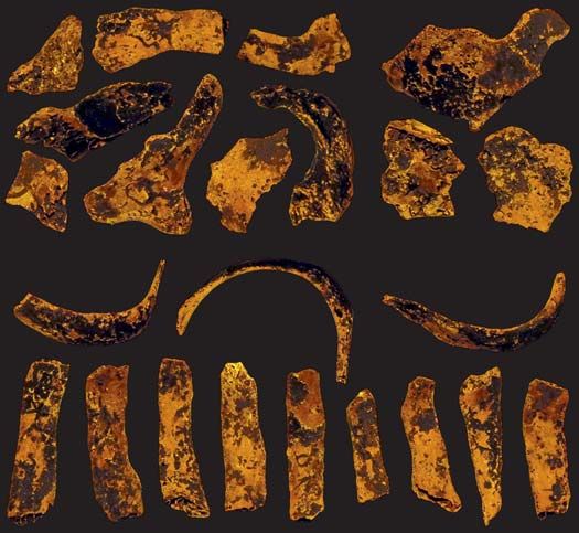
30                                                                                    ACTA PALAEONTOLOGICA POLONICA 65 (1), 2020

                                                                           vitulina Linnaeus, 1758 (Carnivora, Phocidae), and Zalophus
 A                                    B
 34°                                                                       californianus Lesson, 1828 (Carnivora, Otariidae) (Sokolov et
                                                                           al. 1968; Harrison and Denny 1985; Bertassoli and Santos 2013;
          Rí
             o                                                             Moore et al. 2014; Junior et al. 2016; Moreto et al. 2017).
          Pl d e
 Buenos     at la
               a
                           TINA
 Aires
 35°
                         ARGEN

                                  A
                                                                           Systematic palaeontology
 Los Tobianos
                                                                           Family Glyptodontidae
     Farm                                                                  Genus Panochthus
                Salado                                                     Type species: Glyptodon tuberculatus Owen, 1845; Middle Pleisto-
 36°             River
                                                                           cene–late Pleistocene) from the southern and center-northern areas of
                                                                           Argentina, Uruguay, south and central areas of Bolivia and southeast-
  58°       100 km                                                         ern region of Brazil.

Fig. 1. A. Geographic location of studied area within Buenos Aires Prov-   Panochthus sp.
ince, Argentina (inset) near General Belgrano city, Los Tobianos Farm      Material.—MHM-P 87, twenty-three fragments of tracheal
(35°45’5.52” S, 58°37’35.96” W), asterisk indicates where the studied
                                                                           rings, including fragments that probably belong to the crioid
specimen was discovered. B. Photo of the outcroup along the Salado River
right bank, the site where Panochthus sp. (MHM-P 87) was found.            cartilage. The specimen consists of part of the skull (badly
                                                                           preserved), mandibular rami, postcranial bones (both humeri,
paleolagoon, in which several remains of fossil mammals were               radii-ulnae, both femurs, tibiae-fibulae, several elements of the
collected (Hippidion principale [Lund, 1842], Smilodon popu-               carpus and tarsus, thoracic and lumbar vertebrae, cervical ribs)
lator Lund, 1842, Doedicurus clavicaudatus [Owen, 1847], and               and the almost complete dorsal carapace. Because of the ex-
Megatherium americanum Cuvier, 1796, among others), which                  ceptional preservation of this material, fragments of tracheal
corroborated the age of the level.                                         rings, the crycoid cartilage and, some elements of the hyoid
    In general, the material recovered in the area where the re-           apparatus could be identified and were published by Zamorano
mains studied here were collected are exceptionally preserved.             et al. (2018). Late Pleistocene of General Belgrano, Buenos
Fossils of mammals recovered in this area contain large                    Aires, Argentina (35°45’5.52” S, 58°37’35.96” W) (Fig. 1). The
amounts of preserved collagen (Scanferla et al. 2013; Delsuc et            remains were collected in sediments of the Luján Formation
al. 2016; Mitchell et al. 2016), and mummified exoskeletons of             dated between ca 14 and 12 ky.
insects were found (Ramirez and Michat 2016). The high per-
                                                                           Description.—Tracheal cartilages are mostly small and frag-
centage of collagen preserved is a rare condition with respect
                                                                           ile fragments; their thickness does not exceed 3 mm. Twenty-
to the rest of the Pleistocene deposits of the Pampas region
(Scanferla et al. 2013), and this is probably what allowed the
exceptional preservation of the cartilage studied here.

Material and methods
The fossil specimen described here was compared with tra-
cheal rings and cricoid cartilage of domestic mammals, whose
shape has been studied in detail, e.g., Bos primigenius taurus
Linnaeus, 1758 (Artiodactyla, Bovidae), Canis lupus familiaris
Linnaeus, 1758 (Carnivora, Canidae), Capra aegagrus hircus
Linnaeus, 1758 (Artiodactyla, Bovidae), Equus ferus caballus
                                                                            A1
                                                                                                                      20 mm
(Linnaeus, 1758) (Perissodactyla, Equidae), Felis silvestris ca-
tus (Linnaeus, 1758) (Carnivora, Felidae), Ovis orientalis aries
Linnaeus, 1758 (Artiodactyla, Bovidae), and Sus scrofa do-
mestica Linnaeus, 1758 (Artiodactyla, Suidae) (Barone 1984;
Dabanoğlu et al. 2001; Climent et al. 2005; König and Liebich
2005; Martínez and Salvador 2010; Powell et al. 2010). I also
used those wild mammals for comparison, but only through
literature, e.g., Cerdocyon thous (Linnaeus, 1766) (Carnivora,
Canidae), Didelphis sp. (Didelphimorphia, Didelphidae), Hyd-               A2                                             A3
rochoerus hydrochaeris Linnaeus, 1766 (Rodentia, Caviidae),                Fig. 2. Tracheal cartilages of glyptodont mammal Panochthus sp. (MHM-P
Mirounga angustirostris Gill, 1866 (Carnivora, Phocidae),                  87) from the Late Pleistocene of General Belgrano, Argentina, in ventral
Odobenus rosmarus (Linnaeus, 1758) (Carnivora, Odobenidae),                views. Lateral (A1), ventral and ventro-lateral (A2) portions, fragment of
Ursus maritimus Phipps, 1744 (Carnivora, Ursidae), Phoca                   cricoid cartilage (A3) in ventral views.
BRIEF REPORT                                                                                                                                           31

           A1                             A2                                              B                C
 larynx

                                                                                                                            20 mm
                                     thyroid
                                    cartilage
                                                                           tr        tc              mr
                                     cricoid                                    cc        hy
                                   cartilage
                                     anular
                                   ligament
 trachea

                                                                     20 mm
                                    trachea
                                    cartilage

                                    C

                                                                                          D                            E
Fig. 3. Cartilages present in the neck of glyptodont mammal Panochthus sp. compared with Recent California sea lion and domestic pig. A. Thyroid, cri-
coid, and tracheal cartilages in ventral view; generalized mammal (A1), Panochthus sp. (A2). B. Explanatory drawing of the Panochthus sp. skull in lateral
view, with hyoid apparatus and tracheal rings. C–E. Tracheal rings in anterior view. C. Panochthus sp. (MHM-P 87). D. California sea lion Zalophus
californianus Lesson, 1828. E. Domestic pig Sus scrofa domestica Linnaeus, 1758. A, modified from Martínez and Turpín 2015; D, E, modified from
Moore et al. 2014. Abbreviations: cc, cricoid cartilage; hy, hyoid apparatus; mr, mandibular rami; sk, skull; tc, thyroid cartilage; tr, tracheal rings.

three fragments of these tracheal rings were found, twelve of                    mals (in this case S. scrofa domestica and Z. californianus) are
them belong exclusively to lateral sectors 19–54 mm in length                    practically in contact, because the tracheal muscle holds them
and 6–8 mm in width. The three longest of these last frag-                       together, whereas in the fossils of Panochthus sp. these free
ments are fully curved (none of them cover the lateral sector                    ends are more widely separated.
completely), whereas the other nine are almost straight (Fig.
2A1). Eight fragments belong to ventral portions of the rings;                   Concluding remarks
in this category are also included fragments of fused ventral
and lateral portions (Fig. 2A2), three of them, the largest ones,                During inspiration the air pressure is lower in the trachea than
probably belong to the cricoid cartilage (this latter is similar                 the atmospheric pressure, and without the tracheal rings, the
in shape to a tracheal ring, although larger; Drake et al. 2010)                 trachea would collapse (Villee et al. 1971). For this reason the
(Fig. 2A3). A tracheal ring was reconstructed; it is 80 mm high                  tracheal rings are totally indispensable for animals with lung
and 60 mm wide. Its diameter occupies approximately 15%                          breathing. In mammals these rings have the only basic and
of the skull height (without the mandible) (see Fig. 3B). The                    fundamental function of maintaining an open channel that
tracheal rings of Panochthus sp. would belong to a cylindrical                   allows the circulation of air from the larynx to the lungs and
trachea, slightly flattened on the dorsal and ventral sides, the                 vice versa. Moore et al. (2014) studied the structure and rigid-
free ends of each ring (that is, on its dorsal side) overlap, the                ity of the tracheae of marine mammals and observed how the
                                                                                 flow of air circulating through the tracheae behaved during
left one above the right one (Fig. 3C).
                                                                                 diving. The rigidity of the trachea, deduced from the shape of
Remarks.—Among domestic mammals, it is similar to those                          the rings, is expressed in the capacity not to collapse. For some
of Sus scrofa domestica (Fig. 3E), whereas among wild mam-                       authors, this rigidity is an adaptation of marine mammals
mals it is similar to those of Zalophus californianus (Fig. 3D).                 (Scholander 1940; Olsen et al. 1969; Kooyman and Sinnett
Tracheal rings of Sus scrofa scrofa, a wild subspecies similar                   1979; Kooyman and Cornell 1981). However, Denison et al.
to its conspecific, are also similar, but those of Zalophus cal-                 (1971), Bostrom et al. (2008), and Moore et al. (2014) stated
ifornianus resemble even more those of Panochthus sp., since                     that it is similar to the rigidity of the tracheae of terrestrial
these rings are flattened dorsally and ventrally, and not as in                  mammals. In this sense, Moore et al. (2014) concluded that
cross section as those of S. scrofa scrofa. Noteworthy, in S.                    Sus scrofa (S. scrofas crofa and S. scrofa domestica) is the
scrofa domestica and Z. californianus the shape of the rings is                  terrestrial mammal most similar to marine mammals. In turn,
constant along the entire trachea, as well as the overlapping of                 Harrison and Denny (1985) suggested that the influence of the
their free ends (Moreto et al. 2017). It has to be taken into ac-                shape and size of tracheal rings has to be taken into account
count that the free ends of the tracheal cartilage of living mam-                on the running speed reached by mammals. To test this hy-
32 ACTA PALAEONTOLOGICA POLONICA 65 (1), 2020
pothesis, they used the relationship between the area occupied Acknowledgments.—I thank Álvaro Mones ( Augsburg, Germany),
by the glottis lumen, the tracheal lumen (generated by the Hernán Zamorano (Río Turbio, Argentina), Ricardo Bonini (CON-
tracheal ring) and the body mass. In fossil mammals, these ICET, Buenos Aires, Argentina), Néstor Toledo (CONICET), and
Agustín Abba (Centro de Estudios Parasitológicos y de Vectores, La
relationships cannot be calculated as a whole. Although the
Plata, Argentina) for bibliographic support, María Julia Sanchez Ron-
body mass has been estimated in several species of different dini (La Plata, Argentina), Juan Ramón Artigas (La Plata, Argentina),
groups (Fariña et al. 1998; Seebabher 2001; Reguero et al. and Cecilia Krmpotic (CONICET) for taking photographs of the mate-
2010; Cassini et al. 2012; Toledo et al. 2014) the surface of rial, Laureano Raúl González Ruíz (CONICET) for reading and com-
the glottis lumen is unknown, and as for the lumen of the tra- ments on the manuscript, Juan Cruz González (La Plata, Argentina) for
chea, the present contribution is the first to report, figures and the drawings. Leandro M. Pérez, an anonymous reviewer, and editors
describes a tracheal ring of a fossil mammal. Tracheal rings are thanked for comments and suggestions that helped to improve the
manuscript.
have only been published in extinct birds: Llallawavis sca-
gliai Degrange, Tambussi, Taglioretti, Dondas, and Scaglia,
2015 (Cariamiformes, Phorusrhacidae) (Degrange et al. 2015) References
and Vegavis iaai Clarke, Tambussi, Noriega, Erickson, and Acuña Navas, M.J., Arce Rodríguez, E., Baquero Barcenas, A.M., Bonilla
Ketcham, 2005 (Anseriformes, Vegaviidae) (Clarke et al. Mora, W., Coto Chinchilla, K., Guerrero Gamboa, L., Gutiérrez Por-
2016); and other dinosaurs: Scipionyx samniticus Dal Sasso ras, M., Jiménez Delgado, J., Leitón Villagra, C., Madrigal Rojas, J.P.,
and Signore, 1998 (Theropoda, Coelurosauria) (Dal Sasso and Monge Carvajal, C., Morales González, F., Núñez Delgado, N., Penón
Portmann, M., Quirós Castro, A.G., Rivera Calderón, C., and Vargas
Sig nore 1998).
Sanabria, M. 2010. Embriología del desarrollo de los bronquios y el
In the literature, data on cricoid cartilage and tracheal rings parénquima pulmonar. Medicina Legal de Costa Rica 27 (7): 61–74.
of living xenarthrans are very scarce. Naples (1999) states Barone, R. 1984. Anatomie Comparée des Mammifères Domestiques.
that the cricoid cartilage is partially ossified in the skull of Tome III. Splachnologie I. Appareil digestif. Appareil respiratoire. 879
the giant anteater Myrmecophaga tridactyla Linnaeus, 1758, pp. Vigot, Paris.
Bertassoli, B.M. and Santos, A.C. 2013. Morfologia da laringe e traqueia
and that small movements between this and the back of the
de gambás Didelphis sp.). Ciencia Animal Brasileira 14: 222–229.
tracheal rings are likely; according to Borges et al. (2017), in Borges, N.C., Nardotto, J.R., Oliveira, R.S., Rüncos, L.H., Ribeiro, R.G.,
M. tridactyla the trachea begins at the sixth cervical. For the and Bogoevich, A.M. 2017. Anatomy description of cervical region
folivores, Gilmore et al. (2008) report the differences in length and hyoid apparatus in living giant anteaters Myrmecophaga tridactyla
of the trachea between the tree sloths, Choloepus Illiger, 1811, Linnaeus, 1758. Pesquisa Veterinária Brasileira 37: 1345–1351.
Bostrom, B.L., Fahlman, A., and Jones, D.R. 2008. Tracheal compression
in which it is extremely short and Bradypus Linnaeus, 1758,
delays alveolar collapse during deep diving in marine mammals. Re-
in which it is very long (25–28 cm) and would be probably spiratory Physiology, and Neurobiology 161: 298–305.
correlated with the wide range of movement between the neck Burmeister, G. 1866. Lista de los mamíferos fósiles del terreno diluviano.
and the head. Anales del Museo Público de Buenos Aires 1: 121–232.
Thanks to exceptional preservation conditions, three car- Cassini, G.H., Vizcaíno, S.F., and Bargo, M.S. 2012. Body mass estimation
in Early Miocene native South American unulates: a predictive equa-
tilaginous structures of the neck of a glyptodontid referred
tion based on 3D lgandmarks. Journal of Zoology 287: 53–64.
to the genus Panochthus are preserved, corresponding to a Cione, A.L. and Tonni, E.P. 1999. Biostratigraphy and chronological scale
first record in an extinct mammal. Two of these structures of upper-most Cenozoic in the Pampean Area, Argentina. In: J. Rabas-
belong to the laryngeal zone, the thyroid cartilage described in sa and M. Salemme (eds.), Quaternary of South America and Antarctic
Zamorano et al. (2018) and the cricoid cartilage, and the third Peninsula 12: 23–51.
originates in the tracheal zone, the cartilages of the tracheal Cione, A.L., and Tonni, E.P. 2001. Correlation of Pliocene to Holocene
southern South American and European vertebrate-bearing units. In:
rings; the last two structures are presented in this communica- L. Rook and D. Torre (eds.), Neogene and Quaternary continental stra-
tion (Figs. 2, 3A2). tigraphy and mammal evolution. Bollettino della Societá Paleontolog-
In summary, the tracheal rings of Panochthus sp. corre- ica Italiana 40: 167–173.
sponds to a cylindrical trachea, slightly flattened on the dorsal Cione, A.L., Gasparini, G.M., Soibelzon, E., Soibelzon, L. H., and Tonni,
and ventral sides, the free ends of each ring overlap, the left E.P. 2015. The GABI in southern South America. In: A.L. Cione, G.M.
Gasparini, E. Soibelzon, L.H. Soibelzon, and E.P. Tonni (eds.), The
one above the right one. Similar to those of Sus scrofa domes- Great American Biotic Interchange. Springer Netherlands 1: 71–96.
tica and Zalophus californianus, but those of Z. californianus Cione, A.L., Tonni, E.P., Bond, M., Carlini, A.A., Pardiñas, U.F.J., Scillato-
resemble even more those of Panochthus sp., since these rings Yané, G.J., Verzi, D., and Vucetich, M.G. 1999. Ocurrence charts of
are flattened dorsally and ventrally. Pleistocene mammals in the Pampean area, eastern Argentina. In: E.P.
There are currently no other studies on tracheal rings in Tonni and A.L. Cione (eds.), Quaternary of South America and Antarc-
tic Peninsula, Special Volume, Quaternary Vertebrate Paleontology in
extinct mammals and in extant xenartrans; similar studies in South America 12: 53–73.
extant xenartrans would provide valuable information on their Clarke, J.A., Chatterjee, S., Li, Z., Riede, T., Agnolin, F., Goller, F., and
adaptations. Future, more detailed analyses of the MHM-P 87, Novas, F.E. 2016. Fossil evidence of the avian vocal organ from the
and possibly new findings of exceptionally preserved fossils Mesozoic. Nature 538: 502–505.
could provide information on the paleobiological implications Clarke, J.A., Tambussi, C.P., Noriega, J.I., Erickson, G.M., and Ketcham,
R.A. 2005. Definitive fossil evidence for the extant avian radiation in
of this structure in glyptodonts. Likewise, these new data on the Cretaceous. Nature 433: 305–308.
the trachea and its adaptations in glyptodonts would allow to Climent, S., Sarasa, M., Muniesa, P., and Latorre, R. 2005. Manual de
make inferences in other fossil mammals. anatomía y embriología de los animales domésticos: cabeza: aparatoBRIEF REPORT 33
respiratorio: aparato digestivo: aparato urogenital. 433 pp. Editorial Lund, P.W. 1842. Blik paa Brasiliens Dyreverden för sidste jordomvaelt-
Acribia, Zaragoza. ning. Fjerde Afhandling: Fortsaettelse af Pattedyrene. Lagoa Santa
Cuvier, G. 1796. Notice sur le squelette d’une très grande espèce de d. 30 Januar 1841. Copenhague. K. Danske videnskabernes Selskabs
quadrupède inconnue jusqu’à présent, trouvé au Paraquay, et déposé naturvidenskapelige og mathematiske Afhandlinger 9: 137–208.
au cabinet d’histoire naturelle de Madrid. Magasin encyclopédique, ou Martínez, M.E.G. and Salvador, C.R. 2010. Anatomía Veterinaria. 2. Estu-
Journal des Sciences, des Lettres et des Arts 1: 303–310. dio de la tráquea y del pulmón. Morfología y lobulaciones pulmonares.
Dabanoğlu, I., Öcal, M.K., and Kara, M.E. 2001. A quantitative study on Rediuca 2: 21–28.
the trachea of the dog. Anatomia, Histologia, Embryologia 30: 57–59. Martínez, R.A. and Turpín, J.M.I. 2015. Embriología y anatomía de la
Denison, D.M., Warrell, D.A., and J.B. West. 1971. Airway structure and tráquea y el esófago. In: SEORL-PCF (eds.), Libro Virtual de for-
alveolar emptying in the lungs of sea lions and dogs. Respiratory Phys- mación en Otorrinolaringología 134: 1846–1858.
iology and Neurobiology 13: 253–260. Mitchell, K.J., Scanferla, A., Soibelzon, E., Bonini, R., Ochoa, J., and
Dal Sasso, C. and Signore, M. 1998. Exceptional soft-tissue preservation Cooper, A. 2016. Ancient DNA from the extinct South American giant
in a theropod dinosaur from Italy. Nature 392: 383–387. glyptodont Doedicurus sp. (Xenarthra: Glyptodontidae) reveals that
Degrange, F.J., Tambussi, C.P., Taglioretti, M.L., Dondas, A., and Scaglia, glyptodonts evolved from Eocene armadillos. Molecular Ecology 25:
F.A. 2015. A new Mesembriornithinae (Aves, Phorusrhacidae) pro- 3499–3508.
vides new insights into the phylogeny and sensory capabilities of terror Moore, C., Moore, M, Trumble, S., Niemeyer, M., Lentell, B., McLel-
birds. Journal of Vertebrate Paleontology 35: e912656. lan, W., Sostidis, A., and Fahlman, A. 2014. A comparative analysis
Delsuc, F., Gibb, G.C., Kuch, M., Billet, G., Hautier, L., Southon, J., Rouil- of marine mammal tracheas. Journal of Experimental Biology 217:
lard, J.M., Fernicola, J.C., Vizcaíno S.F., MacPhee R.D., and Poinar, 1154–1166.
H.N. 2016. The phylogenetic affinities of the extinct glyptodonts. Cur- Moreto, A.O., Oliveira, F.D., Bertassoli, B.M., and Neto, A.C.A. 2017.
rent Biology 26: R155–R156. Morfologia comparada do aparelho respiratório de capivaras (Hydro-
Drake, R.L., Vogl, A.W., and Mitchell, A.W. 2010. Gray’s Anatomy for Stu- choerus hydrochoeris). Pesquisa Veterinária Brasileira 37: 69–277.
dents. 2nd ed. 1062 pp. Churchill Livingstone/Elsevier, Philadelphia. Naples, V.L. 1999. Morphology, evolution and function of feeding in the
Fariña, R.A. 2001. Física y Matemáticas para reconstruir la vida en el pas- giant anteater (Myrmecophaga tridactyla). Journal of Zoology 249:
ado. Actas de Fisiología 6: 45–70. 19–41.
Fariña, R.A., Vizcaíno, S.F., and Bargo, M.S. 1998. Body mass estimations Olsen, C.R., Hale, F.C., and Elsne, R. 1969. Mechanics of ventilation in
in Lujanian (late Pleistocene–early Holocene of South America) mam- the pilot whale. Respiratory Physiology and Neurobiology 7: 137–149.
mal megafauna. Mastozoología Neotropiocal 5: 87–108. Owen, R. 1839. Note on the Glyptodon. In: W. Parish (ed.) Buenos Ayres,
Fernicola, J.C. 2008. Nuevos aportes para la sistemática de los Glyptodon- and the Provinces of the Rio de la Plata: Their Present State, Trade,
tia Ameghino 1889 (Mammalia, Xenarthra, Cingulata). Ameghinana and Debt, 1–178. John Murray, London.
45: 553–574. Owen, R. 1845. Descriptive and Illustrated Catalogue of the Fossil Organic
Fucks, E., Pisano, M.F., Huarte, R., Di Lello, C.V. Mari, F., and Carbonari, Remains of Mammalia and Aves Contained in the Museum of the Royal
J. 2015. Stratigraphy of the fluvial deposits of the Salado river basin, College of Surgeons of London. 391 pp. R. & J.E. Taylor, London.
Buenos Aires province: lithology, chronology and paleoclimate. Jour- Owen, R. 1847. Notice of some fossil Mammalia of South America. In:
nal of South American Earth Sciences 60: 129–139. Report of the 16th Meeting of the British Association for the Advance-
Gill, T. 1866. Prodome of a monograph of the Pinnipedia. Proceedings of ment of Science, 65–67. Southampton.
the Essex Institute 5: 3–13. Paula Couto, J.C. 1979. Tratado de Paleomastozoología. 590 pp. Aca-
Gilmore, D., Duarte, D.F., Costa, and C.P. 2008. The physiology of two- and demia Brazileira de Ciencias, Rio de Janeiro.
three-toed sloths. In: S.F. Vizcaino and W.J. Loughry (eds.), The Biology Phipps, C.J. 1774. A Voyage Towards the North Pole. 253 pp. J. Nourse,
of the Xenarthra, 130–142. University Press of Florida, Gainesville. London.
Harrison, D.F.N., and Denny., S. 1985. The possible influence of laryngeal Porpino, K.O., Fernicola, J.C., Cruz, L.E., and Bergqvist, L.P. 2014. The
and tracheal size on the running speed of mammals. Acta Oto-Laryn- intertropical Brazilian species of Panochthus (Xenarthra, Cingulata,
gologica 9: 229–235. Glyptodontoidea): a reappraisal of their taxonomy and phylogenetic
Illiger, J.K.W. 1811. Prodromus systematis mammalium et avium additis affinities. Journal of Vertebrate Paleontology 34: 1165–1179.
terminis zoographicis utriusque classis. 301 pp. C. Salfeld, Berolini. Powell, R.J., Du Toit, N., Burden, F.A., and Dixon, P.M. 2010. Morpho-
Junior, P.S., Carvalho, N.C., Mattos, K., Anjos, B.L., and Santos, A.L. logical study of tracheal shape in donkeys with and without tracheal
2016. Morfologia a laringe em Cerdocyon thous (Linnaeus, 1766). obstruction. Equine Veterinary Journal 4: 136–141.
Pesquisa Veterinária Brasileira 36: 45–54. Ramírez, L.C., and Michat, M.C. 2016. First fossil predaceous diving bee-
König, H.E. and Liebich, H.G. 2005. Anatomía de los animales domésticos: tle from the late Pleistocene of Buenos Aires, Argentina. Ameghiniana
texto y atlas en color. Tomo 2: Órganos: sistema circulatorio y sistema 53: 512–517.
nervioso. 385 pp. Editorial Médica Panamericana. Buenos Aires. Reguero, M.A., Candela, A.M., and Cassini, G.H. 2010. Hypsodonty and
Kooyman, G.L. and Cornell, L.H. 1981. Flow properties of expiration and body size in rodent-like notoungulate. In: R.H. Madden, A.A., Carlini,
inspiration in a rained bottlenose porpoise. Physiological Zoology 54: M.G., Vucetich, and R.F. Kay (eds.), The Paleontology of Gran Bar-
55–61. ranca 1: 362–377. Cambridge University Press, Cambridge.
Kooyman, G.L. and Sinnett, E.E. 1979. Mechanical properties of the har- Scanferla, A., Bonini, R., Pomi, L., Fucks, E., and Molinari, A. 2013. New
bor porpoise lung, Phocoena phocoena. Respiratory Physiology and late pleistocene megafaun assemblage with well-supported chronolo-
Neurobiology 36: 287–300. g1y from the Pampas of southern South America. Quaternary Interna-
Lesson, R.P. 1828. Cetaces. Vol. 1 of Complément des oeuvres de Buffon ou tional 307: 97–161.
Histoire Naturelle des animaux rares découverts par les naturalistes et Scholander, P.F. 1940. Experimental investigations on the respiratory func-
les voyageurs depuis la mort de Buffon. 442 pp. Baudouin frères, Paris. tion in diving ammals and birds. Hvalradets Skrifter 22: 1–131.
Linnaeus, C. 1758. Systema naturæ per regna tria naturæ, secundum class- Scillato-Yané, G.J. 1977. Sur quelques Glyptodontidae nouveaux (Mam-
es, ordines, genera, species, cum characteribus, differentiis, synonymis, malia, Edentata) du Déséadien (Oligocene inferieur) de Patagonie
locis. Tomus I. Editio decima, reformata. 824 pp. Laurentius Salvius, (Argentine). Bulletin du Muséum National d’Histoire Naturelle 64:
Holmiae. 249–262.
Linnaeus, C. 1766. Systema naturæ per regna tria naturæ, secundum class- Scillato-Yané, G.J., and Carlini, A.A. 1998. Un Gigantesco Gliptodonte
es, ordines, genera, species, cum characteribus, differentiis, synonymis, en los Alrededores de la Ciudad de La Plata. Revista del Museo de La
locis. Tomus I. 12th ed. 532 pp. Laurentius Salvius, Holmiae. Plata 11 (2): 45–48.34 ACTA PALAEONTOLOGICA POLONICA 65 (1), 2020
Scillato-Yané, G.J., Carlini, A.A., Vizcaíno, S.F., and Ortiz-Jaureguiza, E. dos en elementos óseos del aparato hioides. Aspectos sobre la mono-
1995. Xenarthra. In: M.T. Alberdi, E.P. Tonni, and G. Leone (eds.), filia de gliptodóntidos. Revista Brasileira de Paleontologia 22: 53–66.
Evolución biológica y climática de la región Pampeana durante los Zamorano, M. and Jara Almonte, G. 2018. Primer registro fehaciente de
últimos cinco millones de años. Un ensayo de correlación con el Med- Panochthus (Xenarthra; Cingulata; Glyptodontidae) para Perú. Acta
iterraneo Occidenta. Monografías de la CSIC, 183–209. Madrid. Geológica Lilloana 30: 23–30.
Seebacher, F. 2001. A new method to calculate allometric lengthmass rela- Zamorano, M. and Oliva, C. (in press). Materiales de Panochthus Burmeis-
tionships of dinosaurs. Journal of Vertebrate Paleontology 21: 51–60. ter (Xenarthra; Cingulata; Glyptodontidae) registrados a mayores alti-
Sisson, S. and Grossman, J.D. 1982. Anatomía de los animales domésticos: tudes (msnm). Actas de las VII Jornadas Paleontológicas Regionales.
1a parte. 1022 pp. Salvat, Barcelona. Zamorano, M., Scillato-Yané, G.J., and Zurita, A.E. 2014a. Revisión del
Soibelzon, L.H., Zamorano, M., Scillato-Yané, G.J., Piazza, D., Rodrí- genero Panochthus (Xenarthra, Glyptodontidae). Revista del Museo
guez, S., Soibelzon, E., Tonni, E.P., San Cristóbal, J., and Beilinson, de La Plata 14: 1–46.
E. 2012. Un Glyptodontidae de gran tamaño en el Holoceno tempra- Zamorano, M., Scillato-Yané, G.J., Soibelzon, E., Soibelzon, L.H., Bonini,
no de la región Pampeana. Revista Brasileira de Paleontologia 15: R., and Rodriguez, S. 2018. Hyoid apparatus of Panochthus sp. (Xe-
113–122. narthra; Glyptodontidae) from the Late Pleistocene of the Pampean
Sokolov, A.S., Kosygin, G.M., and Shustov, A.P. 1968. Structure of lungs region (Argentina). Comparative description and muscle reconstruc-
and trachea of Bering Sea pinnipeds [in Rusian]. In: V.A. Arsenev and tion. Neues Jahrbuch für Geologie und Paläontologie Abhandlungen
K.I. Panin (eds.), Lastonogie severnoj časti Tihogo okeana, 250–262. 288: 205–219.
Izdatel’stvo Piŝevaâ Promyšlennost, Moskva. Zamorano, M., Taglioretti, M., Zurita, A.E., Scillato-Yané, G.J., and Sca-
Toledo, N., Cassini, G.H., Vizcaíno, S.F., and Bargo, M.S. 2014. Mass es- glia, F. 2014b. El registro más antiguo de Panochthus (Xenarthra,
timation in Santacrucian sloths from the Early Miocene Santa Cruz Glyptodontidae). Estudios Geológicos 70: e004.
Formation of Patagonia, Argentina. Acta Palaeontologica Polonica 59: Zurita, A.E., Miño Boilini, A.R., Soibelzon, E., Carlini, A.A., and Paredes
267–280. Ríos, F. 2009. The diversity of Xenarthra in the Tarija valley (Bolivia):
Villee, C.A., Dethier, V.G., and Harner, G. 1971. Biological Principles and systematic, biostratigraphic and paleobiogeographic aspects of a par-
Processes. 166 pp. Saunders, Philadelphia. ticular assemblage. Neues Jahrbuch für Geologie und Paläontologie
Zamorano, M. 2012. Los Panochthini (Xenarthra: Glyptodontidae): Siste- Abhandlungen 251/252: 225–237.
mática y evolución. 269 pp. Tesis Doctoral, Universidad Nacional de Zurita, A.E., Scillato-Yané, G.J., Ciancio, M., Zamorano, M., and González
La Plata, La Plata. Ruiz, L.R. 2016. Los Glyptodontidae (Mammalia, Xenarthra): historia
Zamorano, M. 2013. Diagnosis y nueva descripcion de Prpanochthus biogeográfica evolutiva de un grupo particular de mamíferos acoraza-
bullifer (Burmeister) (Xenarthra, Glyptodontidae). Consideraciones dos. In: F.L Agnolin, G.L. Lio, F. Brisson Egli, N.R. Chimento, and
bioestratigráficas y cronológicas de su procedencia. Spanish Journal F.E. Novas (eds.), Historia evolutiva y paleobiogeográfica de los ver-
of Palaeontology 28: 283–292. tebrados de América del Sur, 249–262. Museo Argentino de Ciencias
Zamorano, M. 2019. Análisis filogenético de Xenatros (Mammalia), basa- Naturales, Ciudad autónoma de Buenos Aires.
Martín Zamorano [marzamorano@fcnym.unlp.edu.ar], División Paleontología de Vertebrados, Museo de La Plata, Facultad de Ciencias Naturales y
Museo, Universidad Nacional de La Plata. CONICET. Paseo del Bosque s/n, (1900), La Plata, Buenos Aires, Argentina.
Received 16 July 2019, accepted 27 September, available online 7 February 2020.
Copyright © 2020 M. Zamorano. This is an open-access article distributed under the terms of the Creative Commons Attribution License (for details please
see http://creativecommons.org/licenses/by/4.0/), which permits unrestricted use, distribution, and reproduction in any medium, provided the original
author and source are credited.You can also read