Exposure to Bisphenol A (BPA) and Metabolic Disruption
←
→
Page content transcription
If your browser does not render page correctly, please read the page content below

Digital Comprehensive Summaries of Uppsala Dissertations
                from the Faculty of Medicine 1728

                Exposure to Bisphenol A (BPA)
                and Metabolic Disruption

                LINDA DUNDER

    ACTA
UNIVERSITATIS
 UPSALIENSIS    ISSN 1651-6206
                ISBN 978-91-513-1161-6
  UPPSALA       urn:nbn:se:uu:diva-437856
    2021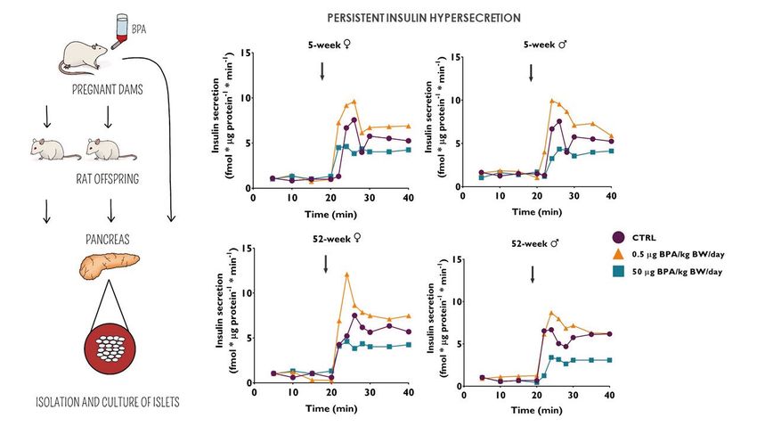
Dissertation presented at Uppsala University to be publicly examined in Sal X,
Universitetshuset, 75310, Uppsala, Friday, 7 May 2021 at 13:00 for the degree of Doctor of
Philosophy (Faculty of Medicine). The examination will be conducted in English. Faculty
examiner: PhD, Senior researcher Anna-Maria Andersson (Department of growth and
reproduction, Rigshospitalet, Denmark).

Abstract
Dunder, L. 2021. Exposure to Bisphenol A (BPA) and Metabolic Disruption. Digital
Comprehensive Summaries of Uppsala Dissertations from the Faculty of Medicine 1728.
98 pp. Uppsala: Acta Universitatis Upsaliensis. ISBN 978-91-513-1161-6.

Metbolic disorders such as obesity, type 2 diabetes, liver lipid disorders and metabolic syndrome
are increasing rapidly and have largely been attributed to genetic background and changes
in diet, exercise and aging. However, there is now considerable evidence showing that other
environmental factors, including environmental chemicals, may contribute to the rapid increase
in the incidence of these metabolic diseases. Of particular growing concern is low-dose
developmental exposure to endocrine disrupting chemicals (EDCs). The developing period is an
extremely sensitive window of exposure to environmental stressors, including EDCs, and early
life exposure has been linked to metabolic disorders later in life. Consistent with hormones,
EDCs can act at very low serum concentrations and even small changes in the endocrine system
may lead to extensive effects.
   The overall aim of this thesis has been to investigate potential metabolic disruption following
exposure to Bisphenol A (BPA), which is a known EDC. The experimental animal study
demonstrated that male and female rat offspring generally exhibited differential susceptibility
to developmental exposure to BPA (0.5 µg/kg BW/day or 50 µg/kg BW/day). The main results
showed that the lowest dose of BPA induced increased plasma triglyceride levels and increased
adipocyte cell density in inguinal white adipose tissue in female offspring. Further, this low
dose increased fatty acid indices and altered the fatty acid composition in male offspring
and enhanced insulin secretion in pancreatic islets from male and female offspring and dams.
Contrastingly, the higher BPA-dose decreased insulin secretion in pancreatic islets from male
and female offspring and dams. The increased fatty acid indices, and the altered fatty acid
composition together with enhanced insulin secretion may be early risk factors for insulin
resistance. Furthermore, depending on the tissue, dose and sex, BPA altered the expression of
genes involved in lipid and adipocyte homeostasis.
   The epidemiological study with a meta-analysis of data from the National Health and
Nutrition Survey (NHANES) did not disclose any associations between urinary BPA and
dyslipidemia. However, considering the cross-sectional nature of the present study, this should
rather be investigated in carefully designed prospective cohort studies with repeated BPA
measurements. Nonetheless, we hope that this paper can encourage researchers to evaluate
NHANES data using meta-analyses instead of pooling of data.
   This thesis concludes that exposure to BPA, which is a known EDC, most likely is a
contributor, along with genetic, social and behavioral factors, to the development of metabolic
disorders.

Keywords: Bisphenol A (BPA), Endocrine disrupting chemicals (EDCs), Developmental
exposure, Metabolic disruption, Adipose tissue, Fatty acids, Insulin secretion, Experimental
rat study, Epidemiology

Linda Dunder, Department of Medical Sciences, Akademiska sjukhuset, Uppsala University,
SE-75185 Uppsala, Sweden.

© Linda Dunder 2021

ISSN 1651-6206
ISBN 978-91-513-1161-6
urn:nbn:se:uu:diva-437856 (http://urn.kb.se/resolve?urn=urn:nbn:se:uu:diva-437856)
“You can’t go back and change the beginning, but you can start where you
                                             are and change the ending"
                                                             ~ C.S Lewis

                                 Till min älskade farfar Henry i himlen ♥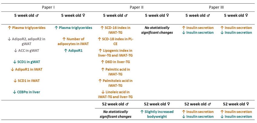
Illustrationer av Isabelle Rönnquist
List of Papers

This thesis is based on the following papers, which are referred to in the text
by their Roman numerals.

      I       Lejonklou, M.H.*, Dunder, L.*, Bladin, E., Pettersson, V.,
              Rönn, M., Lind, L., Waldén, T.B., Lind, P.M. (2017) Effects of
              low-dose developmental bisphenol A exposure on metabolic
              parameters and gene expression in male and female Fischer 344
              rat   offspring.    Environmental     Health    Perspectives.
              125(6):067018.

      II      Dunder, L., Lejonklou, M.H., Lind, L., Risérus, U.*, Lind,
              P.M.* (2018) Low-dose developmental bisphenol A exposure
              alters fatty acid metabolism in Fischer 344 rat offspring. Envi-
              ronmental Research. 166:117-129.

      III     Manukyan, L.*, Dunder, L.*, Lind, P.M., Bergsten, P.*,
              Lejonklou, M.H.* (2019) Developmental exposure to a very
              low dose of bisphenol A induces persistent islet insulin hyper-
              secretion in Fischer 344 rat offspring. Environmental Research.
              172:127-136.

      IV      Dunder, L., Lejonklou, M.H., Lind, P.M., Lind, L. (2019) Uri-
              nary bisphenol A and serum lipids: a meta-analysis of six
              NHANES examination cycles (2003-2014). Journal of Epide-
              miology & Community Health. 73(11):1012-1019.

*The authors contributed equally.

Reprints were made with kind permission from the respective publishers.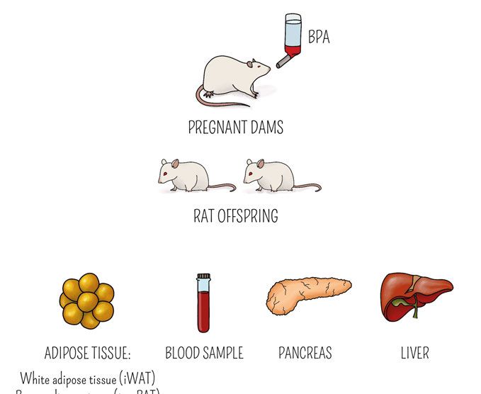
Additional publications (not included in the thesis)

Lind L., Lind, P.M., Lejonklou M.H., Dunder, L., Bergman, Å., Guerrero-
Bosagna, C., Lampa, E., Kyu Lee, H., Legler, J., Nadal, A., Kim Pak, Y.,
Phipps, R.P., Vandenberg, LN., Zalko, D., Ågerstrand, M., Öberg, M., Blum-
berg, B., Heindel, J.J., Birnbaum, LS. (2016) Uppsala Consensus Statement
on Environmental Contaminants and the Global Obesity Epidemic. Environ-
mental Health Perspectives. 124(5):A81-3. doi: 10.1289/ehp.1511115.

Lind, T., Lejonklou, M.H., Dunder, L., Rasmusson, A., Larsson, S., Melhus,
H., Lind, P.M. (2017) Low-dose developmental exposure to bisphenol A in-
duces sex-specific effects in bone and blood of Fischer 344 rat offspring. En-
vironmental     Research.       (159):61-68.    doi:    doi.org/10.1016/j.en-
vres.2017.07.020.

Stubleski, J., Salihovic, S., Lind, P.M., Lind, L., Dunder, L., McCleaf, P.,
Eurén, K., Ahrens, L., Svartengren, M., van Bavel, B., Kärrman, A. (2017)
The Effect of Drinking Water Contaminated with Perfluoroalkyl Substances
on a 10-year Longitudinal Trend of Plasma Levels in an Elderly Uppsala Co-
hort. Environmental Research. (159):95-102. doi: 10.1016/j.en-
vres.2017.07.050.

Mobacke, I., Dunder, L., Lind, L., Lind, P.M. Circulating levels of
perfluoroalkyl substances and left ventricular geometry in the elderly. (2017)
Environment International. doi: 10.1016/j.envint.2018.03.033.

Alavian-Ghavanini, A., Lin, P.I., Risén Rimforms, S., Tang, M., Lejonklou,
M.H., Dunder, L., Lind, P.M., Lindh, C., Bornehag, C.G., Rüegg. J. (2018)
Prenatal Bisphenol A Exposure is Linked to Epigenetic Changes in Glutamate
Receptor Subunit Gene Grin2b in Female Rats and Humans. Scientific Re-
ports. 27;8(1):11315. doi: 10.1038/s41598-018-29732-9.

Spörndly-Nees, E., Boberg, J., Ekstedt, E., Holm, L., Fakhrzadeh, A., Dun-
der, L., Lejonklou, M.H and Lind, P.M. (2018) Low-dose exposure to Bi-
sphenol A during development has limited effects on male reproduction in
midpubertal and aging Fischer 344 rats. Reproductive Toxicology. 81:196-
206. doi: 10.1016/j.reprotox.2018.08.007.

Lind, T., Lejonklou, M.H., Dunder, L., Kushnir, M.M., Öhman-Mägi, C.,
Larsson, S., Melhus, H., Lind, P.M. (2019) Developmental low-dose exposure
to bisphenol A induces chronic inflammation, bone marrow fibrosis and re-
duces bone stiffness in female rat offspring only. Environmental Research.
177:108584. doi: 10.1016/j.envres.2019.108584.

Contents
Introduction ................................................................................................... 11
Background ................................................................................................... 13
Programming during development ........................................................... 13
Tissues involved in metabolism ............................................................... 14
From the obesogen hypothesis to metabolism disrupting chemicals
(MDCs) .................................................................................................... 17
Environmental chemicals – Bisphenol A (BPA) ...................................... 19
Legislation and low level exposure to BPA......................................... 20
Metabolic fate of BPA ......................................................................... 23
CLARITY-BPA ................................................................................... 24
Effects of BPA exposure ..................................................................... 25
Risk of regrettable substitution – BPA analogues ............................... 26
Low doses and non-monotonic dose response (NMDR) .......................... 27
Sensitive test methods and route of administration for endocrine
disruptors .................................................................................................. 29
Objectives ..................................................................................................... 31
Material and methods .................................................................................... 32
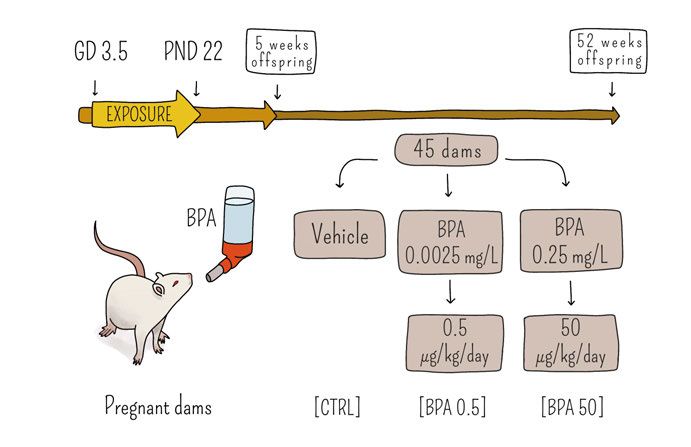
Experimental animal study (papers I, II, and III) ..................................... 32
Ethical statement.................................................................................. 32
Animals and housing ........................................................................... 32
Exposure .............................................................................................. 34
Blood and organ sampling ................................................................... 35
Histological analysis of adipose tissue and liver ................................. 35
Real-time quantitative polymerase chain reaction (RT-qPCR) ........... 36
Plasma lipid analyses ........................................................................... 36
Analysis of fatty acids ......................................................................... 36
Epidemiological study – NHANES (paper IV) ........................................ 37
Ethical statement.................................................................................. 37
Study population .................................................................................. 37
Urinary BPA ........................................................................................ 37
Lipid measures ..................................................................................... 38
Demographic and lifestyle factors ....................................................... 38
Statistical analyses.................................................................................... 39
Papers I and II ...................................................................................... 39Paper III ............................................................................................... 39
Paper IV ............................................................................................... 40
Summary of main results .............................................................................. 41
Low-dose effects from developmental BPA exposure in the
experimental animal study ....................................................................... 41
Paper I – increased plasma lipids and adipocyte cell density, and
altered gene expression ........................................................................ 41
Paper II – increased fatty acid indices and altered fatty acid
composition in various tissues ............................................................. 41
Paper III – Low-dose insulin hypersecretion ....................................... 43
Paper IV – Epidemiological study using NHANES data ......................... 43
Discussion ..................................................................................................... 45
Experimental animal study ....................................................................... 45
Low-dose- and sex-specific effects...................................................... 45
Lipid levels and adipocyte hyperplasia................................................ 46
Altered fatty acid levels ....................................................................... 47
BPA exposure and body weight .......................................................... 48
Insulin hyper- and hyposecretion......................................................... 49
A high fat diet and carbohydrates aggravate the metabolic effects of
BPA ..................................................................................................... 51
Strengths and limitations of the experimental animal study (papers
I–III)..................................................................................................... 52
Epidemiological study – NHANES data .................................................. 56
BPA exposure and measurement ......................................................... 58
Strengths and limitations of the epidemiological study (paper IV) ..... 59
Concluding remarks & future perspectives ................................................... 61
Svensk sammanfattning/Swedish summary .................................................. 63
Metabolismstörande kemikalier ............................................................... 63
Bisfenol A (BPA) – en hormon- och metabolismstörande
plastkemikalie........................................................................................... 64
Exponering och verkningsmekanism ....................................................... 64
Lågdoseffekter och lagstiftning ................................................................ 65
”BPA-fria” produkter ............................................................................... 67
Avhandlingens syfte och resultat ............................................................. 67
Slutsats ..................................................................................................... 69
Acknowledgements ....................................................................................... 70
References ..................................................................................................... 73Abbreviations
AOPs Adverse outcome pathways
BPA Bisphenol A
BPA-G Bisphenol A glucuronide
BPA0.5 0.5 µg BPA/kg BW/day
BPA50 50 µg BPA/kg BW/day
BPF Bisphenol F
BPS Bisphenol S
BW Body weight
CD-SD Charles-River Sprague-Dawley
CLARITY-BPA Consortium Linking Academic and Regulatory Insights
on BPA Toxicity
CTRL Control
DEHP Phthalate di-(2-ethylhexyl)
DES Diethylstilbestrol
DOHaD Developmental origins of health and disease
ECHA European Chemicals Agency
EDCs Endocrine-disrupting chemicals
EFSA European Food Safety Authority
ERα Estrogen receptor alpha
ERβ Estrogen receptor beta
F344 Fischer 344
FDA U.S. Food and Drug Administration
GD Gestational day
GPER G-coupled protein receptor
GR Glucocortiod receptor
GSIS Glucose-stimulated insulin secretion
HDL High-density lipoprotein
IATA Integrated approach to testing and assessment
IPCS International Programme on Chemical Safety
iscpBAT Interscapular brown adipose tissue
iWAT Inguinal white adipose tissue
LOAEL Lowest-observed-adverse-effect-level
MetS Metabolic syndrome
MDC Metabolism-disrupting chemical
NAFLD Non-alcoholic fatty liver disease
NAMs New approach methodologiesNHANES National Health and Nutrition Examination Survey
NMDR Non-monotonic dose response
NOAEL No-observed-adverse-effect-level
PAHs Polycyclic aromatic hydrocarbons
PCR Polymerase chain reaction
PL-CE Plasma cholesterol esters
PND Post natal day
PPARγ Peroxisome proliferator-activated receptor gamma
REACH Registration, Evaluation, Authorisation and Restriction
of Chemicals
RXR Retinoid X receptor
SERM Selective estrogen receptor modulator
SVHC Substances of very high concern
T2DM Type 2 diabetes mellitus
TDI Tolerable daily intake
TG Triglyceride
TR Thyroid hormone receptor
MetS Metabolic syndrome
qPCR Quantitative real-time PCR
UGT Uridine diphosphate glucoronyltransferase
WHO World Health OrganizationIntroduction
The incidences of metabolic diseases such as obesity, fatty liver disorders,
type 2 diabetes mellitus (T2DM) and metabolic syndrome (MetS) have dra-
matically risen over the last few decades. The cornerstones of the development
of these disorders have been and still are largely attributed to genetic heritage,
imbalance in intake and expenditure of calories, aging, and sleep deficits.
However, there is now considerable evidence that these parameters cannot
fully explain the current metabolic disease epidemic. It has been demonstrated
for example that for a given level of activity and caloric intake, individuals in
today’s society weigh more than they did 20–30 years ago (1). Consequently,
other factors are now being implicated in the global deterioration of metabolic
health, such as exposure to environmental factors.
One environmental factor that has gained a lot of attention is exposure to en-
docrine-disrupting chemicals (EDCs) that can interfere with the body’s hor-
mones, including hormones involved in metabolic pathways (2). EDCs can be
found in a wide range of consumer products, which has resulted in ubiquitous
exposure in humans, in wildlife, and in the environment. There are also natural
endocrine disruptors that originate from animal, human, or plant (phytoestro-
gen) sources; however, the main international concern is currently focused on
synthetic chemicals with identified endocrine-disruptive properties. The
World Health Organization (WHO) and International Programme on Chemi-
cal Safety (IPCS) proposed an EDC definition in 2002 which is now broadly
accepted within the scientific community: “An endocrine disruptor is an ex-
ogenous substance or mixture that alters function(s) of the endocrine system
and consequently causes adverse health effects in an intact organism, or its
progeny or (sub)populations. A potential endocrine disruptor is an exogenous
substance or mixture that possesses properties that might be expected to lead
to endocrine disruption in an intact organism, or its progeny, or (sub)popula-
tions.” (3). Scientific criteria for identification of endocrine disruptors, based
on the WHO/IPCS definition, have been implemented within the EU (EU reg-
ulations 2017/2100 and 2018/605) and read as follows: A substance shall be
considered as having endocrine properties if it meets all of the following cri-
teria:
a) it shows an adverse effect in [an intact organism or its progeny]/[on-
target organisms]…;
11b) it has an endocrine mode of action, i.e., it alters the function(s) of
the endocrine system;
c) the adverse effects are a consequence of the endocrine mode of ac-
tion.
The production of synthetic chemicals has increased dramatically since the
1960s, with a peak of 314 million tonnes within the EU in 2007 (4). However,
much of the world’s chemical production has since then been moved outside
the EU, and total chemical production in the world has probably not yet
reached its peak. There are 1,482 chemicals listed on the TEDX list of known
or suspected EDCs (5), and studies that link exposure to EDCs with various
diseases are increasing. Of particular concern is exposure to EDCs during fetal
development and early childhood, when development and organization of en-
docrine organs take place. A mounting body of evidence demonstrates that
such exposure early in development may cause alterations that program risks
for diseases that manifest later in life, possibly via mechanisms of epigenetic
memory (6-8). The increasing concern regarding potential health effects from
EDC exposure has prompted two position statements from the Endocrine So-
ciety, a professional, international medical organization in the fields of endo-
crinology and metabolism (9, 10). Within the EU, EDC exposure has been
reported to contribute to disease and dysfunction across the lifespan, with
costs that exceed EUR 100 billion annually. And this number is most likely
an underestimate, since the estimates represent only a small number of EDCs
and a limited number of diseases (11).
12Background
Programming during development
It has become evident that there are critical periods during early development
(in utero and during the first years of life) where the susceptibility or set point
for diseases such as obesity, diabetes and non-alcoholic fatty liver disease
(NAFLD) is established. These periods are thus extremely sensitive for expo-
sure to environmental stressors, including EDCs (8, 12, 13). The developing
period can be described as a “one-way street” (9), a unidirectional process of
events that occur in an organized manner where there is no chance of going
back to “re-do” the specific developmental events. Hormones act in an organ-
izational manner during this period, whereas, after the onset of puberty, they
enter a more “activational” role. Critical windows in human development have
been identified, and chemical exposure during these vulnerable periods can
lead to subtle changes in gene expression, tissue organization, or other levels
of biological organization that could induce permanent dysfunction leading to
increased susceptibility to disease (14).
Data from epidemiological studies show that exposure to environmental fac-
tors during development is associated with an increased risk of developing
chronic diseases such as cancer, infertility, diabetes, cardiovascular disease,
obesity, and behavioral disorders, such as schizophrenia, later in life (15, 16).
There is growing evidence that disturbances of the central endocrine regula-
tory systems that are developed in early life may cause development of meta-
bolic diseases later in life (17, 18). The developing organism has a number of
underdeveloped mechanisms, which may be the explanation for its suscepti-
bility to exposure; these include DNA repair mechanisms, the immune system,
detoxifying enzymes in the liver, and the blood/brain barrier. Additionally,
compared to adults, children eat and drink more per body weight, which con-
sequently leads to higher chemical exposure.
Yet another important reason for the increased susceptibility of the developing
period to EDCs is the epigenetic signaling that regulates gene expression
which controls development. Even subtle alterations in epigenetic mecha-
nisms during development may lead to persistent changes that have conse-
quences much later in life, or even in the next generation. Epigenetic changes
can be defined as “any long-term change in gene function that persists even
13when the initial trigger is long gone and does not involve a change in gene sequence or structure” (19). The best-understood epigenetic modifications in- clude for example post-translational modification of histone tails and methyl- ation of the DNA at the cytosine base, both of which are important for the structure and accessibility of the DNA for transcription factors (20). There is now compelling evidence from experimental and epidemiological studies showing that EDCs can induce epigenetic changes (21). The best-known study of the “early exposure late effects” phenomenon is the Dutch famine during the winter of 1944-1945 when the World War II was coming to a close. The lack of food for many months led to low birth weight for children born to women that were pregnant during this period. A cohort study later showed that the prevalence of a number of adult-onset diseases such as obesity, diabetes, cardiovascular disease, and renal dysfunction in- creased for these children when they reached adulthood (22). In addition, re- sults from a later historical cohort study display that the effects caused by ma- ternal malnutrition did not stop at the first generation for individuals of the famine cohort, but persists in the next generation as well, leading to the con- clusion that the effects can be transgenerational (23). Yet another cohort-study from Finland has confirmed this; low birth weight is strongly associated with coronary heart disease, T2DM, and hypertension (24). These and similar find- ings have given rise to the “developmental origins of health and disease” (DO- HaD) paradigm (25). These later-life consequences of early-life exposure have also been shown to be true for pharmaceuticals and chemicals, including EDCs. Many environ- mental chemicals have the ability to cross the placenta and produce develop- mental effects in the offspring (26). Among the first and most striking evi- dence of this was the use of diethylstilbestrol (DES) as an estrogenic pharma- ceutical drug for women during pregnancy to prevent miscarriage and prema- ture births. Pregnant women were prescribed DES from 1941 through 1971 and the American Cancer Society has estimated that about 5 to 10 million people were exposed to DES worldwide, including women who took DES while pregnant and women and men whose mothers took DES while pregnant with them. Regrettably, a higher risk for clear cell adenocarcinoma, infertility, miscarriage, ectopic pregnancy, and breast cancer was found for children born by DES-treated mothers (27). Tissues involved in metabolism There are many tissues involved in the regulation of metabolism, including the gastrointestinal tract (ghrelin, cholecystokinin, glucagon-like peptide), en- 14
docrine pancreas (insulin, glucagon), muscle (insulin), liver (insulin, gluca-
gon), immune system (cross-talk between the immune system and adipose tis-
sue in obesity), brain (neuropeptide y, agouti-related protein, pro-opiomelano-
cortin, alpha melanocyte-stimulating hormone) thyroid gland (thyroid hor-
mone), and adipose tissue (leptin, adiponectin and a variety of other factors)
(reviewed in (2)).

Adipose tissue is regarded as one of the most central organs in metabolic ho-
meostasis. The traditional view of adipose tissue is as a depot of superfluous
energy and as a supportive tissue for other organs. Today it has become evi-
dent that the characteristics and properties of adipose tissue are much more
advanced than this. In 1994, the hormone leptin was identified and character-
ized, which firmly established adipose tissue as an endocrine organ (28). It is
now known that the cells of adipose tissue, i.e., adipocytes, express endocrine
hormones such as leptin, adiponectin, apelin, and others which regulate nutri-
ent homeostasis, food intake, cardiovascular function, blood pressure, blood
coagulation and inflammation. These hormones have been given the name ad-
ipokines and scientists are continually discovering more of these adipose-se-
cretory products (29). In addition, adipose tissue is highly connected to me-
tabolism of steroid hormones (estrogens, androgens, and glucocorticoids) by
the expression of various enzymes responsible for activation, interconversion,
and inactivation (30). Adipose tissue also contains a metabolic machinery that
enables communication with distant organs, such as the central nervous sys-
tem (CNS) and the immune system. As a result, adipose tissue is involved in
organizing various biological processes, including energy metabolism and
neuroendocrine and immune function (31). The liver and adipose tissue inter-
act on the regulation of the metabolism of lipid and glucose by secreting var-
ious factors with diverse metabolic regulatory effects. For example, in the
liver, excess energy from the diet is stored in the form of glycogen (main
source of glucose for all tissues), and when glycogen depots are full, any ad-
ditional excess energy is stored in the form of lipids in adipose tissue (32).

Adipose tissue is classified by morphology and function into white, brown, or
beige compartments. White adipose tissue (WAT) maintains energy homeo-
stasis through energy storage in the form of triglycerides (TGs). WAT is fur-
ther classified by location, namely subcutaneous (under the skin) and visceral
(intra-abdominally and around internal organs). Excess visceral adipose tissue
is associated with a much more deleterious metabolic profile than subcutane-
ous fat accumulation (33). In fact, some studies have shown that subcutaneous
adipose tissue may even be protective against metabolic diseases, such as di-
abetes (34). Men have a higher tendency towards visceral fat deposition while
women mainly store adipose tissue in subcutaneous areas (35).

                                                                            15
Brown adipose tissue (BAT) has a different morphology than WAT and con- sists of highly concentrated mitochondria which gives its unique characteris- tics. Mitochondria contain the unique protein uncoupling protein 1 (UCP1), which mediates the process of non-shivering thermogenesis that produces heat. The function of BAT can be described in a very simplified manner in that it transfers energy from food (mainly fatty acids) into heat. This process is particularly important for small mammals during hibernation and for human infants to obtain core body temperature. Active BAT was traditionally thought to be restricted to small mammals and human infants. However, in 2009, stud- ies unambiguously reported that healthy adult humans indeed have significant depots of functionally active BAT (36-38). Beige fat, also called “browned” or “brite” depots is a rather new classification of adipose tissue where classical WAT compartments contain brown adipocytes. There are many similarities between beige fat and BAT; for example, both are highly metabolically active and utilize chemical energy for heath production (39). The thyroid gland regulates diverse important metabolic pathways, mainly via actions of thyroid hormone in the brain, WAT and BAT, skeletal muscle, the liver, and the pancreas. During development, thyroid hormone regulates met- abolic pathways essential for normal growth and development, and in adult- hood it regulates metabolism (40). Specific mechanisms of action of thyroid hormone in metabolic regulation include, for example, positive effects on lipid and lipoprotein metabolism (41), cross-talk with nuclear receptors that re- spond to nutrient signals (42), and regulation of facultative thermogenesis, for which BAT is the major site, thus implying that thyroid hormone plays a per- missive role as a thermogenic hormone for the function of BAT (43). It is well known that levels of thyroid hormone are closely connected to body weight and energy expenditure, as is evident in the effects of the conditions hyperthy- roidism (overactive thyroid) and hypothyroidism (underactive thyroid). Indi- viduals who suffer from hyperthyroidism have increased energy expenditure, weight loss, reduced cholesterol levels and increased lipolysis, and gluconeo- genesis. In contrast, hypothyroidism causes the opposite effects on all of the aforementioned parameters (44). The endocrine pancreas is built up by the pancreatic islets of Langerhans, which is a heterogeneous population of 1000–3000 cells/islets where the in- sulin-releasing β-cell is the main cell type. In rodent pancreases, the cell pop- ulation consists of about 70–80 % β-cells and 20 % α-cells. This differs from the composition of the human pancreas, which is comprised of 40–45% α- cells, 50 % β-cells, and up to 10 % δ-cells. The α-cells are responsible for glucagon secretion, and the δ-cells for somatostatin release, a master control- ler of all gastrointestinal hormones. It is crucial for the body to obtain blood glucose levels within the normal range, and this is accomplished through var- ious processes in different tissues. After food intake, blood glucose levels rise, 16
and the glucose is taken up by β-cells; this glucose metabolism then increases
the ATP/ADP, ratio and insulin can be released. This subsequently closes
plasma-membrane-sensitive K+ (KATP) channels which are responsible for
maintaining the resting membrane potential. This leads to cellular depolariza-
tion and insulin can be released from the cell into circulation. Next, insulin
binds to receptors on the surface of target cells, which allows glucose uptake
and metabolism in that tissue. If this closely regulated β-cell insulin produc-
tion and/or transport is compromised, the result will be increased blood glu-
cose levels that with time can lead to the development of insulin resistance
and ultimately diabetes (2). If glucose levels decrease in the body, the pancreas
will instead respond by releasing glucagon, a process which triggers the liver
to stimulate glucogenolysis and gluconeogenesis to raise circulating blood
sugar levels again (45).

From the obesogen hypothesis to metabolism disrupting
chemicals (MDCs)
Previously, research on EDCs has mainly been implemented in reproductive
biology but has now an established role in the field of metabolic disruption.
In 2002, Paula Baille-Hamilton, a physician, wrote the very first review article
focusing on environmental chemicals and obesity. In the paper entitled
“Chemical toxins: a hypothesis to explain the global obesity epidemic,” she
presented an overview of studies showing associations between exposure to
organochlorine pesticides, solvents, plastic additives such as phthalates and
bisphenol A (BPA), flame retardants and heavy metals, and increased body
weight (46). The studies included in the review had overlooked the effects on
weight gain since the main focus was to study weight loss and other robust
toxic effects from high dose exposures. In the paper she noted that “Therefore
it can be posited that the relatively recent presence of synthetic chemicals in
the environment may be a significant causative factor in the current world-
wide obesity epidemic. These chemicals may be causing weight gain via toxic
effects on the body’s natural weight-control mechanisms.” (46). Baille-Ham-
ilton succeeded, through her background as a medical doctor and her ability
to see the connection between environmental contaminants and obesity, in
putting two scientific fields together. Two of the papers that she cited in the
review were conducted by scientists in the field of reproductive toxicology,
and at the time studying the effects of BPA. The authors serendipitously dis-
covered that developmental exposure to BPA increased body weight in rats
and mice (47, 48).

In 2006, Grun and colleagues observed that the biocide tributyltin induced
differentiation of adipocytes in a murine 3T3-L1 cell model, increased lipid

                                                                              17
accumulation in adipose depots, liver, and testis of neonate mice and for- mation of ectopic adipocytes in and around gonadal tissues in the amphibian Xenopus laevis. These effects were most probably mediated via activation of the peroxisome proliferator-activated receptor gamma (PPARγ) and retinoid X receptor (RXR), which was examined and confirmed in the study (49). Based on this and some other similar studies the term “obesogen” was coined later that year in a paper by Grun and Blumberg (50), and the authors wrote a follow-up review article on the topic three years later with the title “Endocrine disruptors as obesogens.” Obesogens are defined as chemicals that have the ability to promote obesity by either increasing the number of adipocytes and/or the amount of fat stored in existing adipocytes (2). The definition fur- ther expands to the ability of obesogens to act via indirect mechanisms to pro- mote obesity, including altering of the gut microbiota to promote food storage (51) and alteration of levels of hormones involved in the control of appetite and satiety (52). The obesogen hypothesis puts forward two important points: First, susceptibility to the development of obesity starts during development (in utero and the first years of life). Secondly, the susceptibility to the devel- opment of obesity is due in part to exposure to obesogens that alter program- ming, which in turn disrupts the set point for weight gain later in life (2). Prob- able mechanisms for obesogens are via epigenetic modifications. Exposure to environmental chemicals including several known obesogens has been shown to result in epigenetic alterations in vivo and altered epigenetic signatures and obesity phenotypes even in unexposed generations, i.e., transgenerational ef- fects (53-55). Thus it seems that chemical exposure can cause changes in the germline leading to observable phenotypes in subsequent generations, show- ing that these epigenetic alterations can be inherited across generations (56). A number of workshops with a focus on developmental exposures to EDCs and obesity have been organized worldwide. Two of the workshops were held in Uppsala, Sweden in 2010 and 2015, and a consensus statement was pub- lished as a result of the 2nd International Workshop on Obesity and Environ- mental Contamination. The authors, who were the speakers at the workshop, presented recommendations for an action plan in order to restrict the use of contaminants that potentially may induce harmful metabolic-disruptive effects (57). In 2015, the Parma Consensus Statement proposed that the obesogen field should be expanded to also include chemicals that cause or increase the sus- ceptibility to develop T2DM, NAFLD, MetS, and altered lipid metabolism. In line with the Parma statement, a new name for the obesogen hypothesis was proposed, and the research field is from now on termed “the metabolism-dis- rupting chemical (MDC) hypothesis” (58). A number of MDCs have been identified through experimental animal studies with consistent associations in epidemiological studies, including some phthalates (59), which is a class of 18
chemicals mainly used to promote flexibility in plastic products, but also as
fixatives in fragranced household and personal care products. In rats, devel-
opmental exposure to the best-studied phthalate di-(2-ethylhexyl) (DEHP) in-
duces elevated blood glucose and impaired insulin and glucose tolerance,
which are clear signs of β-cell dysfunction (60). Another example of MDCs is
a family of chemicals denoted polycyclic aromatic hydrocarbons (PAHs),
which are byproducts of fossil-fuel burning including diesel exhaust, air pol-
lution, and cigarette smoke. Airway exposure to one PAH, benzo[a]pyrene has
been shown to cause alteration of hepatic lipids in mice, which may be a risk
factor for the developing of NAFLD (61). Yet another chemical group in
which some have suspected metabolism-disruptive properties are bisphenols,
and mainly one bisphenol that has been extensively studied, namely BPA,
which is the environmental chemical in focus in this thesis and will thus be
further discussed below.
Environmental chemicals – Bisphenol A (BPA)
The world’s industrialization has resulted in a massive increase in the produc-
tion of a great number of various chemicals, many of which are used in the
plastics industry. The environmental problem of this is evident, but im-
portantly it has also become a human health problem due to the continuous
and unintentional exposure to monomers and additives in plastic released
mainly from plastic packaging by direct contact with food, beverages and air.
BPA is one example of such a chemical. In the 1930s BPA was investigated
for potential commercial use as a pharmaceutical, and as a result it was con-
firmed that BPA possesses estrogenic qualities. BPA was however compared
to a far more potent estrogen, namely DES, which initiated the use of the latter
substance as a pharmaceutical agent instead (62). In 1954, DES was also ap-
proved as hormone supplementation for growth stimulation in cattle and was
used for this purpose until 1972 when it was taken off the market due to de-
tected residues in edible animal products (63). Another use was found for
BPA, in the plastics industry. The main use of BPA is in the manufacture of
polycarbonate plastics and epoxy resins but also as a non-polymer additive to
other plastics. As a result, BPA can be found in numerous consumer products
such as baby bottles, food containers and food coatings, toys, building mate-
rials, water bottles, CD/DVDs, receipts, and dental sealants (64). BPA has also
been used in epoxy lining in water pipes in the past and has therefore been
found in drinking water in some places in Sweden (65).
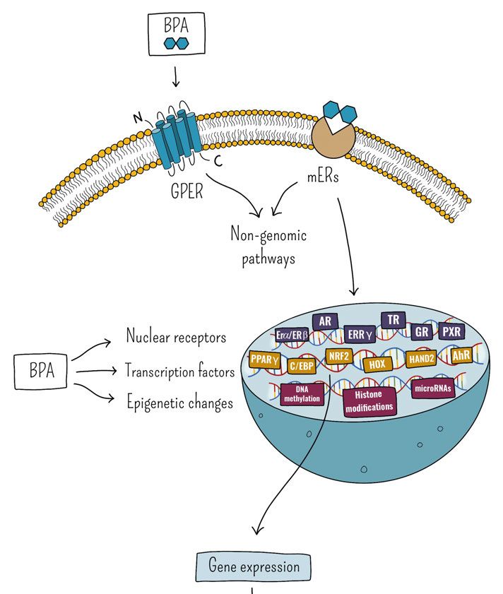
BPA is one of the best-studied chemicals with endocrine-disrupting properties
because of its high-volume production worldwide, its use in a wide range of
products, and as a result, the ubiquitous human exposure (66). The estrogenic
activity of BPA with activation of the nuclear estrogen receptors alpha (ERα)
19and beta (ERβ) both in vivo and in vitro is the most fully studied mechanism of action (67, 68). However, the endocrine activity of BPA is far more com- plex. In addition to being a nuclear ER agonist, it is also an agonist of the membrane ERs (mERs), the G protein-coupled receptor 30 (GPR30/GPER), and can act through these receptors via non-genomic pathways at very low concentrations. BPA is also an antagonist of the thyroid hormone receptor (TR), which together with the GPR30/GPER are important receptors for met- abolic regulation (69). Further, it has been reported that BPA can activate the androgen receptor (AR) and the PPARγ (70, 71). Finally, BPA also binds to some orphan receptors, including estrogen-related receptor γ (ERRγ) (72), and the aryl hydrocarbon receptor (AhR) (73). This displays the multifaceted na- ture of BPA and makes it clear that it is an oversimplification to conceptualize BPA as simply an estrogenic substance (Figure 1). Thus, it seems that BPA can act as a selective estrogen receptor modulator (SERM), meaning that BPA can execute other modes of actions than through classical estrogenic path- ways, and, additionally, that signaling may vary depending on dose, timing of exposure, and cell types and tissues (74). The mimicking of or antagonizing of estrogens and/or androgens by BPA may alter metabolism and the pattern of synthesis of natural hormones involved in for example regulation of appe- tite and hunger, as well as modifying receptor levels. Fetal exposure to BPA in mice has been shown for example to affect food intake during puberty and in adulthood, as well as leptin and insulin levels (75), which together with other hormones, neurotransmitters, and growth factors control pathways in the hypothalamus that regulate food reward mechanisms and food cravings. Legislation and low level exposure to BPA Because of the widespread usage, ubiquitous human exposure, and subse- quently concern regarding human health, BPA has been regulated in some ar- eas of application and products. BPA was banned from baby bottles within the EU in March 2011 (76). The production, import, export and marketing of food containers containing BPA have also been totally banned in France since Jan- uary 2013 (77). In Sweden, the use of BPA for epoxy lining in water pipes was banned in September 2016. BPA was added to the Candidate List of sub- stances of very high concern (SVHC) under the EU regulation REACH (Reg- istration, Evaluation and restriction of Chemicals) due to its toxic properties for reproduction (article 57c in REACH) in January 2017. Entry status for BPA on the candidate list was updated in June 2017 to also include endocrine- disrupting properties for human health and in January 2018 for the environ- ment (Article 57f in REACH) (78). In 2018, new EU legislation resulted in the prohibition of materials and products that can leach BPA to foods intended for children between 0 and 3 years of age. In January 2020 restrictions were adopted within the EU regarding thermal paper (used for receipts and tickets) containing BPA, and the concentration limit is 0.02 % by weight. In January 20
2021, the revised drinking water directive was adopted within the EU, where
limit values were introduced for additional EDCs, including BPA. Sweden has
to implement the directive in Swedish legislation within 2 years (Directive
(EU) 2020/2184).

BPA levels have been detected in human tissues, urine and blood, and BPA
can be transferred from mother to offspring via the placenta and breast milk
(66, 79). Biomonitoring studies show that the serum concentration of bioac-
tive (unconjugated) BPA in humans is within the 1–10 nM range (66). The
main route of exposure is via digestion or oral mucosal absorption. BPA can
leak from food containers, water bottles and into food, especially when the
food/water is heated (80-83). Another earlier unstudied route of exposure has
gained attention in the past few years, namely absorption through the skin
when handling thermal paper such as receipts and tickets. An increased uri-
nary concentration of BPA after continuously handling receipts for 2 hours
was reported in a pilot study from the US (84). Studies from other countries
such as Belgium and China have consistently found that dermal exposure to
BPA is a significant additional route of exposure, especially for workers han-
dling receipts but also for the general population (85, 86).

The European Food Safety Authority (EFSA) has previously established a no-
observed-adverse-effect-level (NOAEL) of 5 mg BPA/kg BW/day, which cor-
responds to a tolerable daily intake (TDI) of BPA of 50 µg/kg BW/day. This
TDI is based on guideline-driven toxicity studies in rats and mice (87, 88), and
the doses are generally higher than doses that have been shown to produce
adverse effects in animals, especially if exposure occurs during development
(89, 90). The sufficiency of this TDI has therefore been questioned, and it was
reduced to 4 µg/kg/day within the EU in 2015. However, this TDI is prelimi-
nary, pending results from a two-year study by the U.S. National Toxicology
Program. The US Environmental Protection Agency (EPA) reference dose for
BPA remains unaltered at 50 µg/kg BW/day. Alarmingly, an exploratory
study, where a single dose of 50 µg/kg BW/day, the estimated safe dose for
humans by EPA, was administered to male and female human volunteers,
showed that BPA may suppress insulin and C-peptide concentrations in re-
sponse to glucose stimulation. Bioactive BPA was measured in serum and was
at levels detected in human biomonitoring studies (91). These results have
been supported by another experimental study where BPA was administered
at 50 µg/kg BW/day to 11 adult human volunteers. The results indicated that
a single dose of the EPA presumed safe dose of BPA of 50 µg/kg BW/day
decreased glucose, insulin, and C-peptide responses to an oral glucose toler-
ance test (92).

                                                                             21
Figure 1. A potential integrative model of molecular mechanisms of BPA. BPA acts in pleiotropic ways to modulate physiological pathways linked to the development of various disturbances and diseases. Inspired by and adopted from (93) and (69). The figure is protected by copyright and permission is required to distribute the con- tent. 22
Metabolic fate of BPA
For humans, the main exposure route of BPA is orally via various dietary
sources, but there are also a number of possible non-dietary sources of BPA,
such as through thermal papers (receipts, tickets), cigarette filters, air, dust,
and personal care products (94). Thus, administration is not limited solely to
the oral route but can also occur via dermal exposure or via inhalation (83).
When BPA enters the gastrointestinal tract, it is absorbed into the mesenteric
blood vessels and transported to the liver where metabolism is thought to oc-
cur rapidly by phase II conjugation by the enzyme uridine diphosphate glu-
coronyltransferase (UGT), resulting in extensive production of the BPA-glu-
curonide (BPA-G) metabolite. The addition of glucuronic acid makes the me-
tabolite more water soluble and excretion can therefore occur via urine. This
process significantly reduces the concentration of unconjugated or “free BPA”
circulating in the blood. However, the metabolism is not complete; data from
both human and animal toxicokinetic studies demonstrate that some unconju-
gated BPA remains and can stay in the circulation even when exposures occur
strictly via the oral route (95-98). A smaller amount of BPA-sulfate is also
formed, and this process is catalyzed by sulfotransferase enzymes (99-101).
These conjugates are thought to be inactive due to their inability to bind to
estrogen receptors; both of the metabolites are instead eliminated via the urine
(102, 103). However, BPA-G has been detected in blood, which indicates that
the metabolites may not be removed from the circulation as efficiently as some
models suggest (104, 105). A third metabolite formed in small amounts upon
metabolism of BPA is 4-methyl-2,4-bis(4-hydroxyphenyl)pent-1-ene (MBP)
which is 250–1000 times more potent than BPA (106-108).

What makes BPA metabolism even more complex is that there is evidence
that enzymes in the body can deconjugate BPA metabolites to the bioactive
form of BPA. This suggests that even conjugated forms of BPA that are pre-
sumed safe may not be inconsequential for human health effects. This may
occur especially during pregnancy, since deconjugating enzymes are abundant
in the placenta and fetal tissues (109, 110), thus making fetuses and babies
especially sensitive to BPA exposure due to their vulnerable underdeveloped
drug-metabolizing system. Since BPA has a short half-life of only a few hours,
and since most of the BPA is conjugated and eliminated, it is not a matter of
any known accumulation of BPA in living organisms, but there are many po-
tential sources for human exposure; thus exposure to BPA is continuous.

If BPA enters the body from a non-dietary source, for example via dermal
absorption or inhalation, its passage will not be through the liver before it en-
ters systemic circulation. Studies in animals suggest that the toxicokinetics of
BPA in these cases can be very different, since phase II-conjugation in the
liver is by-passed. As a result, unconjugated BPA can circulate in the body for

                                                                              23
longer periods before being metabolized (95, 111, 112). Also, important to acknowledge is that oral BPA exposure may result in additional absorption via the tongue and oral mucosa (sublingual absorption). This may result in a different metabolism route compared to absorption solely through the gastro- intestinal tract (if BPA is administered through oral gavage). In a study on dogs, the oral transmucosal passage of BPA exposure was evaluated. Three different routes of exposure were compared: intravenous, orogastric (oral ga- vage) and sublingual, and the results implied that BPA can be rapidly and ef- ficiently absorbed via the oral mucosa after sublingual exposure, and that the bioavailability of BPA was higher after this exposure route compared with the other routes (113). The toxicokinetics of BPA seem to be similar in mice, rhe- sus monkeys, and humans after oral exposure. An oral dose of 400 µg/kg ad- ministered to monkeys and mice yielded an average 24-hour unconjugated se- rum concentration of 0.5 ng/mL. In comparison, multiple human studies report serum BPA concentrations in the range of 0.3-4.4 ng/mL. This indicates that the total daily human exposure cannot come from oral exposure alone and that the exposure is underestimated (114, 115). Indeed, in a study on human vol- unteers, where BPA patterns were studied over a 48-h fasting period, the levels did not decrease as much as anticipated based on knowledge about for exam- ple half-time, and fluctuations in BPA levels were evident. The results of this study further supports the idea that there are other considerable sources of ex- posure to BPA that need to be taken into consideration (116). In risk assessments it is essential to understand how much BPA enters the human body. For BPA, accurate assessment in humans requires the measure- ment of bioactive BPA (unconjugated) in serum and the metabolites in urine. Biomonitoring studies have mainly focused on analyzing BPA in urine over time, which has been thought to give quite good insight to human exposure to BPA over time. However, a recent study in which new direct assay methods were used to measure BPA metabolites show that previous studies using indi- rect techniques have underestimated actual human levels of BPA (117). CLARITY-BPA A lot of controversy exists regarding human health risks from especially low- dose exposure to BPA. The lack of corroboration between the results from academic laboratories and those from traditional regulatory toxicity studies (guideline studies) has contributed to the ongoing debate and prompted an in- ter-agency collaboration between the National Institutes of Environmental Health Sciences (NIEHS), the National Toxicology Program, and the US Food and Drug Administration (FDA). In 2012, this collaboration resulted in the Consortium Linking Academic and Regulatory Insights on Toxicity of BPA (CLARITY-BPA) whose purpose was to combine guideline and academic 24
studies to deliver comprehensive data for accurate risk assessment and ulti-
mately resolve uncertainties on BPA toxicity (118). To date, CLARITY-BPA
is the most comprehensive study investigating a full range of health effects of
different BPA doses in rats. BPA or vehicle-treated rats from an FDA facility
were used to conduct a guideline study and animals and/or tissues were sub-
sequently provided to academic researchers for investigation. In September
2018, the full report consisting the results from the core guideline study of
CLARITY-BPA was released with the conclusion that BPA exposure causes
minimal health effects and single low-dose effects and non-monotonic pat-
terns were dismissed (119, 120). In contrast, academic researchers, some that
have conducted research within the CLARITY-BPA study and some that have
evaluated all of the CLARITY-BPA data, conclude that “the cumulative re-
sults of the CLARITY study provide solid documentation of BPA effects in the
low-dose range, below the current NOAEL, and conclude that the TDI dose is
not reasonably protective of public health” (121).

The goal from the beginning was that an integrative publication of all CLAR-
ITY-BPA data from both the core guideline study and the studies from the
independent researchers should be prepared at the finalization of the studies.
However, it now seems that FDA is not interested in participating in writing
such a publication. Instead, the independent researchers have published, with
help from other experts in the field, a final report of 8 of the independent stud-
ies. In this report they present their data from CLARITY-BPA and put it in
perspective to their previously published data on BPA. One of the conclusions
from this unique publication is “Because the low dose of BPA affected end-
points in the same animals across organs evaluated in different labs, we con-
clude that these are biologically - and toxicologically relevant” (122).

Effects of BPA exposure
Mammalian experimental studies have shown that early life exposure to low
doses of BPA can induce various effects in endocrine-driven systems involved
in growth, metabolism, behavior, fertility, and cancer risk (123). More specific
effects include for example altered spatial learning, disturbed development of
male and female reproductive organs (124, 125), altered hormone levels (126),
reduced immune response (127), increased incidence of female mammary
gland adenocarcinoma (128), and altered bone geometry (129, 130). In addi-
tion, BPA exposure has been reported to result in numerous disturbances of
multiple metabolic outcomes such as augmented glucose-stimulated insulin
secretion (131), insulin resistance (132), elevated blood lipids (133, 134) and
glucose levels (133), increased liver fat (135, 136), and body weight (47, 75,
134, 137) in animal studies. The timing of exposure to EDCs during develop-
ment is critical, and various developing periods have different susceptibility.
In mice, different exposure windows have been evaluated with regard to low

                                                                               25
You can also read