External fixation of airway stents for upper tracheal stenosis and tracheoesophageal fistula
←
→
Page content transcription
If your browser does not render page correctly, please read the page content below
Interactive Cardiovascular and Thoracic Surgery 33 (2021) 442–447                                                                          ORIGINAL ARTICLE
doi:10.1093/icvts/ivab098 Advance Access publication 20 April 2021

  Cite this article as: Niwa H, Oki M, Saka H, Torii A, Yamada A, Shigematsu F et al. External fixation of airway stents for upper tracheal stenosis and tracheoesophageal
  fistula. Interact CardioVasc Thorac Surg 2021;33:442–7.

      External fixation of airway stents for upper tracheal stenosis and
                           tracheoesophageal fistula
        Hideyuki Niwa               *, Masahide Oki , Hideo Saka, Atsushi Torii , Arisa Yamada, Fumie Shigematsu,
                                                  Akane Ishida and Yoshihito Kogure
  Department of Respiratory Medicine, National Hospital Organization Nagoya Medical Center, Nagoya, Japan

                                                                                                                                                                             Downloaded from https://academic.oup.com/icvts/article/33/3/442/6242271 by guest on 01 November 2021
* Corresponding author. Department of Respiratory Medicine, National Hospital Organization Nagoya Medical Center, 4-1-1 Sannomaru, Naka-ku, Nagoya 460-
  0001, Japan. Tel: +81-52-951-1111; fax: +81-52-951-0664; e-mail: nmc.niwahide@gmail.com (H. Niwa).

Received 5 February 2021; accepted 7 March 2021

Abstract
OBJECTIVES: Stent migration is a common complication of airway stent placement for upper tracheal stenosis and tracheoesophageal fis-
tula. Although several researchers have reported that external fixation is effective in preventing stent migration, the usefulness and safety
of external fixation have not been proved because their cohorts were small. We therefore investigated the efficacy and safety of external
fixation during upper tracheal stenting.
METHODS: Records of patients who underwent airway stent placement from May 2007 to August 2018 in a single centre were retrospec-
tively reviewed. We included only patients whose stent had been placed in the upper trachea with external fixation to the tracheal wall.
The primary endpoint of this study was the rate of stent migration.
RESULTS: Altogether, 51 procedures were performed in 45 patients (32 males, 13 females; median age 60 years, range 14–91 years). The
median follow-up period was 9 months (range 0.3–90 months). Among the procedures, 15 were performed for benign disease and 36 for
malignancy. Stents were composed of either silicone (n = 42) or metal (n = 9). Stent migration occurred in 3 (6%) patients. The stents with

C The Author(s) 2021. Published by Oxford University Press on behalf of the European Association for Cardio-Thoracic Surgery. All rights reserved.
V
H. Niwa et al. / Interactive CardioVascular and Thoracic Surgery 443
migration were all composed of silicone. Other sequelae were granulation tissue formation in 10 (20%) patients, sputum obstruction in 6
(12%), cellulitis in 3 (6%) and pneumonia in 1 (2%).
CONCLUSIONS: External fixation was an effective method for preventing migration of airway stents placed for upper tracheal stenosis and
tracheoesophageal fistula. The complications were acceptable in terms of safety.
Keywords: Airway stent • External fixation • Stent migration • Tracheal stenosis • Tracheoesophageal fistula
INTRODUCTION 2019; identifier: 2018–107). The need for informed consent for
this study was waived because of its retrospective nature.
Airway stenting for tracheal stenosis is an effective tool when
providing palliative therapy [1]. Stent placement benefits patients Bronchoscopic procedures
with benign and malignant disease [2]. The effectiveness of tra-
cheal stent placement for tracheoesophageal fistulas has been
Downloaded from https://academic.oup.com/icvts/article/33/3/442/6242271 by guest on 01 November 2021
All bronchoscopic procedures were performed in an operating
VASCULAR
reported [3, 4], although there have been reports of complica- room under general anaesthesia. First, the airway was re-estab-
tions associated with airway stenting. For example, respiratory lished using one or more of the following tools: argon plasma co-
movement, coughing or swallowing may cause stent migration agulation, electrocautery, cryosurgery, high-pressure balloon
[5], which in turn can result in relapse or exacerbation of the tra- dilatation and/or rigid bronchoscopy (using the bevelled edge of
cheal stenosis, causing further patient discomfort. Stent replace- the bronchoscope). Second, to determine the size of the stent,
ment is then required [6]. There have been several reports of the diameter of the stenosis was measured using balloon-type ra-
tracheal stent migration, especially with subglottic or upper tra- dial probe endobronchial ultrasonography and flexible bron-
cheal stenosis and fistula [7, 8]. Migration rates are 16–24% for sil- choscopy. The operator then selected a DumonTM silicone stent
icone stents [5, 9] and 2.2–6.4% for metallic stents [10, 11]. R
(Novatech SA, La Ciotat, France), an UltraflexV metallic stent
Migration rarely occurs with the T-shaped stent. However, be- R
(Boston Scientific Corp., Natick, MA, USA), or an AEROV metallic
cause the placement of a T-shaped stent requires an invasive stent (Merit Medical Systems, South Jordan, UT, USA). The se-
procedure through an incision in the anterior neck and subse- lected airway stent was placed under fluoroscopic guidance.
quent cosmetic problems, the straight stent is the first choice in
our institution.
Several investigators have reported the effectiveness of exter-
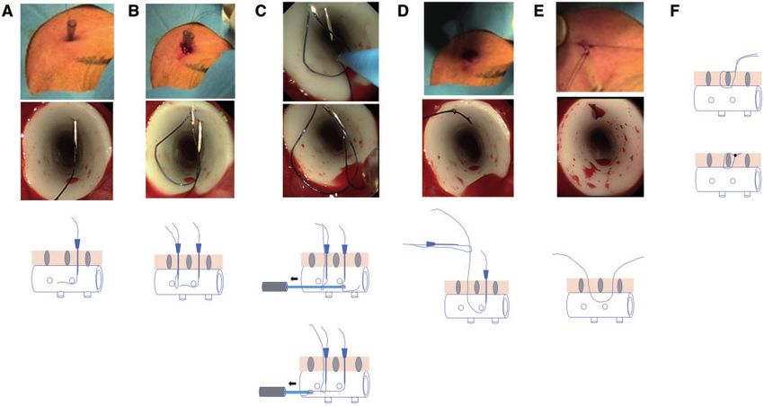
External fixation techniques
nal fixation of straight-type stents in preventing stent migration
The first step of external fixation was to prepare two 22-gauge
[12–15]. To date, however, studies have reported only relatively R
needles with a 3–0 nylon thread or a coated VicrylV suture
small numbers of patients who underwent this procedure.
(Ethicon, Inc., Somerville, NJ, USA). A needle threaded for fixation
Hence, no standard procedure has been established for external
was then inserted into the patient’s anterior neck and through
fixation of airway stents to treat patients who require upper tra-
the anterior aspect of the stent into the lumen (Fig. 1A). Then, a
cheal stenting. We therefore investigated the efficacy and safety
loop using a second thread was made inside the stent through
of external fixation of airway stents in a relatively large cohort of
the other needle (Fig. 1B). The thread for fixation was then
patients undergoing stenting for disease in the upper trachea.
guided through the fixation area with forceps under broncho-
scopic control (Fig. 1C). Next, the thread was pulled out with the
MATERIALS AND METHODS loop (Fig. 1D) and tied to fix the stent to the tracheal wall
(Fig. 1E). Finally, the stent was secured with a buried suture
Patients (Fig. 1F). In some patients, the fixation technique reported by
Miwa et al. [13] was performed with a device initially designed
In total, 739 airway stenting procedures were performed in 649 for gastropexy during endoscopic gastrostomy.
patients at National Hospital Organization Nagoya Medical
Center between May 2007 and August 2018. The medical records Statistical analysis
of all patients who had undergone external fixation of tracheal
stents for upper tracheal stenosis or tracheoesophageal fistula The primary endpoint of this study was the incidence of stent mi-
were retrospectively reviewed. The stents had been externally gration in patients who underwent airway stenting with external
fixed to the tracheal wall. fixation. Secondary endpoints included pulmonary function,
Prior to the stenting procedure, anaesthetic tolerance was con- need for supplemental oxygen and stent-related complications
firmed by the anaesthesiologist’s evaluation and several proce- without migration. The complications included were limited to
dures, including electrocardiography, cardiac ultrasonography, patients who required treatment with bronchoscopy (such as re-
pulmonary function testing and arterial blood gas analysis. The fi- placement of a stent or clearance of sputum).
nal adaptation of airway stenting with external fixation, including Differences in pulmonary function before and after stenting
tolerance to the general anaesthesia, was assessed by the (1–5 days after intervention) were analysed using the Wilcoxon
operator. signed-rank test. The results were considered statistically signifi-
Before stent placement, written informed consent for the pro- cant at 2-tailed P444 H. Niwa et al. / Interactive CardioVascular and Thoracic SurgeryOriginal Article
Downloaded from https://academic.oup.com/icvts/article/33/3/442/6242271 by guest on 01 November 2021
Figure 1: (A) A needle, through which suture material for fixation was threaded, was inserted through the anterior neck of the patient into the stent lumen. (B) Using
another needle, a second loop with suture material was made inside the stent. (C) The suture material for fixation was then threaded through the fixation area with for-
ceps under bronchoscopic control. (D) The suture material was pulled out with the loop. (E) The suture material was tied to fix the stent to the tracheal wall. (F) The 2
threads were fixed using a buried suture.
Table 1: Patients’ characteristics Table 2: Procedural details
Characteristic Data Parameter Data
Number of patients 45 Stent type, silicone/metallic 42/9
Number of procedures 51 Median stent outer diameter, mm (range) 16 (11–20)
Median age, years (range) 60 (14–91) Median stent length, mm (range) 50 (25–80)
Sex, male, number (%) 32 (71) Thread type, nylon/absorbable 50/1
ECOG PS at time of procedure, number (%) Number of stitches, 2/1 45/6
1 25 (49)
2 13 (25)
3 4 (8)
4 9 (18) Median patient age was 60 years (range 14–91 years). Patients’ di-
Airway stenosis/fistula 42/3
Diagnosis, malignant/benign 33/12 agnoses and other characteristics are detailed in Table 1. The me-
Malignant, number (%) dian follow-up period was 9 months (range 0.3–90.0 months). Of
Lung cancer 13 (29) the 45 patients, 33 had malignant disease, with oesophageal can-
Oesophageal cancer 13 (29) cer and lung cancer accounting for 26 of the 33 malignancies.
Thyroid cancer 3 (7)
Tracheal cancer 1 (2) Three patients with tracheoesophageal fistula were included in
Pharyngeal cancer 1 (2) the group of patients with malignant disease. The 12 patients
Lymphoma 1 (2) with benign disease had post-tracheostomy tracheal stenosis or
Colon cancer 1 (2) granulation tissue (such as late effects of tuberculosis).
Benign, number (%)
Post-tracheostomy 4 (9)
Granulation tissue 4 (9) Procedures
Post-intubation 2 (4)
Benign tumour 2 (4)
Table 2 summarizes the procedural details. Among the 51 stents
ECOG PS: Eastern Cooperative Oncology Group Performance Status.
placed, there were 42 silicone stents and 9 metallic stents (8
R R
UltraflexV and 1 AEROV). Nylon thread was used in all but one of
R
RESULTS the procedures (in which coated VicrylV was used). Most of the
procedures required 2 stitches for fixation. External fixation using
Patients a gastrostomy device was performed in 13 of the 51 procedures.
The fixation sutures were placed at the stent site located the
During the study period, 51 airway stent procedures with exter- shortest distance from the anterior neck that the needle was able
nal fixation were performed in 45 patients (32 males, 13 females). to reach, regardless of the lesion site.H. Niwa et al. / Interactive CardioVascular and Thoracic Surgery 445
Efficacy previous studies evaluating external fixation of airway stents.
Stent migration occurred in only 3 of 51 patients (6%), and we at-
Among the 51 patients, 12 (24%) received oxygen therapy, in- tributed this low incidence of stent migration to the external
cluding 4 patients under mechanical ventilation; most of these fixation.
patients (11 of 12, 92%) improved and no longer needed oxygen Previous (including recent) studies have also evaluated the effi-
therapy. Pulmonary function test results were available before cacy and safety of airway stenting with external fixation for upper
and after stenting for 21 patients. As shown in Table 3, the peak tracheal stenosis [12–17]. Colt et al. [12] performed external fixa-
expiratory flow had significantly improved after stenting when tion using 2 angiocatheters to fix the stent to a button. Their re-
compared with before stenting (P = 0.02), although there was no port stated that no migration had occurred in any of the 6
significant difference in the vital capacity or the forced expiratory patients [12]. Granulation tissue had formed in 3 patients and cel-
volume in 1 s. Successful treatment with the stenting led to sub- lulitis in 1 [12]. Majid et al. [14], who improved the method of
sequent stent removal in 4 patients. The stent was removed using Colt et al. [12], threaded an angiocatheter that was used to pene-
R
flexible bronchoscopy with a rigid bronchoscope under general trate the stent and tracheal wall. Next, with an Endo CloseV sutur-
anaesthesia; the nylon thread was first cut with scissors and then ing device (used in endoscopic surgery) from another
Downloaded from https://academic.oup.com/icvts/article/33/3/442/6242271 by guest on 01 November 2021
removed. Stent removal was performed without complications in angiocatheter, the thread was picked up and sewn to the external
VASCULAR
all 4 cases. button. External fixation was performed in 9 patients using this
method [14]. Because the device enables remote suturing, the
procedure was facilitated by passing an angiocatheter through the
Safety R
Endo CloseV device. The reported complications were migration in 1
patient, granulation tissue formation in 2 patients, sputum obstruc-
Stent-related complications occurred in 21 (41%) patients: migra-
tion in 2 patients and cellulitis in 1 patient [14]. In a recent study,
tion in 3 (6%), granulation tissue formation in 10 (20%), sputum-
Andreetti et al. [16] reported on 27 patients who underwent ex-
related obstruction in 6 (12%), cellulitis due to thread infection in
ternal fixation, also with angiocatheters. Their external fixation
3 (6%) and pneumonia in 1 (2%). Table 4 summarizes the data for
method was to tie a knot inside the trachea rather than using an
the 3 patients with airway stent migration identified in our study.
external button [16]. The group did not report any specific com-
The stents with migration were all composed of silicone. Two
plications, including stent migration [16]. Conversely, Miwa et al.
patients required replacement of the stent and 1 underwent tra-
[13] performed external fixation in 6 patients using a gastrostomy
cheotomy. Furthermore, 7 of the 10 patients with granulation tis-
device; a recent report showed that this method resulted in no
sue formation required replacement of the stent.
migration in 11 cases, including metallic stents [17].
The method used in our study (Fig. 1) follows the external fixa-
DISCUSSION tion method of Miwa et al. [13] (Fig. 1). Although a gastrostomy
device is used, the method of applying the suture is the same as
When placing a straight stent in the upper trachea, migration is a in other reported techniques. The difference between previous
major stent-related complication that can lead to relapse of tra- studies using angiocatheters and the technique used for our 45
cheal stenosis, which can cause significant discomfort. The risk of patients is that our procedure is relatively easy and can be per-
migration is high for silicone stents in the upper trachea without formed safely. Furthermore, the 22-gauge needles used in our in-
external fixation [7, 8]. To the best of our knowledge, our study stitution seem to result in a low risk of damage to the stent,
cohort included a larger number of patients than the cohorts in blood vessels and trachea.
Table 4 summarizes the 3 instances of airway stent migration
identified in our study. All 3 patients had benign disease and had
undergone the 2-stitch method for stent fixation. The stents were
Table 3: Pulmonary function test results before and after all composed of silicone. In all patients, stent migration occurred
stent placement (n = 21) within 2 months after stent placement. Patient 2 had post-tra-
cheal intubation tracheal stenosis. In this patient, the suture was
Variable Before stenting After stenting P-value removed 3 weeks after stent placement to prevent cellulitis. One
mean, SD mean, SD
month after the suture was removed, migration developed and
VC, l 2.96, 0.30 2.85, 0.29 0.28 stent replacement was required. The suture used for stent fixation
R
FEV1, l 1.86, 0.18 2.01, 0.18 0.24 in patient 3 was coated with VicrylV, which is an absorbable syn-
PEF, l/s 2.62, 0.30 3.30, 0.30 0.02 thetic material. This patient was the only one in our study for
FEV1: forced expiratory volume in 1 s; l: litre; l/s: litre/second; PEF: peak ex- whom absorbable suture material was used. This suture material
piratory flow; SD: standard deviation; VC: vital capacity. is often used for ligating or fixing soft tissue. It retains its strength
Table 4: Characteristics of the 3 patients with stent migration
Patient Diagnosis Age Stent type Outer diameter Thread Number of Follow-up, Treatment after
/length stitches months migration
1 Post-intubation 64 Silicone 16/50 Nylon 2 1.0 Tracheostomy
2 Post-intubation 19 Silicone 15/50 Nylon 2 2.0 Stent replacement
3 Post-tracheostomy 40 Silicone 14/35 Absorbable 2 1.5 Stent replacement446 H. Niwa et al. / Interactive CardioVascular and Thoracic SurgeryOriginal Article
in the tissue for about 2–3 weeks and is hydrolyzed within 8– from the standpoint of the stent migration rate but also because
R
10 weeks. Because of its absorbency, coated VicrylV does not ap- of the sequelae due to suture infection and other invasive
pear to be suitable for external fixation. On the basis of this re- aspects.
sult, we use nylon suture and do not remove it in clinical Our study had several limitations. One was that it is a single-
practice. The causes of migration in patient 2 and patient 3 were centre, retrospective study. It is unknown whether similar inter-
thought to be the removed and absorbable sutures, respectively. ventions can be undertaken at other institutions. However, the
The exact cause of stent migration in patient 1 was unknown. On external fixation procedure presented in our study is simple and
the basis of these experiences, we believe that the likelihood of does not require special devices. Hence, the potential for its use
stent migration is reduced by using non-absorbable suture mate- at other institutions is promising. Additionally, although our
rial and preventing suture removal by using buried sutures. In the study on external fixation is the largest in comparison with past
present study, all 3 patients with stent migration had benign dis- studies, we think that the number of cases is insufficient to solve
eases; however, as 2 of these cases had clear reasons for stent mi- the subgroup questions of whether external fixation is required
gration (absorbable suture material in one case, and suture for placing a metallic stent and whether the appropriate number
removal in another), it cannot be concluded that stent migration of stitches is 1 or 2.
Downloaded from https://academic.oup.com/icvts/article/33/3/442/6242271 by guest on 01 November 2021
occurs because of benign disease.
In the study of Andreetti et al. [16], no migration was observed
in 27 patients with tracheal stenosis who underwent stent place- CONCLUSION
ment with silicone stents and external fixation using absorbable
thread. Their success may be due to their use of knots for fixa- External fixation of an upper tracheal stent is an effective, safe,
tion, even with the use of absorbable suture. and convenient method for preventing stent migration.
Cellulitis is a stent-related complication specific to external fix- Therefore, we propose that external fixation be performed when
ation. In our study, cellulitis occurred in 6% of patients. Majid placing an airway stent in the upper trachea.
et al. [14] reported an incidence of cellulitis of 11%, which was
similar to our results. All patients with cellulitis in our study were
cured with antibiotics and did not require stent replacement. ACKNOWLEDGEMENTS
Additionally, 10 patients (20%) experienced granulation tissue
formation, 7 of whom required stent replacement. None of the We thank Andrea Baird, MD, from Edanz Group (https://en-author-
other complications required replacement. Although there were services.edanzgroup.com/ac) for editing a draft of this manuscript.
3 cases of cellulitis in our study, there was no skin irritation or ne-
Conflict of interest: none declared.
crosis due to suturing. Cellulitis was not found in the 4 cases in
which the stent was removed. The incidence of stent replacement
is acceptable compared with previous reports (22–50%) [12, 14].
Author contributions
With the exception a recent study, most previous studies on ex-
ternal fixation dealt with silicone airway stents, with a few focusing Hideyuki Niwa: Conceptualization; Formal analysis; Project administration;
on metallic stents [12–16]. In general, metallic stents have a lower Writing—original draft; Writing—review & editing. Masahide Oki:
migration rate than silicone stents in the upper trachea [3, 10, 11], Conceptualization; Investigation; Project administration. Hideo Saka:
although metallic stent migration can occur. Our study included 9 Conceptualization; Investigation. Atsushi Torii: Data curation; Investigation.
R R Arisa Yamada: Data curation; Investigation. Fumie Shigematsu: Data cura-
patients with metallic stents (8 UltraflexV and 1 AEROV). The metallic
tion; Investigation. Akane Ishida: Data curation; Investigation. Yoshihito
stent group experienced no stent migration, and none of the metal- Kogure: Data curation; Formal analysis.
lic stents were broken due to the sutures associated with external
fixation. Others have also shown that, when placed in the upper tra-
chea, metallic stents are less likely to migrate than silicone stents [5, Reviewer information
9–11]. Although we previously encountered cases in which there
was migration of a metallic stent without external fixation [3], there Interactive CardioVascular and Thoracic Surgery thanks Khaled M. Al-Kattan,
was no such migration in the present study. These results, combined Claudio Andreetti, Rajashekara H.V. Reddy and the other anonymous
with ours, suggest that external fixation of a metallic stent may be reviewer(s) for their contribution to the peer review process of this article.
beneficial for patients with upper tracheal stenosis or a tracheoeso-
phageal fistula because of the low risk of stent migration. Although
REFERENCES
the metallic stent is made of monofilament, it was not particularly
damaged and was treated in the same way as the silicone stent, [1] Ost DE, Ernst A, Grosu HB, Lei X, Diaz-Mendoza J, Slade M, AQuIRE
which did not cause any problems in our study. We believe that ex- Bronchoscopy Registry et al. Therapeutic bronchoscopy for malignant
ternal fixation of metallic stents further reduces the risk of migration. central airway obstruction: success rates and impact on dyspnea and
In the present study, it was left to the operator’s discretion as quality of life. Chest 2015;147:1282–98.
to whether external fixation of the stent was accomplished with 1 [2] Puma F, Ragusa M, Avenia N, Urbani M, Droghetti A, Daddi N et al. The
role of silicone stents in the treatment cicatricial tracheal stenoses. J
or 2 stitches. In 6 patients, the operator judged that 1 stitch was Thorac Cardiovasc Surg 2000;120:1064–9.
sufficient to prevent stent migration and, indeed, no migration [3] Ishida A, Oki M, Saka H. Fully covered self-expandable metallic stents for
was observed in these patients. Furthermore, 3 of the 6 patients malignant airway disorders. Respir Investig 2019;57:49–53.
received metallic stents. Although our study was small, and the [4] Han X, Li L, Zhao Y, Liu C, Jiao D, Ren K et al. Individualized airway-cov-
association was difficult to assess, the results suggest that there ered stent implantation therapy for thoracogastric airway fistula after
esophagectomy. Surg Endosc 2017;31:1713–8.
may be little correlation between the number of stitches used for [5] Noppen M, Meysman M, Claes I, D’Haese J, Vincken W. Screw-thread vs
external fixation and the prevention of stent migration. Future Dumon endoprothesis in the management of tracheal stenosis. Chest
studies of external fixation of airway stents are needed, not only 1999;115:532–5.H. Niwa et al. / Interactive CardioVascular and Thoracic Surgery 447
[6] Martı́nez-Balları́n JI, Dı́az-Jiménez JP, Castro MJ, Moya JA. Silicone stents [12] Colt HG, Harrell J, Neuman TR, Robbins T. External fixation of subglottic
in the management of benign tracheobronchial stenoses. Tolerance and tracheal stents. Chest 1994;105:1653–7.
early results in 63 patients. Chest 1996;109:626–9. [13] Miwa K, Takamori S, Hayashi A, Fukunaga M, Shirouzu K. Fixation of sili-
[7] Hervé D, Fabien M, Laroumagne S, Astoul P. Silicone stents, the rigid cone stents in the subglottic trachea: preventing stent migration using a
bronchoscope, and the standard of care in central airway stenosis. Curr fixation apparatus. Ann Thorac Surg 2004;78:2188–90.
Respir Care Rep 2012;1:46–53. [14] Majid A, Fernandez-Bussy S, Kent M, Folch E, Fernandez L, Cheng G et
[8] Plojoux J, Laroumagne S, Vandemoortele T, Astoul PJ, Thomas PA, Dutau al. External fixation of proximal tracheal airway stents: a modified tech-
H. Management of benign dynamic "A-shape" tracheal stenosis: a retro- nique. Ann Thorac Surg 2012;93:167–9.
spective study of 60 patients. Ann Thorac Surg 2015;99:447–53. [15] Spelsberg FW, Wollenberg B, Weidenhagen R, Lang RA, Winter H, Jauch
[9] Olze H, Dörffel W, Kaschke O. Endotracheal silicon stents in therapy K-W et al. Tracheal stents for esophagotracheal fistula in laryngectomy
management of benign tracheal stenoses. HNO 2001;49:895–901. patients. Safe fixation to the tracheostoma. HNO 2009;57:1065–9.
[10] Menna C, Poggi C, Ibrahim M, D’Andrilli A, Ciccone AM, Maurizi G et al. [16] Andreetti C, Menna C, D’Andrilli A, Ibrahim M, Venuta F, Santini M et al.
Coated expandable metal stents are effective irrespective of airway pa- A modified technique to simplify external fixation of the subglottic sili-
thology. J Thorac Dis 2017;9:4574–83. cone stent. Interact CardioVasc Thorac Surg 2018;27:878–80.
[11] Lemaire A, Burfeind WR, Toloza E, Balderson S, Petersen RP, Harpole DH [17] Huang J, Zhang Z, Zhang T. Suture fixation of tracheal stents for the
Jr et al. Outcomes of tracheobronchial stents in patients with malignant treatment of upper trachea stenosis: a retrospective study. J
airway disease. Ann Thorac Surg 2005;80:434–7. Cardiothorac Surg 2018;13:111.
Downloaded from https://academic.oup.com/icvts/article/33/3/442/6242271 by guest on 01 November 2021
VASCULARYou can also read