FINAL ACCEPTED MANUSCRIPT
←
→
Page content transcription
If your browser does not render page correctly, please read the page content below
FINAL ACCEPTED MANUSCRIPT
Morphological and molecular assessment of Lithophyllum okamurae with the
description of L. neo-okamurae sp. nov. (Corallinales, Rhodophyta)
AKI KATO 1, DANIELA BASSO 2, ANNALISA CARAGNANO 2, GRAZIELLA RODONDI 3, LINE LE GALL 4, VIVIANA PEÑA 5, JASON
M. HALL-SPENCER 6,7 AND MASASUKE BABA 8

1 Takehara Fisheries Research Station, Setouchi Field Science Center, Graduate School of Integrated Sciences for Life,
Hiroshima University, Takehara, Hiroshima 725-0024, Japan. E-mail: katoa@hiroshima-u.ac.jp

2 Dipartimento di Scienze dell’Ambiente e della Terra, Università degli Studi di Milano–Bicocca (UNIMIB), CoNISMa
Research Unit at UNIMIB, Piazza della Scienza 4, Milano 20126, Italy

3 Dipartimento di Bioscienze, Università degli Studi di Milano, Via Celoria 26, Milano 20133, Italy

4 Institut de Systématique, Evolution, Biodiversité (ISYEB), Muséum National d’Histoire Naturelle, CNRS, Sorbonne
Université, EPHE, Université des Antilles, Paris, France

5 BIOCOST Research Group, Facultade de Ciencias, Universidade da Coruña, Campus de A Coruña, A Coruña 15071,
Spain

6 School of Biological and Marine Sciences, University of Plymouth, Plymouth PL4 8AA, UK

7 Shimoda Marine Research Centre, Tsukuba University, Shimoda, Shizuoka 415-0025, Japan

8 Central Laboratory, Marine Ecology Research Institute, Iwawada, Onjuku-machi, Isumi-gun, Chiba 299-5105, Japan

ABSTRACT
Lithophyllum okamurae has been widely reported in the Pacific Ocean with identification based on morpho-
anatomical observations. Two infraspecific taxa, L. okamurae f. okamurae and f. angulare, described from
Japan, have been recorded in the temperate region of Japan. We assessed branched Lithophyllum samples
morphologically referable to L. okamurae using morpho-anatomical data and DNA sequences (psbA, rbcL and
partial LSU rDNA) obtained from herbarium specimens, including type material, as well as recently field-
collected material in Japan. The molecular analyses showed that these ‘L. okamurae’ samples contained two
species: L. okamurae and a cryptic new species which we describe as L. neo-okamurae sp. nov. Because the
holotype of L. okamurae f. angulare was conspecific with original material cited in the protologue of L.
okamurae, it is a heterotypic synonym of L. okamurae f. okamurae. Lithophyllum okamurae and L. neo-
okamurae were morphologically similar in having warty, lumpy and fruticose thalli and in often forming
rhodoliths. Lithophyllum okamurae can be morpho-anatomically distinguished from L. neo-okamurae by the
thallus with tapering or plate-like protuberances (knobby protuberances in the latter) and by having smaller
tetrasporangial conceptacle chambers (167–314 μm; 248–380 μm in L. neo-okamurae). Our LSU rDNA
sequence data from L. okamurae f. angulare (=L. okamurae f. okamurae) was identical to that of the type of L.
margaritae, which has nomenclatural priority over L. okamurae. However, considering that psbA and rbcL
sequences of L. margaritae type material could not be generated in the present study, we refrain, for the
moment, from proposing the taxonomic synonymy between these two taxa until the status of L. margaritae
and its synonyms from the type locality (Gulf of California) are clarified.

KEYWORDS
Lithophyllum margaritae; LSU rDNA; Morpho-anatomy; Non-geniculate coralline algae; Northwestern Pacific
Ocean; psbA; rbcL; Rhodolith

INTRODUCTION

Lithophyllum Philippi (Corallinales, Rhodophyta) is the largest genus of non-geniculate coralline
algae and currently includes 130 taxonomically accepted species names (Guiry & Guiry 2021).
However, most of these species have been described based only on morpho-anatomical
characters. An effective method to validate species identities, diversity and distribution has been to
combine DNA sequencing of freshly field-collected specimens and type specimens with traditional
morpho-anatomical characters (Gabrielson et al. 2011). This approach has confirmed that there are
many species of Lithophyllum in Europe (Hernández-Kantúnet al. 2015a; Peña et al. 2018;
Pezzolesi et al. 2019; Caragnano et al. 2020), the warm temperate-tropical western Atlantic ocean
basin (Hernández-Kantún et al. 2016; Richards et al. 2018), the western Indian ocean basin (Basso
et al. 2015; Maneveldt et al. 2019), and the northwestern Pacific ocean basin (Kato & Baba 2019).
Lithophyllum okamurae Foslie (1900, ‘okamurai’), described from Misaki, Kanagawa Prefecture
(previously as Sagami Province), Japan, has been reported worldwide based on morpho-anatomical
characters. This species is also known as one of major components of rhodolith beds in the
northwestern Pacific Ocean (Kato et al. 2017). Moreover, based on growth-form and anatomical
similarities, Basso et al. (1996) suggested that L. okamurae could be the Indo-Pacific vicariant of
the Mediterranean Lithophyllum racemus (Lamarck) Foslie. However, the application of the species
name L. okamurae has not been confirmed based on sequences from the type specimen.
Lithophyllum okamurae has a complicated taxonomic history. The species was described by Foslie
(1900) from Japan based on material collected by K. Yendo (Yendo specimens no. 80, 120, 270,
379, 382, 389, 408). No infraspecific taxa were proposed in Foslie (1900). One year later, Foslie
(1901) proposed in reference to L. okamurae the forms ‘japonica’ and ‘angularis’ as manuscript
names (‘Fosl. mscr’), without description or diagnosis; these names were therefore not validly
published.
Foslie (1904) validated both L. okamurae f. angulare (‘angularis’ in the text, ‘angulata’ in the legend
of the figure) and L. okamurae f. japonicum (‘japonica’) by providing illustrations of specimens
(Foslie 1904, pl. 11, fig. 12 for f. angulare and pl. 11, figs 13–19 for f. japonicum; see; Turland et al.
2018, ICN, Art. 38.8 and 38.10); and his illustration of the species (Foslie 1904, legend to pl. 11, fig.
11) has been taken as indicating the illustrated specimen as the (lecto) type of L. okamurae
(Woelkerling 1993, p. 163). However, in the text Foslie (1904, p. 60) states, concerning his original
description of L. okamurae: ‘I then possessed specimens only of the form of the species which I
have afterwards named f. angularis (pl. XI, figs 11, 12)’.
Later, Foslie (1909, p. 30) used the designation ‘L. okamurae f. typica’ for the typical form of the
species (not validly published; Turland et al. 2018, Art. 24.3, 26.2), and listed as a synonym L.
okamurae f. japonicum, thereby considering it to represent the typical form. Herbarium specimens
labelled L. okamurae f. japonicum should then be referred by the autonym L. okamurae f.
okamurae, as correctly proposed by Woelkerling et al. (2005).
The lectotype of L. okamurae, illustrated by Foslie (1904, pl. 11, fig. 11), corresponds to Yendo
specimen no. 408 (TRH A21-1318) while the rest of Yendo’s specimens quoted in the protologue
were divided into TRH A21-1325 (no. 270) and TRH A21-1326 (no. 80, 120, 379, 382, 389). All
these specimens were regarded as the main form (autonym) L. okamurae f. okamurae (Woelkerling
et al. 2005). In Foslie (1904), among figs 13–19 of pl. 11 of the form japonicum (currently as f.
okamurae), only fig. 13 was from the Pacific coast of Japan. Unfortunately, Woelkerling et al. (2005)
did not find the specimen corresponding to that figure.
The holotype of L. okamurae f. angulare, indicated by Foslie (1904, pl. 11, fig. 12), corresponds to
specimen TRH A21-1327, quoted by Woelkerling et al. (2005). Foslie (1909) mentioned that f.
angulare was characterized by less closely spaced branches than the typical form, and more or less
angular branches. This holotype was not included in the material cited in the protologue of L.
okamurae (Foslie 1900).
Five more infraspecific taxa of L. okamurae have been described: Lithophyllum okamurae f.
ptychoides Foslie [currently as Lithophyllum ptychoides (Foslie) Foslie], Lithophyllum okamurae f.
trincomaliense Foslie (‘trincomaliensis’), Lithophyllum okamurae f. validum Foslie (‘valida’) [currently
as Lithophyllum validum (Foslie) Foslie], Lithophyllum okamurae f. subplicatum Foslie (‘subplicata’)
[currently as L. subplicatum (Foslie) D. Basso, Caragnano, L. Le Gall & Rodondi] and Lithophyllum
okamurae f. contiguum Foslie (‘contigua’) [currently as Sporolithon australasicum (Foslie)
Yamaguishi-Tomita ex M.J. Wynne]. Of these, only f. subplicatum was revised using both DNA
sequences and morpho-anatomical characters from the type material (Basso et al. 2015).
Molecular analyses of Japanese coralline algae morpho-anatomically referable to L. okamurae
reveal that they belong to two distinct species: L. okamurae (including L. okamurae f. angulare,
which is here demonstrated to be synonymous with the typical form) and L. neo-okamurae sp. nov.
Here we report vegetative, sexual and asexual reproductive characters of these species, based on
genetic and morpho-anatomical data of type and herbarium archival material, in addition to recently
collected material from Japan.

MATERIAL AND METHODS

Sample collection
Lithophyllum specimens used in this study (n = 37, Table S1) were collected at 18 sites from less
than 10 m depth in the temperate region of Japan. Voucher specimens (n = 33) used for
morphological and molecular studies were deposited in the Herbarium of Graduate School of
Science, Hokkaido University, Japan (SAP); the remaining four specimens were used for molecular
studies and were deposited in the Herbaria of Muséum National d’Histoire Naturelle, Paris, France
(PC) and University of Santiago de Compostela, Spain (SANT). Additionally, we borrowed and
examined type specimens and herbarium archival specimens of Lithophyllum okamurae f.
okamurae and the holotype of L. okamurae f. angulare from the Herbarium of Norwegian University
of Science and Technology, Norway (TRH) and the Herbarium of the Laboratory of Marine Botany,
Faculty of Fisheries, Hokkaido University, Hakodate, Hokkaido, Japan (HAK; Table S2). The
lectotype of L. okamurae f. okamurae (TRH A21-1318) was studied by T. Masaki in 1969–1970,
who stored at HAK a small fragment and two preparations of the lectotype as isolectotype (HAK M-
179). Both the lectotype (TRH A21-1318) and isolectotype (HAK M-179) were very small fragments,
and therefore no destructive investigation was undertaken following the recommendations of the
curators. The herbarium archival specimens of Lithophyllum okamurae are specimens collected
from the type locality in 1899 when the protologue specimens were collected by the same collector,
K. Yendo. The type specimens of Lithophyllum margaritae (Hariot) Heydrich and L. veleroae E.Y.
Dawson (one of the synonyms of L. margaritae) were borrowed from PC and University of
California, USA (UC) for molecular studies (Table S2). In addition, nine recent collections of L.
margaritae preserved in GALW (National University of Ireland) and FBCS (Universidad Autónoma
de Baja California Sur, Mexico) were used for the molecular studies (Table S1). Herbarium
abbreviations follow Thiers (2021).
Molecular analyses
Total DNA was extracted from the herbarium specimens and field-collected specimens dried by
silica gel, using a DNeasy Blood & Tissue Kit (QIAGEN, Hilden, Germany) or a NucleoSpin® 96
Tissue kit (Macherey-Nagel, GmbH and Co. KG, Germany), according to the manufacturer’s
instructions. PCR of the following gene fragments were carried out using a Blend Taq -Plus- or KOD
FX NEO Reaction Kit (TOYOBO, Osaka, Japan), except for four vouchers (JHS0012, JHS0014,
JHS0029B, VPF00887a) and herbarium specimens of L. okamurae f. okamurae and type material
of L. okamurae f. angulare, L. margaritae and L. veleroae, which followed Basso et al. (2015) and
Peña et al. (2015), and seven specimens (E52, E57, E334, E108, E110, E116, E118) with previous
GenBank records that followed Hernández-Kantún et al. (2014, 2015b).
The primer pairs for the PCR and sequencing of the chloroplast psbA were psbA-F/psbA-R2 and
psbA-F/psbA600R in addition to psbA-F/psbA500F for sequencing (Yoon et al. 2002), while the
primer pairs for PCR and sequencing of the chloroplast rbcL were F-57/R-1150 and F-753/R-rbcS
start (Freshwater & Rueness 1994). When the rbcL primer pair F-57/R-1150 did not amplify, the F-
57/R-753 pair was used instead (Freshwater & Rueness 1994). The PCR and sequencing primer
pairs for the nuclear-encoded LSU (28S) rRNA gene were T01N (Harper & Saunders 2001) and
TR273 (Basso et al. 2015) or T16N (Saunders & McDevit 2012) and the reverse primer designated
T99R (5′TGGTCCGTGTTTCAAGACGG3′).
The PCR products were purified and sequenced by Macrogen Japan (Kyoto, Japan) or by Eurofins
(Eurofins Scientific, France). Three data sets for phylogenetic analyses, psbA, rbcL and LSU
sequences, were assembled, which comprised novel sequences from this study and previously
published sequences for the genus Lithophyllum (Tables S1, S2, S3). The rbcL and LSU rDNA
sequences of L. okamurae and L. neo-okamurae were obtained from a subset of examined
specimens in psbA analyses. Specimens with identical sequences were represented by a single
specimen in the data sets. Moreover, sequences with less than 1% pairwise divergence estimated
in MEGA X (v10.1.8., Kumar et al. 2018) were also combined into a single sequence for the psbA
dataset. Sequences were aligned using MAFFT v7 (Katoh & Standley 2013). Chamberlainium
tumidum (Foslie) Caragnano, Foetisch, Maneveldt & Payri and C. decipiens (Foslie) Caragnano,
Foetisch, Maneveldt & Payri were used as outgroups for psbA and rbcL data sets. Phylogenetic
relationships for psbA and rbcL data sets were inferred by maximum likelihood (ML) using
RAxMLGUI 1.5b1 (Silvestro & Michalak 2012), and Bayesian inference (BI) using MrBayes 3.2.6
(Huelsenbeck & Ronquist 2001). ML analyses were performed using the general-time-reversible
model with gamma distribution and invariant sites (GTR+G+I) and 1000 rapid bootstrap (BS)
replicates. BI analyses were performed using the GTR+G+I model. Four Markov chains were used.
Analyses were run for 300,000 generations for the psbA data set, for 1,000,000 generations for the
rbcL data set, and sampling was performed every 100 generations. The number of generations of
run was chosen to ensure the attainment of an average and standard deviation of split frequencies
lower than 0.01. The burn-in was determined after convergence of the tree samples using Tracer
v1.7.1 (Rambaut et al. 2018) after satisfactory convergences of the tree samples were obtained:
750 in psbA; 2,500 in rbcL.
Consensus topology and posterior probability (PP) values were calculated using the remaining
trees. Neighbour-joining (NJ) analysis for the LSU data set was conducted in MEGA X using the
Maximum Composite Likelihood model with 1,000 BS replicates. In the phylogenetic tree inferred
from ML and NJ analysis, nodes with BS values ≥90% were considered strongly supported; those
between 89% and 70% moderately supported and those
In the psbA analyses (Fig. 1), Lithophyllum neo-okamurae was resolved as an independent lineage
(A) in a strongly-supported clade (100% in ML bootstrap, 1.00 in PP) that included L. atlanticum
Vieira-Pinto, M.C. Oliveira & P.A. Horta, ‘L. margaritae’, and also ‘L. okamurae’ from China, the
latter two specimens appearing distantly related to the type specimens of both species. This lineage
comprised 21 specimens of L. neo-okamurae that shared nearly or completely identical sequences
(0.0%–0.6% sequence divergences in 852 bp; Table S4) with an archival specimen of L. okamurae
f. okamurae (TRH A21-1322) from the type locality.
Lithophyllum neo-okamurae formed a supported clade with L. margaritae from the Gulf of California
(E334) (90% in ML, 1.00 in PP) and the sequence divergences between them were 1.3%–1.7%.
However, the specimen of L. margaritae (E68) from the type locality, which was nearly identical to
three other specimens (E52, E59, E64) from the Gulf of California (0.0%–0.7% sequence
divergences), differed from the specimen E334 by 3.1%–3.2%. Lineage B (Fig. 1) consisted of
Lithophyllum okamurae, ‘L. margaritae’ and the isotype of L. veleroae, which is a synonym of L.
margaritae, with strong support (100% in ML, 1.00 in PP) and the sequence divergences among
them were 0.0%–2.6% (Table S4). Lineage B was distantly related to other species in the
northwestern Pacific Ocean or the temperate regions. The total sequence divergences among the
three archival specimens (see below) and 16 recently collected ones of L. okamurae from Japan
were 0.0%–1.3% (837–852 bp).
The holotype of L. okamurae f. angulare (TRH A21-1327) and an archival specimen from the type
locality of L. okamurae f. okamurae (TRH A21-1321) shared identical sequences, and differed from
the nearest haplotype of recently collected L. okamurae specimens from the type locality (HU39 and
six specimens; Table S4) by 0.4%, whereas the holotype of L. okamurae f. angulare (TRH A21-
1327) differed from the other archival specimen cited in the protologue of L. okamurae (TRH A21-
1326) by 0.7%. A haplotype of L. margaritae from Taiwan was identical to the haplotype of L.
okamurae (HU39 and six specimens), although it differed from specimens from the type locality
(E57, E108, E110) and the other specimens (E116, E118) of L. margaritae from the Gulf of
California by 0.2%–2.4%.
The isotype of L. veleroae formed a moderate to strongly supported subclade with L. margaritae
(E118) from the Gulf of California (73% in ML, 1.00 in PP) and the sequence divergence between
them was 0.9% (524 bp). The subclade differed from other taxa in lineage B by 1.3%–2.6%
sequence divergences. In lineage A of the rbcL trees (Fig. 2), eight Japanese specimens of L. neo-
okamurae showed very similar sequences with 0.0%–0.9% divergences, whereas they differed from
‘L. okamurae’ from China by 4.7%–5.1% (1350 bp; Table S5). Lineage B consisted of Japanese L.
okamurae specimens. The sequence divergences among six specimens of this species were 0.0%–
2.2% (1301–1350 bp; Table S5).
In the LSU rDNA analysis (Fig. 3), the holotypes of L. okamurae f. angulare (TRH A21-1327) and L.
margaritae shared identical sequences with recently collected specimens of these species and
grouped with the isotype of L. veleroae with moderate support (87% in NJ). Lithophyllum neo-
okamurae was distantly related to L. okamurae, L. margaritae and L. veleroae. The LSU rDNA
sequence of the holotype of L. neo-okamurae was identical to an archival specimen of L. okamurae
(TRH A21-1319) from the type locality and L. margaritae (E334) from the Gulf of California.
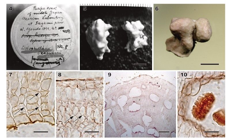
Fig. 1. ML phylogeny inferred from the psbA sequences of Lithophyllum spp. In bold face names of species
sequenced in the present study. Species denoted by single quotes have not been confirmed by comparison
with the DNA sequences of type specimens. GenBank accession or specimen numbers and collection sites
provided. Numbers at nodes represent bootstrap values >70% and Bayesian posterior probabilities >0.90.
Fig. 2. ML phylogeny inferred from the rbcL sequences of Lithophyllum spp. In bold face names of species
sequenced in the present study. Species denoted by single quotes have not been confirmed by comparison
with the DNA sequences of type specimens. GenBank accession or specimen numbers and collection sites
provided. Numbers at nodes represent bootstrap values >70% and Bayesian posterior probabilities >0.90.
Fig. 3. Neighbour-joining phylogeny inferred from the LSU rDNA sequences of Lithophyllum spp. In bold face
names of species sequenced in the present study. Species denoted by single quotes have not been
confirmed by comparison with the DNA sequences of type specimens. GenBank accession or specimen
numbers and collection sites provided. Numbers at nodes represent bootstrap values >70%.

Lithophyllum okamurae Foslie 1900, pp 4, 5
Figs 4–24, S1–S4; Table 1, S6

LECTOTYPE: TRH! A21-1318 (Yendo specimen no. 408), collected 1899 (no habitat data) by K. Yendo.
Lectotype designated in Foslie (1904) (Woelkerling 1993, p. 163; Woelkerling et al. 2005, p. 178). Illustrated in
Foslie (1904, pl. 11, fig. 11) and the present study (Figs 4, 5, S1).
ISOLECTOTYPE: HAK! M-179 (a fragment of Yendo specimen no. 408), present study, illustrated by Figs 6–
10.
TYPE LOCALITY: Misaki, Miura City, Kanagawa Prefecture, Japan (as Marine Laboratory at Sagami
Province; Woelkerling 1993).
HETEROTYPIC SYNONYM: Lithophyllum okamurae f. angulare Foslie 1901 (‘angularis’) (Woelkerling 1993,
p. 26). Holotype TRH A21-1327 (Woelkerling et al. 2005, p. 179). Illustrated in Foslie (1904, pl. 11, fig. 12, as
‘angulata’ in the legend to figure), Printz (1929, pl. 64, fig. 7), and the present study (Figs 11–15).
SPECIMENS EXAMINED: See Table S2. Sequences of two DNA markers were obtained from the holotype of
L. okamurae f. angulare, TRH A21- 1327 (psbA, MZ128805; LSU rDNA, MZ129208). DNA sequences
obtained from two archival specimens of L. okamurae from the type locality, TRH A21-1321 (Yendo specimen
no. 327; Fig. S2) and TRH A21-1326 (Yendo specimen no. 120; Fig. S4), the former not belonging and the
latter belonging to protologue specimens, included two respective psbA and two LSU rDNA sequences.
OTHER SPECIMENS EXAMINED: See Table S1. DNA sequences determined from recently collected
Japanese material including specimens from the type locality contained 16 psbA sequences, six rbcL
sequences and two LSU rDNA sequences.
HABITAT: Plants epilithic or epizoic, or free-living in the upper subtidal zone.
DISTRIBUTION: Lithophyllum okamurae occurs on the temperate coasts of the Pacific Ocean, the Japan Sea
and the Seto Inland Sea in Japan. Based on the psbA sequences, L. margaritae from Taiwan and some
material from the Gulf of California are considered conspecific or very closely allied to L. okamurae
(Hernández-Kantúnet al. 2015b; Liu et al. 2018).
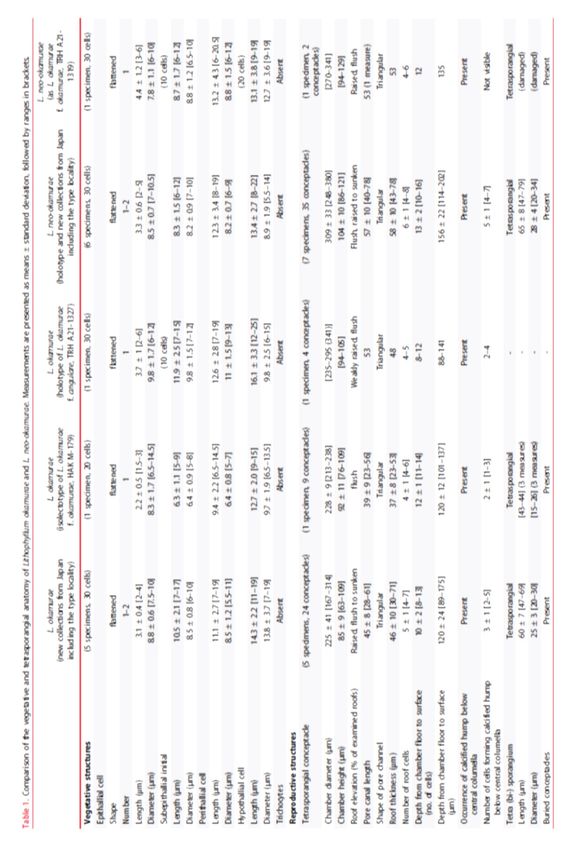
Morphology and vegetative anatomy
Thalli ranged from encrusting, warty, lumpy, foliose to fruticose, and often formed free-living
rhodoliths (Figs 5, 11, 16, 17, S2, S3). The protuberances were up to 10 mm long, tapering or plate-
like (up to 10 mm wide, 1–2 mm thick) or apically enlarged (2–5 mm in diameter), and
dichotomously branched or anastomosing. Colour of living plant was greyish-pink to light purple.
Thalli were dimerous with unistratose hypothallus composed of approximately isodiametric or
slightly elongate or wide cells, non-palisade (Figs 7, 12, 18), 11–19 μm long and 7–19 μm in
diameter. Cells of perithallial filaments were 7–19 μm long and 5.5–11 μm in diameter. Secondary-
pit connections were common. Cell fusions were not observed. Subepithallial initials were 7–17 μm
long and 6–10 μm in diameter. One to two layers of epithallial cells were flattened, 2–4 μm long and
7.5–10 μm in diameter (Figs 8, 13, 19). Medullary regions in branches (protuberances) were coaxial
(Fig. 20). Trichocytes were not observed.

Reproductive anatomy
Gametophytes are dioecious. Gametangial conceptacles were slightly raised above or flush with
surrounding thallus surface. Spermatangial conceptacle chambers were 86–116 μm in diameter and
20–35 μm high, with roofs 18–30 μm thick. Simple spermatangial systems were restricted to the
conceptacle floor (Fig. 21). Carpogonial conceptacle chambers were 94–139 μm in diameter and
18–30 μm high, with roofs 56–96 μm. Carposporangial conceptacle chambers were 207–243 μm in
diameter and 76–109 μm high, with roofs 35–63 μm thick. Carposporangia were cut off from
gonimoblast filaments borne at periphery of a large continuous flattened fusion cell (Fig. 22).
Tetrasporangial conceptacles were uniporate with roofs raised above (Fig. 23) or flush with
surrounding thallus surface or sunken below thallus surface. Buried conceptacles were observed
(Figs 9, 14). Conceptacle chambers were 167– 314 μm in diameter and 63–109 μm high. Pore
canals were triangular and tapering towards surface, 28–61 μm long. Conceptacle roofs were 4–7
cell layers, 30–71 μm thick. Tetrasporangial conceptacle chamber floors were situated 8 to 13 cells
below surrounding thallus surface. A central columella was present or absent; when present, it was
comprised of sterile filaments. A calcified hump (two to five cell layers) below central columella was
absent (Fig. 15) or present (Fig. 24). Tetrasporangia were zonately divided, 47–69 μm long and 20–
30 μm in diameter, and peripherally arranged in the conceptacle chamber (Figs 10, 24). Data on
measured vegetative and reproductive features in the above descriptions were based on recently
collected specimens, because those of the holotype of L. okamurae f. angulare and the isolectotype
of L. okamurae were overlapping within the ranges of the recent material (summarized in Table 1,
S6).
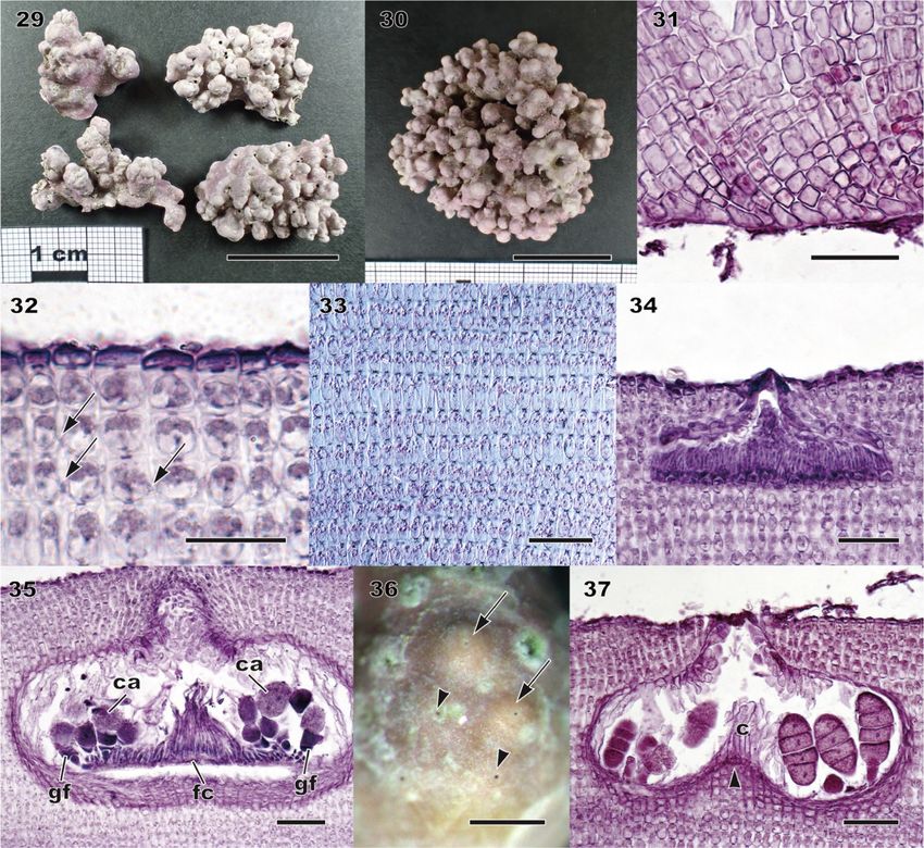
Lithophyllum neo-okamurae A. Kato, D. Basso, Caragnano, Rodondi, V. Peña & M. Baba sp.
nov.
Figs 25–37, S5; Table 1, S6

DIAGNOSIS: Thalli encrusting, warty, lumpy to fruticose, free-living rhodoliths (Figs 25, 29, 30, S5).
Protuberances up to 12 mm long, knobby, columnar (up to 20 mm long) or apically enlarged (2–6 mm in
diameter), dichotomously branched or anastomosing. Colour of living plant greyish-pink to light purple. Distinct
from both L. okamurae and the related species L. margaritae by DNA sequences.
HOLOTYPE: SAP 115594, collected 9 June 2013 by A. Kato and M. Baba; epilithic on rock in the upper
subtidal zone (Fig. 29).
TYPE LOCALITY: Misaki, Miura City, Kanagawa Prefecture, Japan.
ETYMOLOGY: Greek ‘neos’ (in compounds ‘neo-’), new (Stearn 1992); ‘okamurae’, dedicated to the
Japanese phycologist Dr. Kintaro Okamura.
OTHER SPECIMENS EXAMINED: See Tables S1, S2. Sequences of three DNA markers were obtained from
the holotype (psbA, LC620629; rbcL, LC624951; LSU, LC624957). In addition, DNA sequences determined
from Japanese material contained 20 psbA sequences, seven rbcL sequences and two LSU rDNA
sequences. DNA sequences obtained from two archival specimens of L. okamurae f. okamurae collected from
the type locality, TRH A21-1319 (Yendo specimen no. 445; Figs 25–28) and TRH A21-1322 (Yendo specimen
no. 377; Fig. S5), are an LSU rDNA and a psbA sequence, respectively. The respective sequence was
identical to the holotype and recently collected specimens of L. neo-okamurae from the type locality.
DISTRIBUTION: Based on DNA sequences, L. neo-okamurae is widely distributed in the temperate coasts of
the Pacific Ocean, the Japan Sea and the Seto Inland Sea in Japan.
HABITAT: Plants grow on bedrock, small stones, shells, or free-living in the upper subtidal zone.

Vegetative anatomy
Dimerous construction with unistratose hypothallus composed of approximately isodiametric or
slightly elongate or wide cells, non-palisade, 8–22 μm long and 5.5–14 μm in diameter (Figs 26, 31).
Cells of perithallial filaments were rectangular, 8–19 μm long and 6–9 μm in diameter. Secondary-pit
connections were common. Cell fusions were not observed. Subepithallial initials were shortened to
rectangular cells, 6–12 μm long and 7–10 μm in diameter. One to two layers of epithallial cells were
flattened, 2–5 μm long and 7–10.5 μm in diameter (Figs 27, 32). Medullary regions in branches
(protuberances) were coaxial (Fig. 33). Trichocytes were not observed.

Reproductive anatomy
Gametophytes are dioecious. Gametangial conceptacles were slightly raised above or flush with
surrounding thallus surface. Spermatangial conceptacle chambers were 119–182 μm in diameter
and 15–46 μm high, with roofs 25–46 μm thick. Simple spermatangial systems were restricted to the
conceptacle floor (Fig. 34). Carpogonial conceptacle chambers were 94–200 μm in diameter and
23–53 μm high, with roofs 68– 104 μm thick. Carposporangial conceptacle chambers were 218–339
μm in diameter and 73–144 μm high, with roofs 48–89 μm thick. Carposporangia were cut off from
gonimoblast filaments borne at periphery of a large continuous flattened fusion cell (Fig. 35).
Tetrasporangial conceptacles were uniporate with roofs flush with or raised above surrounding
thallus surface (Fig. 36) or sunken below thallus surface. Buried conceptacles were observed.
Conceptacle chambers were 248–380 μm in diameter and 86–121 μm high. Pore canals were
triangular and tapering towards surface, 40–78 μm long. Conceptacle roofs comprised of 4–8 cell
layers, 43–78 μm thick. Conceptacle chamber floors were situated 10 to 16 cells below surrounding
thallus surface. A central columella was present or absent; when present, it was comprised of sterile
cells. A calcified hump (four to seven cell layers) below central columella was present (Figs 28, 37).
Tetrasporangia were zonately divided, 47– 79 μm long and 20–34 μm in diameter, and peripherally
arranged in conceptacle chamber. Data on measured vegetative and reproductive features in the
above descriptions were based on recently collected specimens, because those of the archival
material were overlapping within the ranges of the recent material (summarized in Table 1, S6).
DISCUSSION

Our molecular analyses show that branched Lithophyllum specimens morphologically referable to L.
okamurae belong in fact to two species: Lithophyllum okamurae and L. neo-okamurae. The two
species sometimes occur together in the upper subtidal zone in the temperate region of Japan and
often form rhodoliths. Our molecular analyses indicate that L. okamurae and L. neo-okamurae are
distantly related to branched Lithophyllum species in the Western Pacific Ocean, namely L. kaiseri
(Heydrich) Heydrich, L. kuroshioense A. Kato & M. Baba, L. longense Hernández-Kantún, P.W.
Gabrielson & R.A. Townsend, and L. subtile (Foslie) A. Kato & M. Baba, which have been confirmed
by their type sequences (Kato & Baba 2019; Maneveldt et al. 2019). In contrast, L. okamurae and L.
neo-okamurae are closely related to separate lineages of L. margaritae from its type locality, the
Gulf of California. Each of these three species is discussed below.

Lithophyllum okamurae
The protologue of L. okamurae cited seven specimens, none of which was designated as the
holotype (Foslie 1900). Subsequently, a lectotype (TRH A21-1318) was set apart from the other
specimens (TRH A21-1325 and A21-1326) (Woelkerling et al. 2005, pp 176–179). In the present
study, the gross morphology of the lectotype (TRH A21-1318) and isolectotype (HAK M-179) of L.
okamurae could not be confirmed because of their very small sizes. However, a photograph of the
lectotype taken in 1969–1970 by T. Masaki (Fig. 5) showed that the specimen was sparsely
branched and had somewhat pointed apices, consistent with fig. 11 (TRH A21-1318) and fig. 12
(the holotype of L. okamurae f. angulare, TRH A21-1327) in Foslie (1904, pl. 11). The voucher
collection TRH A21-1325 (Fig. S3) represented a single 3-cm-diameter specimen and had sharp-
pointed protuberances. The collection TRH A21-1326 (Fig. S4) contains six small fragments (less
than 2 cm in diameter) which were mutually similar encrusting to warty thalli. One of them
(specimen no. 120) was conspecific with L. okamurae f. angulare based on the similarities to psbA
sequences of the holotype of this species. Considering these results, we concluded that all
specimens in the L. okamurae protologue were conspecific with L. okamurae f. angulare which we
regard as a heterotypic synonym of L. okamurae f. okamurae.
Verheij (1994) observed old buried male and female conceptacles of the lectotype of L. okamurae.
However, the isolectotype of L. okamurae (HAK M-179) has only tetrasporangial conceptacles.
Verheij (1994, figs 10, 11) showed remnants of spermatangia remaining both on a wall and a floor
of the male conceptacle, whereas spermatangia were formed only on the conceptacle floor in our
observation of L. okamurae and L. neo-okamurae (Figs 21, 34). Therefore, we think that Verheij’s
(1994) observations of the old buried conceptacles are doubtful.
The holotype of L. margaritae, a species name with nomenclatural priority over L. okamurae, was
identical to the holotype of L. okamurae f. angulare in the short LSU rDNA sequence (214 bp),
indicating that L. okamurae was closely related to L. margaritae, not to L. neo-okamurae. However,
we consider that the conspecificity between L. okamurae and L. margaritae is not convincingly
demonstrated solely on the basis of this short and weakly variable LSU rDNA sequence. We refrain
from proposing the taxonomic synonymy between these two taxa because more variable psbA and
rbcL sequences of the L. margaritae type material could not be generated.
The minimum threshold of the interspecific divergences of psbA sequences among closely related
Lithophyllum species was less than 2%; e.g. 1.7%–2.2% in psbA between L. platyphyllum (Foslie)
Foslie and L. pseudoplatyphyllum Hernández-Kantún, W.H. Adey & P.W. Gabrielson (Hernández-
Kantún et al. 2016); 1.5%–3.0% between L. racemus and L. pseudoracemus Caragnano, Rodondi
& Rindi (Caragnano et al. 2020); and mostly 2%–5% among 13 phylogenetic species of L.
stictiforme (Areschoug) Hauck (Pezzolesi et al. 2019). In the present study, the pairwise
divergences of psbA sequences of L. margaritae in lineage B including the isotype of L. veleroae,
one of synonyms of L. margaritae, were 0.0%–2.6%, indicating that it is likely to contain some
cryptic species. A haplotype of L. okamurae (HU39 and six specimens) showed up to 0.5% (4 bp
out of 849 bp) sequence divergence among two haplotypes of L. margaritae from the Gulf of
California (E108) and Taiwan, which were similar to the intraspecific sequence divergence of L.
longense (up to 0.59%, representing 5 bp out of 841 bp; Maneveldt et al. 2019). This means that at
least one genetically circumscribed species is widely distributed in the North Pacific Ocean. The
pairwise divergences of psbA sequences of L. okamurae used in the present study (up to 1.3%)
were similar to or lower than the above minimum threshold of the species-level divergence. In
contrast, the pairwise divergence of rbcL sequences of L. okamurae (2.2%) was similar to the
divergence between L. platyphyllum and L. pseudoplatyphyllum (1.9%; Hernández- Kantún et al.
2016). The pairwise sequence divergences of L. okamurae are at or near the minimum threshold of
the species level. Therefore, species delimitation analyses using multiple genes are needed to
confirm whether more than one species are included under a similar morphology.

Figs 4–10. Morphology and anatomy of herbarium specimens of Lithophyllum okamurae. Figures 4, 5 were
taken by T. Masaki in 1969–1970. The isolectotype L. okamurae f. okamurae (HAK M-179) is a fragment of
the lectotype of L. okamurae f. okamurae (TRH A21-1318). Fig. 4. Box of the lectotype of L. okamurae f.
okamurae (TRH A21-1318). Note the label ‘No. 5ʹ placed on the box by T. Masaki for his reference. Fig. 5.
Habit of the lectotype of L. okamurae f. okamurae (TRH A21-1318). Scale bar = 5 mm. Fig. 6. Habit of the
isolectotype of L. okamurae f. okamurae (HAK M-179). Scale bar = 3 mm. Fig. 7. Vertical section of inner
thallus with dimerous construction comprised of non-palisade cells (HAK M-179). Arrows indicate secondary
pit-connections. Scale bar = 20 μm. Fig. 8. Vertical section of outer thallus showing secondary pit-connections
(arrows) between cells of adjacent filaments (HAK M-179). Scale bar = 20 μm. Fig. 9. Vertical section of
protuberance showing buried conceptacles (HAK M-179). Scale bar = 200 μm. Fig. 10. Vertical section
through tetrasporangial conceptacle with peripherally arranged, divided tetrasporangium (HAK M-179). Scale
bar = 20 μm.
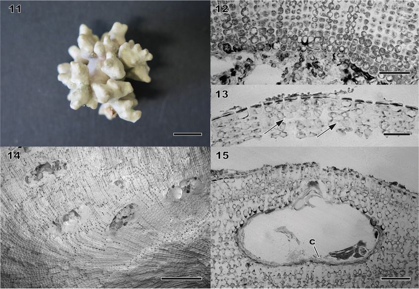
Figs 11–15. Morphology and anatomy of herbarium specimens of Lithophyllum okamurae. Fig. 11. Habit of
the holotype of L. okamurae f. angulare (TRH A21-1327). Scale bar = 1 cm. Fig. 12. Vertical section of thallus
showing dimerous construction (TRH A21-1327). Note non-palisade basal layer of cells. Scale bar = 60 μm.
Fig. 13. Vertical section of outer thallus with secondary pit-connections (arrows) between cells of adjacent
filaments (TRH A21-1327). Scale bar = 30 μm. Fig. 14. Vertical fracture face of inner thallus showing
ascending filaments and buried conceptacles (TRH A21-1327). Scale bar = 200 μm. Fig. 15. Vertical section
through tetrasporangial conceptacle with central columella (c) (TRH A21-1327). Note absence of calcified
hump below the central columella. Scale bar = 50 μm.
Figs 16–24. Morphology and anatomy of recently collected specimens of Lithophyllum okamurae. Figures 16,
21 and 22 taken from specimens from the type locality. Fig. 16. Habit of a warty specimen (SAP 115621).
Scale bar = 1 cm. Fig. 17. Habit of a rhodolith-shaped specimen (SAP 115616). Scale bar = 1 cm. Fig. 18.
Vertical section of inner thallus with dimerous construction comprised of non-palisade cells (SAP 115608).
Scale bar = 50 μm. Fig. 19. Vertical section of outer thallus showing secondary pit-connections (arrows)
between cells of adjacent filaments (SAP 115608). Scale bar = 50 μm. Fig. 20. Longitudinal section of a
branch showing a coaxial medulla (SAP 115615). Scale bar = 50 μm. Fig. 21. Vertical section through a
spermatangial conceptacle with simple spermatangial systems restricted to conceptacle floor (SAP 115621).
Scale bar = 50 μm. Fig. 22. Section through carposporangial conceptacle with large continuous flattened
fusion cell (fc) with peripheral gonimoblast filaments (gf) bearing terminal carposporangia (ca). (SAP 115621).
Scale bar = 50 μm. Fig. 23. Surface view of tetrasporangial conceptacles with raised roofs (arrows) (SAP
115612). Scale bar = 500 μm. Fig. 24. Vertical section through a tetrasporangial conceptacle with peripherally
arranged tetrasporangia around a central columella (c) with calcified hump (arrowhead) (SAP 115608). Scale
bar = 50 μm.
Lithophyllum neo-okamurae
Lithophyllum neo-okamurae differs from L. okamurae in having mostly knobby protuberances, not
tapering nor plate-like (foliose thalli), while L. okamurae shows a wide range of morphologies
including these protuberances. Although L. okamurae can also be anatomically distinguished from
L. neo-okamurae by smaller tetrasporangial conceptacle chambers (167–341 μm vs 248–380 μm;
Table 1), the conceptacle chamber sizes overlap considerably between two species. Thus, DNA
sequences are needed for reliable identification.
In the psbA analyses of the present study, L. neo-okamurae was closely related to the other two ‘L.
margaritae’ lineages from the type locality (E334 and E68). The intraspecific divergence of L. neo-
okamurae (up to 0.6%, representing 5 bp out of 852 bp) in psbA was similar to that of L. longense
(up to 0.59%, representing 5 bp out of 841 bp; Maneveldt et al. 2019). In contrast, the sequence
divergences between ‘L. margaritae’ and L. neo-okamurae were 1.3%–3.2%, which means that ‘L.
margaritae’ includes at least one species different from L. neo-okamurae.

Taxonomic relationships among Lithophyllum okamurae, L. margaritae and related species
Riosmena-Rodríguez et al. (1999) merged five species described from La Paz, BCS, Mexico,
namely Lithophyllum diguetii (Hariot) Heydrich, L. lithophylloides Heydrich, L. margaritae, L.
pallescens (Foslie) Foslie and L. veleroae, within L. margaritae based on their morpho-anatomical
similarity. Subsequently, Schaeffer et al. (2002) indicated that foliose and fruticose growth forms of
L. margaritae were genetically distinct using amplified fragment length polymorphism (AFLP)
analyses. Following Schaeffer et al. (2002), Norris (2014) recognized three species out of the five
synonyms of L. margaritae: L. diguetii for the foliose form; L. pallescens for the fruticose form; and
L. margaritae for the intermediate form. Based on the morphological similarities, Norris (2014)
tentatively treated L. veleroae and L. lithophylloides as synonyms of L. diguetii and L. pallescens,
respectively, until molecular analyses could be done on the type material of these species.
Furthermore, it should be noted that, although the species epithet diguetii has the same taxonomic
priority as margaritae, Riosmena- Rodríguez et al. (1999) chose the species epithet margaritae for
that species because the type material is in better condition and shows the characteristics of the
species more clearly. Therefore, in addition to type material of L. margaritae [DNA sequences of
material ascribed to it have also been reported from Brazil and Taiwan (Vieira-Pinto et al. 2014; Liu
et al. 2018)] and its synonyms, fresh material of each species is required to assess the genuine
boundary of L. margaritae and the phylogenetic relationships among the taxa that were proposed to
be synonyms of this species.
In conclusion, the present study reassessed the complicated taxonomic history of L. okamurae and
showed that L. okamurae f. angulare is a synonym of the autonym L. okamurae f. okamurae, which
takes precedence according to the rules of nomenclature. It also indicated that the diversity of
species morphologically referable to L. okamurae is underestimated in the northwestern Pacific
Ocean, as the present study described a new species, L. neo-okamurae sp. nov., and also showed
that L. okamurae and L. neo-okamurae were genetically different entities from other specimens
reported as ‘L. okamurae’ from the tropical region of China (Hu et al. 2020). Further taxonomic
revisions of L. okamurae and L. margaritae are needed in order to reveal the species diversity and
distribution of their related species.
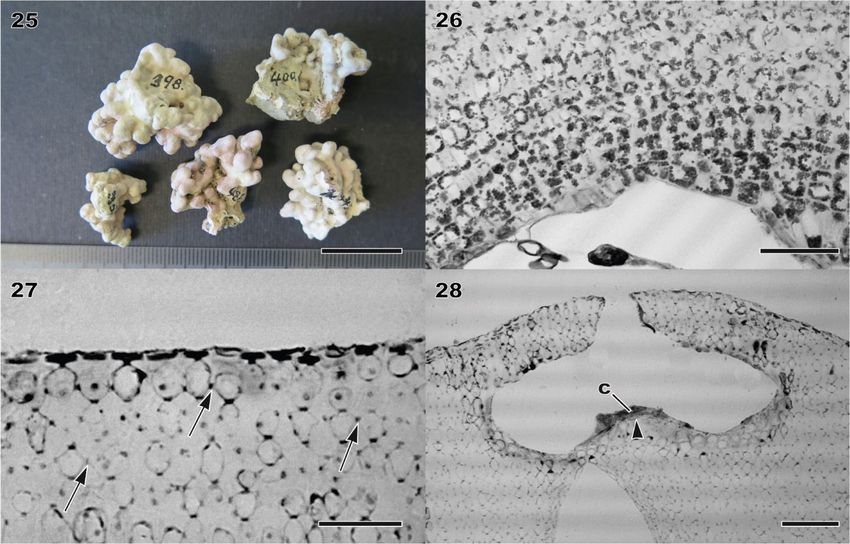
Figs 25–28. Morphology and anatomy of herbarium specimens of Lithophyllum neo-okamurae. Fig. 25. Habit
of warty specimens (TRH A21-1319). Scale bar = 2 cm. Fig. 26. Vertical section of thallus showing dimerous
construction (TRH A21-1319). Note non-palisade basal layer of cells. Scale bar = 60 μm. Fig. 27. Vertical
section of outer thallus with secondary pit-connections (arrows) between cells of adjacent filaments (TRH
A21-1319). Scale bar = 30 μm. Fig. 28. Vertical section through a tetrasporangial conceptacle and a central
columella (c) with calcified hump (arrowhead) (TRH A21-1319). Scale bar = 60 μm.
Figs 29–37. Morphology and anatomy of recently collected specimens of Lithophyllum neo-okamurae.
Specimens from Misaki, Kanagawa, Japan (type locality), except for Fig. 30. Fig. 29. Habit of the warty
holotype specimens (SAP 115594). Scale bar = 2 cm. Fig. 30. Habit of a rhodolith-shaped specimen (SAP
115601). Scale bar = 2 cm. Fig. 31. Vertical section of inner thallus with dimerous construction comprised of
non-palisade cells (SAP 115599). Scale bar = 50 μm. Fig. 32. Vertical section of outer thallus showing
secondary pit-connections (arrows) between cells of adjacent filaments (SAP 115599). Scale bar = 20 μm.
Fig. 33. Longitudinal section of a branch showing a coaxial medulla (SAP 115593). Scale bar = 50 μm. Fig.
34. Vertical section through a spermatangial conceptacle with simple spermatangial systems restricted to
conceptacle floor (SAP 115595). Scale bar = 50 μm. Fig. 35. Section through carposporangial conceptacle
with large continuous flattened fusion cell (fc) with peripheral gonimoblast filaments (gf) bearing terminal
carposporangia (ca). (SAP 115598). Scale bar = 50 μm. Fig. 36. Surface view of tetrasporangial conceptacles
with raised (arrows) and flush (arrowheads) roofs (SAP 115594). Scale bar = 500 μm. Fig. 37. Vertical section
through a tetrasporangial conceptacle with peripherally arranged tetrasporangia around a central columella (c)
with calcified hump (arrowhead) (SAP 115596). Scale bar = 50 μm.
ACKNOWLEDGEMENTS
AK thanks Jazmin J. Hernández-Kantún for providing DNA sequences of L. margaritae and Akira
Kurashima for providing some examined specimens. The authors are grateful to the Herbarium of
TRH (Kristian Hassel, Tommy Presto) PC, UC and HAK (Hajime Yasui). The manuscript was
improved by the comments from two anonymous reviewers.

DISCLOSURE STATEMENT
No potential conflict of interest was reported by the authors.

FUNDING
This research was mainly supported by Japan Society for the Promotion of Science (JSPS
KAKENHI Grant Number 26850123, 17K07908) to AK. Acquisition of some molecular data was
carried out at the CNRS-UMS 2700 (Service de Systematique Moleculaire) with funds from the
Action Transversale du Museum National d’ Histoire Naturelle. VP acknowledges support by
Universidade da Coruña (Contrato programa-Campus Industrial de Ferrol). JH-S was supported by
the International Education and Research Laboratory Program, University of Tsukuba.

REFERENCES
Adey W.H. & Adey P.J. 1973. Studies on the biosystematics and ecology of the epilithic crustose
Corallinaceae of the British Isles. British Phycological Journal 8: 343–407. DOI:
10.1080/00071617300650381.
Adey W.H., Masaki T. & Akioka H. 1974. Ezo epiyessoense, a new parasitic genus and species of
Corallinaceae (Rhodophyta, Cryptonemiales). Phycologia 13: 329–344. DOI: 10.2216/i0031-8884- 13-4-
329.1.
Basso D., Caragnano A., Le Gall L. & Rodondi G. 2015. The genus Lithophyllum in the north-western Indian
Ocean, with description of L. yemenense sp. nov., L. socotraense sp. nov., L. subplicatum comb. et stat. nov.,
and the resumed L. affine, L. kaiseri, and L. subreduncum
Kato et al.: Lithophyllum okamurae and L. neo-okamurae 13(Rhodophyta, Corallinales). Phytotaxa 208: 183–
200. DOI: 10.11646/ phytotaxa.208.3.1.
Basso D., Fravega P. & Vannucci G. 1996. Fossil and living corallinaceans related to the Mediterranean
endemic species Lithophyllum racemus (Lamarck) Foslie. Facies 35: 275–292. DOI: 10.1007/BF02536965.
Basso D. & Rodondi G. 2006. A Mediterranean population of Spongites fruticulosus (Rhodophyta,
Corallinales), the type species of Spongites, and the taxonomic status of S. stalactitica and S. racemosa.
Phycologia 45: 403–416. DOI: 10.2216/04-93.1.
Caragnano A., Rodondi G., Basso D., Peña V., Le Gall L. & Rindi F. 2020. Circumscription of Lithophyllum
racemus (Corallinales, Rhodophyta) from the western Mediterranean Sea reveals the species Lithophyllum
pseudoracemus sp. nov. Phycologia 59: 584–597. DOI: 10.1080/ 00318884.2020.1829348.
Foslie M. 1900. Five new calcareous algae. Kongelige Norske Videnskabers Selskabs Skrifter 1900: 1–6.
Foslie M. 1901. Den botaniske samling. Kongelige Norske Videnskabers Selskabs Skrifter 1900: 18.
Foslie M. 1909. Algologiske notiser VI. Kongelige Norske Videnskabers Selskabs Skrifter 1909: 1–63.
Foslie M. 1904. I. Lithothamnioneae, Melobesieae, Mastophoreae. In: The Corallinaceae of the Siboga
expedition, Siboga Expeditie 61 (Ed. by A. Weber van Bosse, and M. Foslie), pp 10–77, pls 113. E.J. Brill,
Leiden, Netherlands.
Freshwater D.W. & Rueness J. 1994. Phylogenetic relationships of some European Gelidium (Gelidiales,
Rhodophyta) species, based on rbcL nuceotide sequence analysis. Phycologia 33: 187–194. DOI:
10.2216/i0031-8884-33-3-187.1.
Gabrielson P.W., Miller K.A. & Martone P.T. 2011. Morphometric and molecular analyses confirm two distinct
species of Calliarthron (Corallinales, Rhodophyta), a genus endemic to the northeast Pacific. Phycologia 50:
298–316. DOI: 10.2216/10-42.1.
Guiry M.D. & Guiry G.M. 2021. AlgaeBase. World-wide electronic publication, National University of Ireland,
Galway. http://www.algaebase. org; searched on 27 February 2021.
Harper J.T. & Saunders G.W. 2001. The application of sequences of the ribosomal cistron to the systematics
and classification of the florideophyte red algae (Florideophyceae, Rhodophyta). Cahiers de Biologie Marine
42: 25–38.
Hernández-Kantún J.J., Gabrielson P., Hughey J.R., Pezzolesi L., Rindi F., Robinson N.M., Peña V.,
Riosmena-Rodríguez R., Le Gall L. & Adey W. 2016. Reassessment of branched Lithophyllum spp.
(Corallinales, Rhodophyta) in the Caribbean Sea with global implications. Phycologia 55: 619–639. DOI:
10.2216/16-7.1.
Hernández-Kantún J.J., Rindi F., Adey W.H., Heesch S., Peña V., Le Gall L. & Gabrielson P.W. 2015b.
Sequencing type material resolves the identity and distribution of the generitype Lithophyllum incrustans, and
related European species L. hibernicum and L. bathyporum (Corallinales, Rhodophyta). Journal of Phycology
51: 791–807. DOI: 10.1111/jpy.12319.Hernández-Kantún J.J., Riosmena-Rodríguez R., Adey W.H. & Rindi F.
2014. Analysis of the cox2-3 spacer region for population diversity and taxonomic implications in rhodolith-
forming species (Rhodophyta: Corallinales). Phytotaxa 190: 331–354. DOI: 10.11646/ phytotaxa.190.1.20.
Hernández-Kantún J.J., Riosmena-Rodríguez R., Hall-Spencer J.M., Peña V., Maggs C.A. & Rindi F. 2015a.
Phylogenetic analysis of rhodolith formation in the Corallinales (Rhodophyta). European Journal of Phycology
50: 46–61. DOI: 10.1080/09670262.2014.984347.
Hu Q., Yang F., Wei Z., Mo J., Long C., Tian X. & Long L. 2020. Detail description of Lithophyllum okamurae
(Lithophylloideae, Corallinales), a widely distributed crustose coralline alga in marine ecosystems. Acta
Oceanologica Sinica 39: 96–106. DOI: 10.1007/ s13131-019-1470-y.
Huelsenbeck J.P. & Ronquist F. 2001. MRBAYES: Bayesian inference of phylogenetic trees. Bioinformatics
17: 754–755. DOI: 10.1093/bioinformatics/17.8.754.
Irvine L.M. & Chamberlain Y.M. 1994. Seaweeds of the British Isles, Volume 1. Rhodophyta. Part 2B.
Corallinales, Hildenbrandiales. HMSO, London, UK. 276 pp.
Kato A., Baba M., Kawai H. & Masuda M. 2006. Reassessment of the little-known crustose red algal genus
Polystrata (Gigartinales), based on morphology and SSU rDNA sequences. Journal of Phycology 42: 922–
933. DOI: 10.1111/j.1529-8817.2006.00238.x.
Kato A., Baba M., Matsuda S. & Iryu Y. 2017. Western Pacific. In: Rhodolith/maërl beds: a global perspective
(Ed. by R. Riosmena- Rodríguez, W. Nelson & J. Aguirre), pp 335–347. Springer International Publishing,
Basel, Switzerland. DOI: 10.1007/978- 3-319-29315-8.
Kato A. & Baba M. 2019. Distribution of Lithophyllum kuroshioense sp. nov., Lithophyllum subtile and L.
kaiseri (Corallinales, Rhodophyta), but not L. kotschyanum, in the northwestern Pacific Ocean. Phycologia 58:
648–660. DOI: 10.1080/00318884.2019.1643200.
Katoh K. & Standley D.M. 2013. MAFFT multiple sequence alignment software version 7: improvements in
performance and usability. Molecular Biology and Evolution 30: 772–780. DOI: 10.1093/molbev/mst010.
Kumar S., Stecher G., Li M., Knyaz C. & Tamura K. 2018. MEGA X: Molecular Evolutionary Genetics
Analysis. Molecular Biology and Evolution 35: 1547–1549. DOI: 10.1093/molbev/msy096.
Liu L.C., Lin S.M., Caragnano A. & Payri C. 2018. Species diversity and molecular phylogeny of non-
geniculate coralline algae (Corallinophycidae, Rhodophyta) from Taoyuan algal reefs in northern Taiwan,
including Crustaphytum gen. nov. and three new species. Journal of Applied Phycology 30: 3455–3469. DOI:
10.1007/s10811-018-1620-1.
Maneveldt G.W., Gabrielson P.W., Townsend R.A. & Kangwe J. 2019. Lithophyllum longense (Corallinales,
Rhodophyta): a species with a widespread Indian Ocean distribution. Phytotaxa 419: 149–168. DOI:
10.11646/phytotaxa.419.2.2.
Norris J.N. 2014. Marine algae of the northern Gulf of California II: Rhodophyta. Smithsonian Contributions to
Botany Number 96: 1–555. DOI: 10.5479/si.19382812.96.
Peña V., De Clerck O., Afonso-Carrillo J., Ballesteros E., Bárbara I., Barreiro R. & Le Gall L. 2015. An
integrative systematic approach to species diversity and distribution in the genus Mesophyllum (Corallinales,
Rhodophyta) in Atlantic and Mediterranean Europe. European Journal of Phycology 50: 20–36. DOI:
10.1080/09670262.2014.981294.
Peña V., Hernández-Kantún J.J., Adey W.H. & Le Gall L. 2018. Assessment of coralline species diversity in
the European coasts supported by sequencing of type material: the case study of Lithophyllum nitorum
(Corallinales, Rhodophyta). Cryptogamie, Algologie 39: 123–137. DOI: 10.7872/crya/v39.iss1.2018.123.
Pezzolesi L., Peña V., Le Gall L., Gabrielson P.W., Kaleb S., Hughey J.R., Rodondi G., Hernández-Kantún
J.J., Falace A., Basso D. et al. 2019. Mediterranean Lithophyllum stictiforme (Corallinales, Rhodophyta) is a
genetically diverse species complex: implications for species circumscription, biogeography and conservation
of coralligenous habitats. Journal of Phycology 55: 473–492. DOI: 10.1111/jpy.12837.
Printz H. 1929. M. Foslie – ‘Contributions to a monograph of the Lithothamnia’. Det Kongelige Norske
Videnskabers Selskab Museet, Trondhjem, Norway. 60 pp.
Rambaut A., Drummond A.J., Xie D., Baele G. & Suchard M.A. 2018. Posterior summarization in Bayesian
phylogenetics using Tracer 1.7. Systematic Biology 67: 901–904. DOI: 10.1093/sysbio/syy032.
Richards J.L., Gabrielson P.W., Hughey J.R. & Freshwater D.W. 2018. A re-evaluation of subtidal
Lithophyllum species (Corallinales, Rhodophyta) from North Carolina, USA, and the proposal of L. searlesii
sp. nov. Phycologia 57: 318–330. DOI: 10.2216/17-110.1.
Riosmena-Rodríguez R., Woelkerling W.J. & Foster M.S. 1999. Taxonomic reassessment of rhodolith-forming
species of Lithophyllum (Corallinales, Rhodophyta) in the Gulf of California, Mexico. Phycologia 38: 401–417.
DOI: 10.2216/i0031-8884-38-5-401.1.
Saunders G.W. & McDevit D.C. 2012. Methods for DNA barcoding photosynthetic protists emphasizing the
macroalgae and diatoms. In: DNA barcodes. Methods in molecular biology (methods and protocols), vol. 858
(Ed. by W. Kress & D. Erickson), pp 207–222. Humana Press, Totowa, New Jersey, USA. DOI: 10.1007/978-
1-61779-591-6_10.
Schaeffer T.N., Smith G.J., Foster M.S. & DeTomaso A. 2002. Genetic differences between two growth-forms
of Lithophyllum margaritae (Rhodophyta) in Baja California Sur, Mexico. Journal of Phycology 38: 1090–1098.
DOI: 10.1046/j.1529-8817.2002.01108.x.
Silvestro D. & Michalak I. 2012. RaxmlGUI: a graphical front-end for RAxML. Organisms Diversity & Evolution
12: 335–337. DOI: 10.1007/ s13127-011-0056-0.
Stearn W.T. 1992. Botanical Latin, ed. 4. Timber Press, Portland, Oregon, USA. 560 pp.
Thiers B. 2021. Index Herbariorum. A global directory of public herbaria and associated staff. New York
Botanical Garden’s Virtual Herbarium. http://sweetgum.nybg.org/science/ih/; searched on 27 February 2021.
Turland N.J., Wiersema J.H., Barrie F.R., Greuter W., Hawksworth D.L., Herendeen P.S., Knapp S., Kusber
W.-H., Li D.-Z., Marhold K. et al. [Eds] 2018. International Code of Nomenclature for algae, fungi, and plants
(Shenzhen Code) adopted by the Nineteenth International Botanical Congress Shenzhen, China, July 2017.
Koeltz Botanical Books, Glashütten, Germany. xxxviii + 254 pp. [Regnum Vegetabile 159] DOI:
10.12705/Code.2018.
Verheij E. 1994. Nongeniculate Corallinaceae (Corallinales, Rhodophyta) from the Spermonde Archipelago,
SW Sulawesi, Indonesia. Blumea 39: 95–137.
Vieira-Pinto T., Oliveira M.C., Bouzon J., Sissini M., Richards J.L., Riosmena-Rodríguez R. & Horta P.A.
2014. Lithophyllum species from Brazilian coast: range extension of Lithophyllum margaritae and description
of Lithophyllum atlanticum sp. nov. (Corallinales, Corallinophycidae, Rhodophyta). Phytotaxa 190: 355–369.
DOI: 10.11646/phytotaxa.190.1.21.
Woelkerling W.J., Gustavsen G., Myklebost H.E., Prestø T. & Såstad S.M. 2005. The coralline red algal
herbarium of Mikael Foslie: revised catalogue with analyses. Gunneria 77: 1–625.
Woelkerling W.J., Irvine L.M. & Harvey A.S. 1993. Growth-forms in non-geniculate coralline red algae
(Corallinales, Rhodophyta). Australian Systematic Botany 6: 277–293. DOI: 10.1071/ sb9930277.
Woelkerling W.J. 1988. The coralline red algae: an analysis of the genera and subfamilies of nongeniculate
Corallinaceae. British Museum (Natural History) & Oxford University Press, London & Oxford, UK. 268 pp.
Woelkerling W.J. 1993. Type collections of Corallinales (Rhodophyta) in the Foslie Herbarium (TRH).
Gunneria 67: 1–289.
Yoon H.S., Hackett J.D. & Bhattacharya D. 2002. A single origin of the peridinin- and fucoxanthin-containing
plastids in dinoflagellates through tertiary endosymbiosis. Proceedings of the National Academy of Sciences
of the United States of America 99: 11724–11729. DOI: 10.1073/pnas.172234799.
You can also read


