Gemmology TheJournal of - GEMMOLOGICAL ASSOCIATION OF GREAT BRITAIN - Gem-A
←
→
Page content transcription
If your browser does not render page correctly, please read the page content below
Volume 21 No. 4.                   October 1988

                   The
                    Journal of
                   Gemmology

GEMMOLOGICAL ASSOCIATION OF GREAT BRITAIN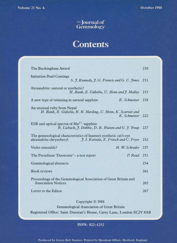
OFFICERS AND COUNCIL

                President: *Sir Frank Claringbull, Ph.D., E l n s t P , FGS
                          Vice-President: R. K. Mitchell, FGA
                           Chairman: *D. J. Callaghan, FGA
                          Vice-Chairman: *N. W Deeks, FGA
                        Honorary Treasurer: *N. B. Israel, FGA

                               Members elected to Council:
*A. J. Allnutt, M.Sc,              J. W Harris, B.Sc,               *J. B. Nelson, Ph.D.,
 Ph.D., FGA                            M.Sc, Ph.D.                 FRMS, E l n s t E , FGA
*E. M. Bruton, FGA             J. A. W Hodgkinson, FGA                W Nowak, CEng.,
*C. R. Cavey, FGA                  D. Inkersole, FGA                      ER.Ae.S., FGA
 E J. E. Daly, B.Sc,                 B. Jackson, FGA                  M. J. O'Donoghue,
 FGA                          *E. A. Jobbins, B.Sc, G Eng.,               MA, FGS, FGA
*A. E. Farn, FGA                       FIMM, FGA                     *P G. Read, CEng.,
 A. J. French, FGA             *G. H. Jones, B.Sc, Ph.D.,          MIEE, MIERE, FGA
 G.Green, FGA                              FGA                         *K. Scarratt, FGA
*R. R. Harding, B.Sc,                D. G. Kent, FGA                       E. Stern, FGA
 D. Phil, FGA                      D. M. Larcher, FGA                *C. H. Winter, FGA
                               A. D. Morgan, FIBF, FGA

^Members of the Executive Committee

                                  Branch Chairmen:
                           Midlands Branch: J. Leek, FGA
                         North-West Branch: R. Perrett, FGA
                 South Yorkshire & District Branch: G. A. Massie, FGA

                                   Examiners:
A. J. Allnutt, M.Sc., Ph.D., FGA                                        D. G. Kent, FGA
E. M. Bruton, FGA                                             P Sadler, B.Sc, FGS, FGA
A. E. Farn, FGA                                                         K. Scarratt, FGA
R. R. Harding, B.Sc, D.Phil., FGA                           M. Virkkunen, M.Phil., FGA
E. A. Jobbins, B.Sc, C.Eng., FIMM, FGA                        C. Woodward, B.Sc, FGA
G. H. Jones, B.Sc, Ph.D., FGA

                   Editor: E. A. Jobbins, B.Sc, C.Eng., FIMM, FGA
                          Editorial Assistant: Mary A. Burland
                               Curator: C. R. Cavey, FGA
                     Secretary: Jonathan P Brown, FGA, Barrister

               Saint Dunstan's House, Carey Lane, London EC2V 8AB
              (By Goldsmith's Hall)            Telephone: 01-726 4374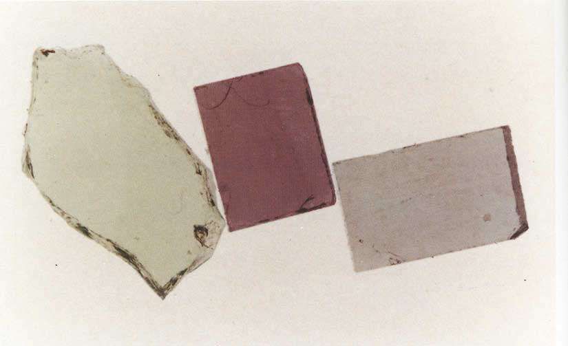
TheJournal of
Gemmology
VOLUME 21
NUMBER FOUR OCTOBER 1988
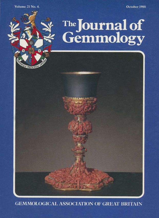
Cover Picture
The Airoldi Chalice; silver and gold plate, decorated with red
coral carved as angel and cherub heads and leaf motifs. Sicilian
workmanship, XVII century; height 230mm.
Photograph courtesy CISGEM, Milan
ISSN: 0022-1252
210

          The .
          Buckingham
             Award
             Award
         Mr   w.e. Buckingham,
         Mr W.C.    Buckingham, FGA,  FGA, hashas very
                                                  very generously
                                                         generously
      donated his
      donated    his fine
                      fine collection    of zircon
                             collection of   zircon rough
                                                       rough to
                                                              to the
                                                                 the
      Gemmological       Association of
      Gemmological Association          of Great
                                           Great Britain    to mark
                                                   Britain to  mark
      his retiral
      his  retiral after  fifty years
                   after fifty  years from
                                       from thethe firm   of George
                                                    firm of  George
      Lindley &
      Lindley       Co. Ltd.
                 & Co.   Ltd. HeHe is
                                    is also
                                       also offering
                                             offering anan award
                                                           award toto
      newly-qualified
      newly-qualified Fellows of the Association who
      carry  out research
      carry out   research on on samples
                                 samples from      the collection.
                                            from the    collection.
         The criteria for the research are:
      1. The rough specimens originate from various
      1.
         localities, mostly Indo-China, and the research
         might be
         might   be directed
                     directedtowards
                                towardsdetermining
                                           determining     any
                                                         any    vari-
                                                             variation
         ation in properties from the different
                                              different localities.
         However, other
                      otherresearch
                               researchtopics
                                          topics   would
                                                would       be con-
                                                        be considered.
         sidered.
      2. Having carried out the research programme, the
         Fellow must present the results in the form of a
         paper which would, in the opinion of the Editor,
         be worthy of    of publication in The Journal   Journal ofof
         Gemmology.
         Gemmology.

                                             3. A Fellow whose research and paper satisfy
                                                                                        satisfy these
                                                criteria will be awarded the sum of £100 or books
                                                and/or instruments to that value.
                                             4. The Fellow must first apply in writing to the
                                                Secretary ofof the Association, setting out his
                                                proposed research and methodology and the
                                                instruments he proposes to use. The time to be
                                                taken must also be specified.
                                                                      specified.
                                                Research materials provided by the Association
                                             must be returned within the time stipulated.
                                                The Association reserves the right to authorize or
                                             reject research projects at its sole discretion and will
                                             not enter into the reasons for any decision made.
                                                Those interested in the Award are invited to write
                                             to the Secretary of of the Gemmological Association,
                                             Saint Dunstan's House, Carey Lane, London EC2V
                                             8AB, giving the information
                                                               information set out in item 4 above.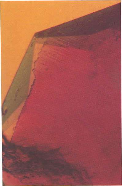
J. Gemm., 1988,21,4
1988,21,4 211
211
Imitation pearl coatings
S.J.
S.J. Kennedy*,J.G.
Kennedy* J.G. Francis**
Francis** andG.C.Jones**
and G.C. Jones**
27 Greville Street, London ECIN
*Gem Testing Laboratory of Great Britain, 27 E O N 8SU.
**Dept. of Mineralogy, British Museum (Natural History), London SW7 SW7 5BD.
5BD.
Abstract that these were not nacreous pearls. The radio-
The coating on an imitation pearl was studied by a graph (Figure 2) shows that the 'pearl' consists of a
variety
variety of techniques.
techniques. The
Thenacreous
nacreouseffect
effectof
ofthe
thecoating
coat- bead that is partly transparent to X-rays, sur-
was found
ing was to to
found be bedue
duetotominute
minute platy
platy hexagonal
hexagonal rounded by an X-ray opaque coating. This
This coating
crystals of basic lead carbonate suspended in a clear shows up as a lighter rim to the greyish disc of the
nitrocellulose lacquer. The form of the crystals was bead.
studied by scanning electron microscopy while their
composition was revealed by infrared
infrared spectroscopy and information would normally be sufficient,
This information sufficient,
electron microprobe analysis. and a report would be issued to the effect
effect that the
beads were imitation pearls. However, in this case,
because questionable claims had been made about
Introduction the composition ofof the imitation pearls, it was
ungraduated bracelet of
A single-row, ungraduated of 24 'pearls' identify the materials used in their
necessary to identify
and 6 round, black beads with 3 colourless, stone- subjected to further
manufacture. One 'pearl' was subjected further
set metal spacers (Figure 1) was submitted to the examination.
examination.
Gem Testing Laboratory
Laboratory of of Great Britain by a
trading standards authority with the request to test Glass bead
both beads and 'pearls'.
'pearls: formed the body of
The bead which formed of the 'pearl'
Identification of the black beads as onyx was not
Identification of diffraction and
was found to be glass by X-ray diffraction
difficult
difficult - the X-ray powder diffraction
diffraction pattern of
electron microprobe techniques. The only point of
obtained was that of of quartz. A cursory examination interest here was that the glass was semi-
of
of the pearls with a lOx loupe showed a form of of the transparent to X-rays, whereas coated imitation
transparent
granular structure typical of of imitation
imitation pearls. A pearls have tended to be made ofof a glass that is
radiograph ofof the necklace demonstrated
demonstrated clearly opaque
opaque to X-rays. This difference
difference can be
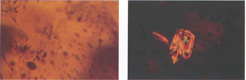
Fig. 1.
I. Imitation
Imitation pearl and onyx bead
bead bracelet
bracelet - the milky
milky Fig. 2. Radiograph of
of imitation
imitation pearl from bracelet.
white bead next
next to an onyx bead (top right)
right) is the
the glass
bead from
from which the nacreous coating
coating has been
been re-
re-
moved.
© Copyright
© Copyright the Gemmological
Gemmological Association ISSN: 0022-1252
N
                                                                                                                                                                                       .....
                                                                                                                                                                                       N

                                                                                ,   ~.       ~
                                                                                         '"."'
                                                                                 •
                                                                                                                                                                                       --
                                                                                                                                                                                        c;"l
                                                                                                                                                                                        "3
                                                                                                                                                                                       13
                                                                                                                                                                                       'J:)
                                                                                                                                                                                       00
                                                                                                                                                                                       JO
                                                                                                                                                                                       N

                                                                                                                                                                                       .j>.
Fig . 3. X-ray powder diffraction patterns of: (top) material coating imitation pearl ; (middle) commercial grade 'white lead'; (bott om) hydrocerussitc ; Ashover, Derbyshire, UK .
                                                                                                                                                                                       -
':-'
                                                                                                                              ':-
                                                                                                                              Q
                                                                                                                              C'l
                                                                                                                              (1)

                                                                                                                              93'"
                                                                                                                              ?
                                                                                                                              \0
                                                                                                                              00
                                                                                                                              -'"
                                                                                                                              yo
                                                                                                                              JO
                                                                                                                              N
                                                                                                                              r:
                                                                                                                              :::-
                                                                                                                              ~
                                                                                                                              ...

                                                                                                                              N
                                                                                                                              ......
                                                                                                                              .....
        Scanning
Fig. 4. Scan      electron
             ning electro n microscope image of                      imitation
                                             of nacreous filler from imit              coating (left)
                                                                          ati on pearl coaling                       SOOOx.
                                                                                               (left) 2000x, (right) 5000x.   W
                                                                                                                              W
214                                                                                             J. Gemm., 1988,21,4

accounted for by the relatively low lead content         Further background
(approx. 1% PbO) as found by electron microp-               After these investigations were completed, M.
robe analysis.                                           Jean Paul Poirot presented a paper at the Interna-
                                                         tional Gemmological Conference in Brazil in 1987
Coating                                                  entitled 'Imitation pearls and their coatings'. He
   The X-ray powder diffraction pattern of the           noted the following crystalline materials as being
pearl coating obtained at the Gem Testing Labora-        present in the nacreous coatings of imitation pearls
tory (Figure 3a) was close to that of the mineral        and visible on microscopic examination of an
hydrocerussite (Figure 3c), but did not match it         acetone extract of the coating:
exactly. The use of hydrocerussite as an imitation
pearl coating has been documented1, but the              (1) rods of guanine approx. 5 x 30 micrometres -
anomaly in the diffraction photograph led us to          this is a component of the well-known 'essence of
pursue the matter further, using a variety of techni-    orient' extracted from fish scales and one of the
ques available at the Department of Mineralogy,          longest-used pearl simulants; or
BM(NH).                                                  (2) square plates of bismoclite (bismuth oxychlor-
   An infrared spectrum of the coating material          ide) approx. 10 micrometres across - this com-
showed a mixture of nitrocellulose and another           pound is also used in 'pearl' cosmetics such as nail
component. During an unsuccessful attempt to             varnish, etc.; or
dissolve the coating in dichloroethane, it separated     (3) hexagonal plates of hydrocerussite approx. 15
into two layers, i.e. a nitrocellulose 'sandwich' with   micrometres across; or
nacreous inner surfaces. Acetone dissolved the           (4) fragments of mica crystals, sometimes coated
nitrocellulose lacquer completely and allowed            with titanium dioxide.
further infrared spectra to be run on the separated
soluble and insoluble materials (approx. 56 wt%          Conclusion
insol.). These spectra confirmed the identification         The imitation pearls in question are coated with
of nitrocellulose and a hydrocerussite-like filler.      a synthetic, basic lead carbonate in the form of
Under the optical and scanning electron micro-           minute hexagonal plates, suspended in and coated
scopes, the acetone-insoluble nacreous filler            by clear nitrocellulose lacquer.
appeared as minute hexagonal plates of average              The anomalies in the X-ray powder diffraction
size 15 x 0.25 micrometres (Figure 4). These             patterns and infrared spectra are the result of the
plates were also examined in the electron microp-        variable nature of the basic lead carbonate, the
robe, which revealed lead as the only detectable         exact composition of which depends on its method
element (elements lighter than sodium are not            of production.
detectable by this instrument).
   Hydrocerussite or lead dihydroxydicarbonate
has been produced synthetically by many different
methods, some of which are quoted as giving rise
to hexagonal nacreous plates2.
   The exact chemical composition of these synthe-
tic products is in doubt, as is the composition of       References
the pigment 'white lead' which is another form of        1. Webster, R., 1983, Gems, their sources, descriptions and
hydrocerussite3. A specimen of white lead gave an           identification. Butterworths, London. 557 pp.
X-ray diffraction pattern and an infrared spectrum       2. Mellor, J.W ., 1930. A comprehensive treatise on inorganic and
                                                            theoretical chemistry, VII, 836-9.
both of which matched much more closely those of         3. Ibid. 846-7.
the pearl material than the mineral hydrocerussite
(see X-ray patterns, Figures 3b & 3c).                   [Manuscript received 3 July 1988.]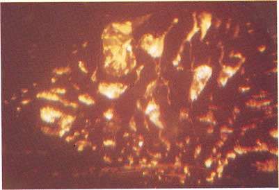
J. Gemm.,1988,21,4                                                                                                          215

                      Alexandrite: natural or synthetic?
                            H. Bank*, E. Gübelin**,              U. Henn* and J. Malley†

                                              *Idar-Oberstein, West Germany
                                                  **Meggen, Switzerland
                                                  †Mainz, West Germany

   Since the appearance on the market of great                    the presence of chromium lines. Yet the question
quantities of rough and faceted alexandrites from                 of natural versus synthetic still remained unsolved.
Brazil (Bank et al, 1987a; Bank et al, 1987b;                        Microscopically, step-like growth striations were
Gübelin and Schiffmann, 1988), the differentiation                observed - which are also no indication of natural
between these (to a great extent relatively inclu-                origin - as well as 'fingerprint-feathers' of bizarre-
sion-free) gemstones, and synthetic alexandrites,                 shaped cavities (Figure 1). Some of the latter
has naturally been pushed into the foreground of                  contained a solid substance which between crossed
gemmological investigations. Recently a dark                      polars displayed interference colours, and was thus
green-to-violet changing to red-violet stone of 1.34              founded to be doubly-refractive. With overhead
ct (faceted, oval, 7.2 x 6.4 x 3.5 mm) arrived for                illumination, these inclusions reflect strongly (Fi-
investigation, which had been identified by a                     gure 2) and are thus reminiscent of the flux
laboratory as a synthetic alexandrite. Yet the owner              residues in synthetic alexandrite (Gübelin and
of the stone doubted this outcome, since he had                   Koivula, 1986, Trossarelli, 1986, Henn, 1987).
purchased it personally in the rough at the mine in                  Several inclusions were exposed to the surface of
Brazil and had also cut it himself (which, however,               the faceted stone, allowing further investigations as
does not prove that it is genuine).                               to their identity with the help of more sophisti-
   The stone was doubly-refractive on the polari-                 cated methods. Qualitative energy-dispersive
scope and biaxial on the conoscope. The standard                  analyses with the aid of a scanning electron micro-
gemmological values were as follows:                              scope (SEM), identified very diverse substances in
n x = 1.745, n y = 1.748, n z = 1.754, A n = 0.009,               the fissures surrounding the exposed inclusions.
D = 3.71 g/cm3.                                                   Figure 3 shows such an area of fissures; the white
These all indicate the mineral variety chrysoberyl                streak measures 50 /mi. The white spherical grain
(BeAl 2 0 4 ), which crystallizes in the orthorhombic             could be identified as tin. Similar solid substances
system. With the aid of spectroscopic analyses, the               present in these fissures proved to be copper,
stone could be identified as an alexandrite through               nickel and lead. Lead-oxide was detected by means

    1. Bizarre-shaped cavities, partly filled with solid subst    Fig. 2. Bizarre-shaped cavities, partly filled with solid subst-
       ance. 15x.                                                         ance. Reflected light. 20x.

© Copyright the Gemmological Association                                                                      ISSN: 0022-1252
0\
.....
-
216 J. Gemm., 1988,21. 4
:c
IV
S'"
00
N
........... -.....
.... ... :.. ':. -, :'. ·: 1
. . . . . . . . . , ., .!
•• • • • ••• • • • • • - · •• 1 t
... . ::::.:::::>::.:: : : :::':::; ~»).:~;:~~:: j
:::: :::: :::::: ::: : ; : : : :::
Fig. 4. Energy-spectrum of alkali-fe ldspar inclu sion.
:: :::: :::.::: ::: : ••• •••••••• 0 • •• •• ••
::: :: . .:.: . : :: : :: ::: :: : :::: :::: . :: :: ::: :: : : .: : :: : . :: : :::: . :: . : :: ::: ::::::: :: .. ..
,,0 •• • , • ••••
;: ~ ~:; ~ \i: ~ ~: l\j:;: j j: j;:::;::::::::::;;:;: ::::::::: :;::::: :: :,::.::::: :.:',' ~ :i~: : : ~ : ·.
I
·· ·· ····::·::. ·: ::::Fe
I
I
_.
II
.
::::: :: K:
II
~ . ' -"
...; .. ..
:::;::i\::::::~ ::::;:::;:;:::::::~ :::::::::::...:.::::...::::;....:::....::;:::.::.... "I
r~ ~ ~ ~ ~
I
::. :· A I : :
;:::::~:::::::::::: ;J.J.Gemm.,
Gemm., 1988,21,4
1988,21,4 217
217
discs, which
discs, which were
were forced
forced into
into the
the fissures
fissures during
during
theseprocesses.
these processes.
AAmore
more precise
precise investigation
investigation ofofthe
the solid
solid fillings
fillings
of these
of these fît
fitsures revealed the
sures revealed the presence
presence ofof potas-
potas-
sium-rich aluminium
sium-rich alarninium silicates
silicates -- probably
probably potas-
potas-
siumfeldspar
sium feldspar(Figure 4). This
(Figure4). Thisdefinitely
definitelyproves
proves thethe
naturalorigin
natural originofofthe
thestone.
stone.
Acknowledgements
Acknowledgements
Scanning electronic
Scanning electronic microscope
microscope analyses
analyses were
were
carried
carriedout
outatatthe
theMax-Planck-Institut
Max-Planck-Institutfür fur Chemie,
Chernie,
Abt. Kosmochemie,
Abt. Kosmochernie, Mainz,
Mainz, West-Germany.
West-Germany.
Financial support
Financial support waswas given
given by by grants
grants ofof the
the
Wirtschaftsrninisterium des
Wirtschaftsministerium des Landes
Landes Rheinland-
Rheinland-
Pfalz,
Pfalz,FRG,
FRG,within
withinaaproject
projectfor
forapplied
applied research.
research.
References
References
Bank,H.,
Bank, H., Henn,
Henn, U. &U.Bank,
& Bank, F.H., 1987a.
EH., 1987a. EinAlexandritvorkommen
Ein neues neues Alexan-
dritvorkommenininBrasilien.
Brasilien.Goldschmiede
GoldschmiedeZeitung, Heft 9,9,
Zeitung, Heft
90-1.
90-1.
Bank,F.H.,
Bank, F.H., Bank,
Bank, H., H., Gube1in,
Gübelin, E., Henn,
E., Henn, U. Alexandrite
U. 1987b. 1987b. Alexan-
von einem
dritevon einemneuen
neuen Vorkommen
Vorkommenbei beiHematita
Hematitainin Minas
Minas
Gerais, Brasilien.
Gerais, Brasilien. Zeitschrift
Zeitschrift der
derDeutschen
Deutschen Gemmologischen
Gemmologischen
36,121-31.
Gesellschaft,36,
Gesellschaft, 121-31.
Gube1in, E.,
Gübelin, E., Koivula, J.I., 1986.
Koivula, J.I., 1986. Photoatlas
Photoatlas ofof inclusions
inclusions inin
gemstones.ABC
gemstones. ABCEdition,
Edition, Zürich.
Zurich.
Giibelin, E.,
Gübelin, E., Schiffmann,
Schiffmann, C.A.,
C.A., 1988.
1988. Alexandrite
Alexandrite from
from aa
Newly Discovered
Newly Discovered Occurrence
Occurrence in in Brazil.
Brazil. Schweizerische
Schweizerische
Uhrmacher und
Uhrmacher und Goldschmiede
Goldschmiede Zeitung, International issue,
Zeitung, International issue,
2/ 1988.
2/1988.
Fig. 3.3. SEM-photograph:
Fig. SEM-photograph: fissure
fissurewith
withsolid
solid substances.
substances. Henn, U.,
Henn, U., 1987.
1987. Inclusions
Inclusionsinin yellow
yellowchrysoberyl,
chrysoberyl, natural
natural and
and
syntheticalexandrite.
synthetic alexandrite.Australian
AustralianGemmologist,
Gemmologist, 16,16,217-20.
217-20.
Trossarelli,C.C.(1986):
Trossarelli, (1986): Synthetic
Synthetic alexandrite
alexandrite fromfrom
USSR. USSR. Gem-
Gemmologia,
ofthe
of the Raman
Raman spectroscope.
spectroscope. The The latter
latter was
was quoted
quoted mologia, ll, 6--22.
11, 6-22.
as proof
as proofthat
that the
the investigated
investigated stone
stone was
was aa synthetic
synthetic
alexandrite. These
alexandrite. These spherical
spherical metal
metal grains,
grains, however,
however,
were not
were not securely
securely lodged
lodged in in the
the fissures,
fissures, and
and are
are
probably remnants
probably remnants left by the
left by the cutting
cutting or
or polishing
polishing received 33May
[Manuscript received
[Manuscript May /988.]
1988.]218 J. Gemm., 1988,21,4
A
A new type of twinning in natural
natural sapphire
Dr Karl
Dr Karl Schmetzer
Schmetzer
Institute of Mineralogy and Petrography, University of Heidelberg, West Germany
Abstract never in synthetic rubies or sapphires of different
different
A new type of twin structure in natural sapphire producers.
from Sri
from Sri Lanka
Lanka isisdescribed.
described.TheThesamples
samplesreveal
revealinserted
in- The new type of twin structure was observed in
serted irregularly
irregularly shaped
shaped bodies
bodies of subordinate
of subordinate corun-
corundum the course of microscopical examination of some
dum individuals, which are confined to intercalated
hundreds of light yellowish or bluish untreated
lamellae parallel to rhombohedral faces r (lOll) (1011) and
related to the dominant crystal by reflection across (i.e. non-heat treated) natural sapphires from Sri
(1011).
(1011). Lanka. In about 50 of these cabochon cut samples,
bodies of corundum crystals were found to occur
in an orientation different
different from the dominant
In some cases, the recognition of certain types of different crystal-
sapphire individual. Due to their different
twinning in ruby and sapphire is applicable to the lographic orientation, these corundum crystals in-
distinction of natural and synthetic corundum. In serted into the dominant individual are clearly
general, aadetailed
detailed knowledge
knowledge about about
twin twin struc-
structures recognizable under crossed polarizers, but not in
tures occurring in natural ruby and sapphire as 1-6). Part of these
plane polarized light (Figures 1-6).
well as in different
different types of synthetic corundum is inserted bodies reveal only irregular surfaces as
necessary in order to avoid misinterpretations of boundaries between dominant and subordinate
structuralproperties
structural propertiesduring
during microscopic
microscopic examina-
examination corundum individuals (Figures 1, 2). 2). Both crystals
tion of samples of unknown origin. A general differing in orientation, in general, are not related
differing
survey dealing with twin structures in natural by reflection
reflection across the positive rhombohedron r
rubies fromdifferent
rubies from localitiesis isgiven
differentlocalities givenbyby Schmet-
Schmetzer (lOTl) and, at present, it is unknown to the author
(lOTI)
(1987), and the results described in the paper
zer (l987), if both parts of the crystals are connected by an
cited are also valid for natural sapphires without unknown twin law or not-not.
any restriction.
restriction.Twinning
Twinningininflux-grown
flux-growngem gem quali-
quality Samples of the second part of sapphire crystals
ty synthetic ruby and sapphire was described in with inserted bodies of corundum reveal at least
detail byby Schmetzer
Schmetzer(1987) andKiefert
(l987)and Kiefert&& Schmet-
Schmetzer one plane surface as boundary between dominant
zer(1988).
(l988). and subordinate individuals (Figure 3). A thor-
In natural corundum, three types of twinning ough microscopic examination indicates that, in all
are observable: contact twins on the basal plane c cases, these contact planes are parts of intercalated
(0001) or on the positive rhombohedron r (lOTI) (lOTl) lamellae on r (lOTI)
(1011) [Figures 4, 5, 6].
6]. The remain-
with two macroscopically developed individuals individuals ing boundaries between main crystals and inserted
are rare. Repetitive twinning on r (lOTI), (lOTl), on the irregular bodies, i.e. those boundaries which are
other hand, is common in natural ruby and sap- confined to an intercalated lamella on r, may
not confined
phire but, in general, only thin lamellae of corun- consist of either irregular surfaces or of plane
dum in twin position are intercalated parallel to 3-6).
crystal faces (Figures 3--6).
one, two or three rhombohedral faces of the domi- In part of the crystals investigated, up to five
nant ruby or sapphire crystal. In some samples, inserted bodies of corundum were observed, which
intercalated lamellae were found to end irregularly are confined
confined to several intercalated lamellae para-
within the dominant corundum individual. The llel to one rhombohedral face r (lOTI).(lOTl). In two
new type of twin structure to be described in this samples, inserted bodies of corundum in twin
confined to intercalated lamellar twinning
paper is confined confined to interca-
position were found which are confined
(lOTl). Up to now, twinning of this particular
on r (lOll). lated lamellae parallel to two rhombohedral faces r
type was observable only in natural corundum, but andr'r' {lOTI}.
and {lOTl}.
©
© Copyright the Gemmological Association ISSN:
ISSN: 0022-1252J. Gemm., 1988,21,4 219
Figs. 1, 2. Natural sapphire from Sri Lanka; inserted bodies of corundum revealing irregular surfaces as boundaries between
subordinate crystals and the dominant individual. Fig. 1, plane polarized light; Fig. 2 crossed polarizers. lOOx.
Fig. 3. Natural sapphire from Sri Lanka; inserted bodies of Fig. 4. Natural sapphire from Sri Lanka; inserted body of
corundum revealing plane boundaries between domi- corundum [below] confined to an intercalated lamella
nant and subordinate individuals. Crossed polarizers. on the positive rhombohedron r (1011) [above] as
30x. boundary between dominant and subordinate indi-
viduals. View almost perpendicular to the intercalated
lamella, crossed polarizers. 20x.
Fig. 5. Natural sapphire from Sri Lanka; inserted body of Fig. 6. Natural sapphire from Sri Lanka; inserted bodies of
corundum confined to an intercalated lamella on the corundum confined to an intercalated lamella on the
positive rhombohedron r (10Ï1) as boundary between positive rhombohedron r(1011)as boundary between
dominant and subordinate individuals. View parallel to dominant and subordinate individuals. View almost
the intercalated lamella, crossed polarizers. 20x. parallel to the intercalated lamella, crossed polarizers.
40x.220 J. Gemm., 1988,21,4
According to its properties, the new type of twin References
structure in corundum
corundum described combines both Kiefert, L., Schmetzer, K., 1988. Morphology and twinning in
single types of rhombohedral twinning, i.e. contact Chatham synthetic blue sapphire. Journal
Chatham Journal of
of Gemmology,
Gemmology,
21, 16-22.
21,16-22.
twinning on r (lOTI)
(lOTl) [consisting of two macrosco- Schmetzer, K. 1987. On twinning in natural and synthetic
pically developed individuals] and lamellar twin- flux-grown ruby. Journal
flux-grown of Gemmology, 20, 294-305.
Journal of
ning on r (lOTI) of intercalated thin
(1011) [consisting of
lamellae]. Consequently, this type of twinning is
classified as combined rhombohedral twinning.
classified [Manuscript received 22 February
[Manuscript received February 1988.]
1988.]
DUOTESTER*
PRESIDIUM DUOTESTER*
Thermal testing and reflectivity measurement
measurement
The Presidium Duotester uses thermal
properties to distinguish between
diamond and its simulants, and also
unit.
has an independent reflectance unit.
It is therefore simple to test a stone
by the two most popular methods
instrument.
combined in one instrument.
It works on either battery or mains
(110 or 220/240v - please specify).
specify).
Supplied with
a set of diamond
diamond
simulants for
reference.
£310.00
+
+ VAT,
VAT, postage
postage and
and packing
packing
**For
For test
test report,
report, see p251.
see p251.
Gemmological
Gemmological Associa tion of
Association of Grea
Greatt Britain
Britain
Saint
Saint Dunstan's House, Carey Lane,
Dunstan's House, Carey Lane, London
LondonEC2V
EC2V8AB
8AB
Telephone:
Telephone: 01-7264374
01-726 4374 Fax:
Fax: 01-7264837
01-726 4837J. Gemm., 1988,21,4
GEMDATA
A computer program for gem identification
Now available in an expanded version with colour-enhanced text,
GEMDATA will run on any IBM PC-compatible computer. It is
designed to help with both appraisal identifications and gemmological
studies. A full report of the program was given in the Journal of
Gemmology, 20, 7/8,467-73.
Optional yearly update of GEMDATA will be available.
GEMDATA is supplied on a SVi-inch double-sided, double-density disk,
and contains the following three sections :-
1. Gem Identification from a databank of over 220 gems
2. Gem Comparisons (side-by-side display of the constants of selected
gems)
3. Gem Calculations (S.G., reflectivity, critical angle, Brewster angle)
The GEMDATA package, complete with disk, operating notes and gem
index, costs £75.00 (plus postage and VAT).
To order your package please use the coupon given on p. 206.
Gemmological Association of Great Britain
Saint Dunstan's House, Carey Lane, London EC2V 8AB
Telephone: 01-726 4374 Fax: 01-726 4837
Cables: Geminst, London EC2222 J. Gemm., 1988,21,4
An unusual ruby from Nepal*
H. Bank1, E. Gübelin2, R.R. Harding3, U. Henn1, K. Scarratt4 andK. Schmetzer5
1
Deutsche Stiftung Edelsteinforschung, Idar-Oberstein, West Germany
2
Meggen, Switzerland
3
British Museum (Natural History), London
4
The Gem Testing Laboratory of Great Britain, London
5
Institute of Mineralogy and Petrography, University of Heidelberg, West Germany
Abstract Introduction
A high quality ruby from Nepal is described. The A purely gemmological routine investigation can
stone, weighing 1.288ct, revealed extraordinary growth sometimes result in a false diagnosis, or at least
structures connected with colour zoning as well as create difficulties, especially when the problem
mineral inclusions (phlogopite), feathers consisting of
two- and most probably three-phase inclusions and concerns the differentiation between natural gems
ultra-fine fluid films, as diagnostic characteristics. and synthetic stones. This is particularly so if the
inclusions observed are not clearly indicative, but
Figs 1 and 2. Growth structures and colour zoning in a natural ruby from Nepal; view aimost perpendicular to the c-axis; broad
alternate colourless and red bands parallel to the basal pinacoid c (0001) forming the lower edge of the sample,
colourless parts confined to growth structures parallel to the hexagonal prism a (1120) on the left side of the sample
and parallel to the hexagonal dipyramid v (4481) on the right of the sample, spindle-like growth structures in the
dark red central part of the stone. Transmitted light using methylene iodide as immersion liquid. 22x. (Photos by K.
Schmetzer.)
*The Editor received two papers on this subject on the same day. They
have been combined to form this paper.
© Copyright the Gemmological Association ISSN: 0022-1252J. Gemm., 1988,21,4 223
Fig. 3. Growth structures and colour zoning in a natural ruby Fig. 4. Spindle-like growth structures in the dark red central
from Nepal; view almost perpendicular to the c-axis; part of a natural ruby from Nepal; view almost perpen-
growth structure parallel to the hexagonal prism a dicular to the c-axis. Transmitted light using methylene
(1120) visible as boundary between colourless edge and iodide as immersion liquid. 25x. (Photo by K.
dark red central part, spindle-like growth structures in Schmetzer.)
the central part, parallel to the basal face c (0001).
Transmitted light using methylene iodide as immersion
liquid. 30x. (Photo byK. Schmetzer.)
ambiguous, i.e. if they could be found in both which had not been observed previously in
natural and synthetic stones and are not typical of Nepalese rubies. So the stone was examined in
either. This happened recently during the inves- detail using spectroscope, microprobe and further
tigation of a faceted red stone, whereby the ques- microscopic investigations.
tion was raised whether it was a natural or a
synthetic stone, and whether it originated from the Investigation
Kingdom of Nepal. The faceted ruby weighs 1.288 ct and is cut as an
Ruby, as well as pink, violet and purplish sap- almost equilateral octagon (6.00 x 6.00 x 4.15
phires, from Nepal were recently described by mm). The physical properties of the sample are
Harding and Scarratt (1986) and Kiefert and within the range known for both natural and
Schmetzer (1986, 1987). Most of the material synthetic ruby, i.e. n 0 = 1.770, n e = 1.762, An =
available until now has been of cabochon quality 0.008, D = 3.98 g/cm3, and with the hand spectro-
and any faceted samples of notable transparency scope the normal chromium spectrum of ruby was
have been few. Thus, the authors were surprised to detected.
receive a faceted sample of more than one carat, The absorption spectrum of the sample in the
with excellent purity, a good 'Burmese red' colour, visible and ultraviolet regions, as examined with
and which was said to originate from Nepal. Under the aid of a UV/VIS spectrophotometer, is similar
the microscope the ruby revealed characteristics to the spectra already published for ruby and
which closely resembled some of the properties sapphire from Nepal by Harding and Scarratt
seen in Ramaura and Kashan synthetic rubies and (1986) and Kiefert and Schmetzer (1986, 1987),224 J. Gemm., 1988,21,4
but does not
but not contain significant Fe 2 ++ /Ti
contain a significant 4+
ITi4+ charge observed
observed previously
previously byby the authors
authors in Ramaura
transfer
transfer absorption
absorptlon in the red
red region of of the visible synthetic rubies. However, in such
synthetic such stones the
spectrum. Due Due to the absence of of ironiron and/or growth
growth zones forming
forming angles ofof 86° are made
made by
titanium
titanium inin distinct
distinct amounts, the sample reveals a different rhombohedral
two different rhombohedral faces rrand
and rr'' (lOTI).
(lOTl).
good
good ruby
ruby colour
colour without
without any purplish
purplish hue, i.e. Many
Many ofof the microscopic
microscopic observations
observations disclosed
without
without anan additional
additional sapphire
sapphire component. ambiguous features
ambiguous features which
which could
could neither
neither be attri-
immersion liquid,
Using methylene iodide as an immersion buted clearly to a natural
buted natural nor a synthetic ruby.
a microscopic examination of of the ruby, in a direc- Amongst the most confusing
Amongst confusing characteristics
characteristics of
of this
tion normal to the table facet, revealed a dark dark red 1.288 ct ruby are the spindle-like growth struc-
1.288
well-defined and near-colourless
central area, two well-defined tures in the darkdark red central part
part of
of the stone.
situated close to the girdle and on opposite
areas situated These are parallel to the basal pinacoid
pinacoid c (0001)
and another
sides and another area, also bounded
bounded at one edge connected with the
(Figures 1 and 4), and are connected
by the girdle, in which there was strong colour colour zoning. These structural
structural characteristics
characteristics re-
zoning (Figures 1, 2 and 3). In the latter area the semble features
features often
often observed
observed in synthetic flux-
broad colourless and red zones (Fig-
alternating broad grown rubies.
ures 1 and 2) are parallel to the basal pinacoid Both at the girdle and near the culet ofof the stone
c (0001), and the two near-colourless areas form several small, solid inclusions are exposed at the
of 90° and 85° respectively, with the growth
angles of surface. Examination
Examination by electron
electron microprobe both
structures
structures connected
connected with this colour zoning. Con- in London
London and Heidelberg
Heidelberg indicated
indicated that these
sequently, these colour zones are confined confined to inclusions are phlogopite (Figure 5), a mica which
growth structures parallel to the hexagonal prism has already been identified
identified in the paragenesis of of
a (1l20)
(1120) and parallel to the hexagonal dipyramid ruby and rose and violet sapphire from Nepal.
v (4481). Similar almost rectangular growth struc- Further
Further microscopic examination
examination revealed the
tures connected
connected with colour zoning have been presence ofof dark 'feathers' consisting of
of small more
A/IV.., SiK,;..
MglJ. Gemm., 1988,21,4 225
Fig. 6. 'Feather' consisting of irregular cavities and negative Fig. 7. 'Feather' consisting of liquid, two- and most probably
crystals with liquid and two-phase filling. Transmitted three-phase inclusions in natural ruby from Nepal.
light using methylene iodide as immersion liquid. 75x. The solid components (probably margarite) show in-
(Photo by U. Henn.) terference colours. Transmitted light using methylene
iodide as immersion liquid, crossed polarizers. 80x.
(Photo by K. Schmelzer.)
Fig. 8. 'Feather' consisting of small irregularly shaped cavities Fig. 9. 'Feather' consisting of small irregularly shaped cavities
and negative crystals with multi-phase filling in natural and negative crystals with multi-phase filling (lower
ruby from Nepal. Darkfield illumination. 40x. (Photo left part) and ultra-thin liquid and two-phase inclu-
byK. Scarratt.) sions showing interference colours under suitable illu-
mination (central and upper right part). Transmitted
light using methylene iodide as immersion liquid,
crossed polarizers. lOOx. (Photo by K. Schmelzer.)
Fig. 10. Ultra-fine liquid films, partly also two-phase (liquid/ Fig. 11. Fine and bright dust-like 'fog' particles in natural
gaseous) in natural ruby from Nepal; these fluid ruby from Nepal. Reflected light. 60x. (Photo by K.
inclusions reveal interference colours under suitable Scarratt.)
illumination. Darkfield illumination. 50x. (Photo by
E. Giibelin.)226 1988, 21 , 4
J. Gemm., 1988,21,4
irregularly shaped cavities as well as small
or less irregularly ultra-fme fluid
as ultra-fine fluid inclusions in this high quality,
crystals (Figures 6 to 9). TheThe filling
filling of
of the small 1.288 ct ruby, on the one hand, proves the sample
1.288
(solid/liquid or liquid/
cavities is liquid, two-phase (solid/liquid of natural origin, and, on the other, confirms
to be of
gaseous) and, most probably, also three-phase its locality as Nepal. Until now the exceptional
(solid/liquid/gaseous). The solid parts of of the inclu- growth structures
growth structures of of this ruby
ruby had not
not been
displayed interference
sions displayed interference colours under
under crossed observed in natural rubies either
observed either from
from this or any
polarizers (Figure 4). Such feathers, which have other locality.
other
observed previously in Nepalese rubies of
been observed of a
much lower quality, closely resemble residual flux Acknowledgement
Acknowledgement
in flux-grown synthetic rubies. In addition, ultra- thank Ms FF. Wall, Department
We would like to thank
fine liquid films, sometimes also as two-phase of Mineralogy, BM(NH), for Microscan
of Microscan IX micro-
(liquid/gaseous) were observed in the
inclusions (liquid/gaseous) probe analyses ofof the ruby
ruby and its phlogopite
natural ruby fromfrom Nepal (Figures 9, 10). Under inclusions.
suitable illumination, these filmy inclusions glow
interference colours. The ultra-fine
with interference ultra-fine films References
testify
testify - together with the phlogopite inclusions Harding, R.R.,
Harding, R.R., Scarran,
Scarratt, K., 1986. A description
description of
of ruby
ruby from
from
examined by electron microprobe - to the natural J ournal of Gemmology, 20, 3-10.
Nepal. Journal
Kiefert, L. , Schmetzer,
Kiefert, L., Schmetzer, K., 1986. Rosafarbene
Rosafarbene und violette
origin of of the ruby. Howeyer,
However, another type of of Sapphireaus ausNepal.
Nepal. Zeitschrift der Deutschen Gemmologi-
Sapphire Zeitschrift der Deutschen Gemmologischen
inclusion was quite ambiguous at first sight; these schen Gesellschaft,
Gesellschaft, 35, 113-25.
are dust-like 'fog' striations (Figure 11), which are Kiefert,L.,L.Schmetzer,
Kiefert, , Schmetzer, K., 1987.
K., 1987. Pink
Pink and andsapphires
violet violet sapphires
reminiscent
reminiscent of of Kashan synthetic rubies. from Nepal. Australian
from Australian Gemmologist, 16, 225-30.
Conclusion
In summary, the presence of of phlogopite, two-
and, most probably, three-phase inclusions, as well [Manuscript
[Manuscript received
received 28 April,
April, /988.]
1988.]
·:~~!h:l' ,.~. NEW GEMMOLOGY COURSE
~...;:~:.::~~- ~ '- The Gemmological Association of Great Britain is proud to
r /r' - announce that it has introduced a new home study course in
examinations
gemmology. This prepares students for the examinations
leading to the award of the Association's Fellowship
Diploma.
The new course is radically different
different from other
gemmological courses, and presents a new, friendly, step-
by-step approach to learning that should be welcomed by
students all over the world.
F or further details,
For details, contact
contact the
the Education
Education Department,
Department,
Gemmological Association of Great Britain,
Saint Dunstan's House, Carey Lane, London EC2V 8AB.
Tel: 01-7264374. Cables:
Tel: Cables: GEMINST.
GEMINST.J. Gemm., 1988,21,4
1988,21,4 227
2
ESR
ESR and
and optical
optical spectra
spectra of Mn2++ sapphire
of Mn sapphire
R. Liebach, Jill
R. Liebach, Jill Dobbie, Hutton and G.].
D.R. Hutton
Dobbie, D.R. G.J. Troup
Physics Department, Monash University, Clayton 3168, Victoria, Australia
Introduction Synthesis of Mn 2 + sapphire
of Mn2+
In our studies of the Electron Spin Resonance Crystals of Mn sapphire were grown in PbO-Pb
(ESR) spectra of natural sapphires (Troup and Fz
F 2 flux (Chase and Osmer, 1970). Analytical re-
Hutton, 1983) we observed in many cases, a large agent grade chemical compounds were used, and
number of small lines covering a large magnetic the composition was: 17 mol % of A1 A1 z200 33,, 30 mol %
field range: an example is given in Figure 1. The of PbFz,
PbF 2 , 53 mol % of PbO and 0.05 mol % of of
hypotheses we put forward to explain these lines MnOz.
Mn0 2 . These amounts of the compounds were
Cr 3+ or
were: (a) that they could be due to pairs of Cr3+ mechanically mixed in an alumina container by
3+
Fe + ions; (b) that they could be due to radiation shaking with a mixing pulsator for two hours.
Mn 2 + .
damage; and (c) that they could be due to Mnz+. Subsequently the mixture was placed in a 60ml
Although the Spin-Hamiltonian
Spin-Hamiltonian (ESR spectral platinum crucible which was then closed with a
2+
parameters and behaviour) of Mn Mn2+ in sapphire platinum plug. The filled crucible was placed in a
have been published previously (Low and Suss, closed-end alumina tube and covered with alumina
1960; Folen, 1962), no illustrations of spectra were bubbles. A ceramic cap was used to close the open
given. It would have been possible to calculate the end of the tube. (Figure 2).
appearance of the spectra, but this involves The alumina tube containing the Pt crucible was
assumptions about line-shapes. Accordingly, it was placed in the furnace and heated to 1270°C, held
decided to synthesize some Mn2+ Mn 2 + sapphire, in for 4 hours and cooled at 4°C/h to 900°C.
order to record the ESR spectra, and compare the The crucible was then cooled with the furnace.
appearance and line positions with the extended, The crystals which grew on the melt surface were
small line spectra mentioned above. removed from the solidified
solidified flux by leaching in hot
1 3 5
t Fe (Z )
f
t Fe ( 2" ) 1 t Fe ( "2 ) 3
_(a)
_ _ _. . ~ ••__ tcr(z) ~____ _ _ _ _ _{'.AtCr(i)
_ _ _ _ _--, _ _B_l_ue
Sapphire
_(b) A__________~ ~~ _________. ~ow
V' - \j --- V -sapphire
I I I I I
o Q2 Q4 Q6 0.8
Magnetic Field l Tesla )
Fig. I.
Fig. 1. ESR spectra of blue and yellow sapphire at -~ 33 cm
cm wavelength
wavelength with
with the
the steady
steady magnetic
magnetic field
field perpendicular
perpendicular to
tothe
the trigonal
trigonal
axis.
axis.
©
© Copyright the Gemmological Association
Association ISSN: 0022-1252228 J. Gemm., 1988,21,4
25% H N 0 3 . They were in the form of pink
pseudo-hexagonal or irregular platelets, which
proved to have the large faces perpendicular to the
c-axis. Most crystals had flux inclusions, and a
somewhat irregular distribution of the pink colora-
tion. Some of them are shown in Figure 3. ^ c e r a m i c cap
A
f 51
11
Optical spectrum m ^ c e r a m i c tube
The optical (visible) spectra to be presented and
discussed below were taken with a Varian DMS100
UV-Visible spectrophotometer. Because the Mn 2 + Alumina
bubbles
tube
sapphire crystals were thin basal pinacoids, only ^ ^ furnace
the ordinary ray spectrum, shown in Figure 4,
could be obtained. A comparison spectrum of Cr 3+
sapphire ('pink ruby') is shown in Figure 5. Be-
cause the spectrophotometer has an unpolarized
light source, and because of the cut of the synthetic
Cr 3+ sapphire sample available to us, its spectrum
1 L-" crucible
is a 'mixture' of ordinary and extraordinary ray
spectra.
However, the familiar absorption bands in the
blue and green are clearly displayed, as is the 1
ultraviolet absorption edge. The feature labelled 1
'D', in the red, results from the usually fluorescent
'ruby doublet'; in this case, because the dispersive
element in the spectrophotometer comes im-
mediately after the source, so that monochrome light Fig. 2. Details of the arrangement used in the furnace in order
falls on the sample, the lines are in absorption. to synthesize Mn 2 + sapphire.
Fig. 3. Some crystals of Mn 2 + sapphire. The largest crystal is ~ 1 cm across.':-'
~
oC')
"8o
~f
I:J
'~
00
00
-
!?"
~
N
N
.....
j>.
.~
-
3.200
3200
2.560
2.560 2.400
-V
V)I
r
......
.....N
W
o
F
(a)
( b)
F
~
0.2 0.3 0.4 0.5 o
(b
Magnetic Field ( Tesla) 3
P
I-"
\0
00
.YJ
N
Fig. 6. ESR spectrum of Mn 2 + sapphire at --- 3 ern wavelength. Curve (a): static magnetic field parallel to the trigonal axis. Curve (b): static magnetic field perpendicular to trigonal I-"
axis. ~J. Gemm.,
Gemm., 1988,21,4
1988,21,4 231
231
Mn 2 + sapphire shows little absorption in
The Mn2+ Thus the lines arising in many natural sapphires sapphires
the visible:
visible: what there is,is, occurs in a region not must be due to some other impurity ion, to to
occupied by the ruby absorption bands. bands. Further,
Further, radiation damage centres, to close pairs of Fe3+ Fe 3 +
the ultraviolet (UV) absorption edge is shifted,shifted, ions, or a combination of these three.
three.
considerably.
towards longer wavelengths, quite considerably. However, there may be a good case for the
Because of the reduction process used to synthe- natural yellow sapphire of Figure lea) 1(a) containing
containing
Mn 2 + ,
size this material, it will contain not only Mn2+, Mn 2 + , since the small lines appear approximately
Mn2+, approximately
but charge compensation centres as well, and also at equal strength on either side of the g == 2 Fe3+ Fe 3 +
some Mn H 3+
. Any or all of these may be the cause of line, for the appropriate field spread. The Fe H 3+
the shift of the UV absorption edge. lines are very broad in this particular specimen,
The Mn sapphire fluoresces under UV light, and magnetic interaction (known as 'anisotropic
'anisotropic
appearing pink to the eye. eye. A gemmological hand- exchange interaction') is possible between the Fe3+ Fe 3 +
held spectroscope showed a broad fluorescent line and Mn22 + +
ions.
ions. This would broaden the Mn2+ Mn 2 +
spectrum.
on the yellow-green edge of the spectrum. lines, thus including the small lines (labelled F in
Figure 6) under the broadened large lines. lines. The
The
ESR spectrum Mn 2 + field lines of Figure 6(b) would simply
lower Mn2+
The ESR spectrum of Mn 22 + +
sapphire is shown be smeared out by this broadening, and thus would
field
in Figure 6: curve (a) for the static magnetic field not be easily detected. However, the breadth of the
parallel to the c-axis, curve (b) for the field perpen- Fe H
3+
lines could indicate a high Fe3+ Fe 3 + concentra-
dicular to the c-axis. The spectrum is complicated, tion, in which case the small lines in the yellow
and spread over quite a large region of of magnetic sapphire could be due to close Fe 33++ pairs. More
field, in comparison to the Fe 33++-and CR 3+ -
-and CRH- work, including quantitative analysis, is necessary
sapphire spectra (static magnetic field perpendicu- to resolve this question.
lar to the c-axis) shown in Figures lea) 1(a) and (b). It is clear that either optical or ESR spectra will
This spread comes about because the nucleus of of discriminate easily between (synthetic) Mn2+ Mn 2 + and
Mn has a spin of 5/2, and this interacts with the Fe 3+ or Cr33++ sapphire. While Mn22 +
Fe3+ +
sapphire is a
total electron spin of 5/2. The phenomenon is pleasing pink colour, different
different from the colour of
splitting,. The small lines in
'hyperfine splitting;
known as 'hyperfine (Cr 3+ ) sapphire~
'pink (Cr3+) sapphire', it is unlikely to become a
Figure 6, labelled F, in between the large lines, are competitor on the synthetic sapphire market, be-
'forbidden transitions;
due to 'forbidden transitions'. So is the group of cause it is much more difficult
difficult to make.
lines, at comparatively low field, labelled T 'L' in
Figure 6(b).
6(b ). References
Discussion Chase,A.B.,
Chase, A.B.,Osmer,
Osmer, Judith
Judith A., 1970.
A., 1970. Habit Habit
changeschanges of sap-
of sapphire
phire grown
grown from
from PbO-PbF
PbO-PbF,2 and and MoOMoO,-PbF,
3 -PbF 2 fluxes.
To our knowledge, pure Mn 2 2+
+ sapphire does not AmericanCeramic
American CeramicSociety
Society Journal,
Journal, 53,53, 343-5.
343-5.
fluxes.
occur naturally. Our hypothesis, that the ESR lines Folen, V.J.,
Folen, V.J.,'Forbidden'
'Forbidden' transitions
transitions in theinparamagnetic
the paramagnetic
resonancereso-
in the g = 2 region (near 0.3 Tesla) might be due to of Mn 2+
nance of Mn'·
in A1 in Al,O,.
2O3. Physical Physical
Review, 125, Review,
1581-3. 125, 1581-3.
2 Low, W.,
W, Suss,
Suss, J.T.,
J.T., 1960. Paramagnetic
Paramagnetic Resonance
Resonance Spectrum
the presence of of Mn
Mn2+ ^ in natural sapphire is, for the Low,
of Manganese in Corundum. Physical Review, 119, 132-3.
of
most part, not supported, because the spread of of the Troup, G.J.,
G.}., Hutton, D.R. 1983. The
Hulton, D.R. The useuse of
of electron
electron spin
Troup,
Mn 22 ++ lines about this region is almost symmetric- resonancespectroscopy
resonance spectroscopy to to distinguish
distinguish synthetic
synthetic fromfrom natu-
natural
al. For reasons of of space, we do not reproduce the ral sapphires.
sapphires. Journal ofJournal of Gemmology
Gemmology, XVIII, 5,, XVIII,
421-31.5, 421-31.
spectra given in Troup and Hutton, 1983 here:
2+
these spectra, we believe, show that Mn Mn2+ is absent.
absent. [Manuscript received
[Manuscript 23 December
received23 December1987.J
1987.]232 J. Gemm., 1988,21,4
The gemmological characteristics of Inamori
synthetic cat's-eye alexandrite chrysoberyl
John Koivula*,*, Dr
John I. Koivula Fritsch * and
Emmanuel Fritsch*
Dr Emmanuel and Chuck
Chuck Fryer **
Fryer**
*Gemological Institute of America, Research Department, 1660 Stewart Street, Santa Monica, California 90404,
USA
**GIA Gem Trade Laboratory Inc., Santa Monica, Los Angeles and New York
Abstract
Kyocera
Kyocera Corporation
Corporation of of Kyoto,
Kyoto, Japan,
Japan, has successfully
success- to sophisticated
sophisticatedtesting
testing equipment,
equipment, the the internal
internal charac-
characteristics
fully synthesized,
synthesized, andand is currently
is currently marketing,
marketing, a cha-
a chatoyant teristics are the only universally available means of
toyant colour change material that gemmologically identifying thisnew
identifying this newsynthetic
synthetic product.
product.
tests as cat's-eye alexandrite chrysoberyl. With the
exception of microscopic
exception microscopic characteristics,
characteristics, all
all the gemmological
gem- Introduction
mological
properties properties
shown by shown by this material
this material are essen-
are essentially Since late 1986 Kyocera America Corporation's
tially
the the
same same as those
as those encountered
encountered in natural
in natural alexan-
alexandrite 'Inamori' gemstone and jewellery division has been
drite cat's-eyes.
cat's-eyes. ForFor those
those gemmologistswithout
gemmologists withoutaccess
access
marketing, as *'Inamori~
Inamori', aa new
new chatoyant
chatoyant colour
colour
change material that gemmologically tests as alex-
andrite cat's-eye chrysoberyl. This new synthetic is
manufactured
manufactured by their parent company, Kyocera
Corporation, which has headquarters in Kyoto,
Japan.
In an effort
effort to provide the gemmological com-
munity with information
information on their new product,
Kyocera recently loaned the Gemological Institute
of America, in Santa Monica, California, some
samples of these new colour change cat's-eyes for
gemmological examination. The results of this
detailed examination comprise the body of this
report.
Description
The two largest stones supplied by Kyocera
(Figure 1) were semi-transparent, well polished,
oval double cabochons that weighed 3.27 and 3.31 3.31
carats respectively, with corresponding measure-
mentsof9.00
ments of 9.00 x 7.01 x 5.55 mmand mm and 8.92 x 7.11 7.11
x 5.61 mm. The remaining bulk of the test sample
consisted of ten smaller uniform-cut
uniform-cut 6 xX 55 mm
double cabochons with a total weight of 10.86
carats.
With the aid of a single overhead incandescent
light source all the stones displayed a relatively
sharp, moderately intense, bluish-white chatoyant
band running across their length (Figure 1).
The stones showed a moderate change of colour
that complemented their near transparency. The
1. The two largest synthetic Kyocera alexandrite chry-
Fig. I.
body-colour in incandescent light (Figure 1) was a
soberyl
so beryl cat's-eyes described in this report. Incandes- vivid, slightly-dark, purplish-red. Under the sun,
cent fibre-optic illumination. or in fluorescent light, the colour changed to a very
©
© Copyright the Gemmological Association
Association ISSN: 0022-1252J. Gemm., 1988,21,4 233
slightly brownish purple-green. In addition to' the stones were no exception. They showed brownish
colour change, under all lighting conditions, the green, brownish yellow and slightly brownish red.
stones possessed a somewhat greyish milky over-
tone which is also shown in Figure 1. Reaction to ultravioletradiation
In transmitted incandescent light these cat's- When exposed to long-wave ultraviolet radiation
eyes showed a columnar cone of milky pink light the cat's-eyes fluoresced a uniform dull, chalky red
(Figure 2). Its diameter was controlled by the size colour of moderate intensity. The short -wave reac-
of the aperture placed between the light source and tion appeared to be a slightly stronger, very chalky,
the stone. brownish-orange. Phosphorescence was not
observed in any of the stones.
Gemmological properties
The properties listed by Kyocera in the prom- Specific gravity
otional brochure for their new 'Inamori Created' Using the hydrostatic method the specific grav-
alexandrite cat's-eye are provided, for reasons of ity of the two largest stones was determined. The
comparison, in the table below. average value for six tests was calculated as 3.74.
Colourfilter reaction
As expected, the colour of these synthetic colour
Classification Chrysoberyl
change cabochons appeared red when viewed
Chemical composition BeAlz0 4
through the Chelsea colour filter.
X-ray diffraction Same as natural
alexandrite cat's-eye
Spectroscopy
Spectograph Same as natural
The visible light spectrum, obtained by trans-
alexandrite cat's-eye
mitting white light through the domes of the
Crystal system Orthorhombic
cabochons, was typical of those recorded previous-
Hardness (Mohs) 81/ 2
ly for alexandrite (Liddicoat, 1981). The observed
Specific gravity 3.72
lines were located at 680, 650, 625, 616 and 471
Melting point 1,870°C
nanometres. In addition there was a smudged band
Transparency Transparent/ semi
between 590 and 535 nanometres, and a cut-off in
transparent
the blue at 445 nanometres. It was also noticed that
Refractive index 1.743-1..752
the largest of the stones showed a weak cat's-eye in
Double refraction 0.008
transmitted light.
Change of colour Distinct
Averagedispersion 0.015
Pleochroism Microscopy
Daylight Strong green/yellowish When microscopically examining these synthe-
green/dark red tic cat's-eyes the first thing noticed is the transmit-
Incandescent light Reddish purple ted light appearance of a multitude of apparently
Chelsea colour parallel colour zones (Figure 3) that run perpen-
filter reaction Red dicular to the chatoyant band (Figure 1). At first
Inclusion Solidus these zones appear to be perfectly straight, but
close scrutiny, in combination with shadowing,
shows that they are very slightly undulating. This
The results of the laboratory testing done by the suggests that these cat's-eyes are crystallized from
authors on Inamori's alexandrite cat's-eyes are re- a high temperature melt rather than grown as
ported as follows: euhedral crystals by a flux or hydrothermal pro-
cess.
When incident illumination is used, numerous
Refractive index thin, purplish blue-white, milky zoned bands
Using the largest possible 'spot' contact area on appear where the colour zones are (Figure 4). The
the refractometer, and sodium light, the refractive precise directional relationship between these mil-
index of these cat's-eyes was read as 1.747 to 1.753. ky bands and the colour zones is revealed when the
Because the stones' surfaces were curved, more stones are examined, directly through the
precise readings and accurate birefringence deter- cabochon's dome, using both fibre optic and sha-
mination were not possible. dowed transmitted light in combination (Figure 5).
These zoned bands are composed of tiny white
Pleochroism particles which are far too small to be individually
Alexandrite chrysoberyls are trichroic and these resolved microscopically. They are the cause of theYou can also read