Getting a good rate of exchange - the mitochondrial ADP-ATP carrier
←
→
Page content transcription
If your browser does not render page correctly, please read the page content below
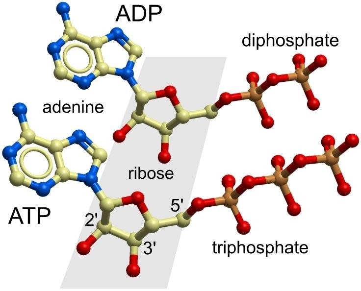
Getting a good rate of exchange – the
mitochondrial ADP-ATP carrier
The ATP-ADP mitochondrial carrier
ATP, adenosine triphosphate, is the energy currency of our cells. It is produced by the enzyme ATP
synthase in the mitochondria. These are organelles that oxidise sugars and lipids from food and
conserve the energy by synthesising ATP (the triphosphate form) from ADP (its diphosphate form).

Structure of ADP (top) and ATP (bottom). The ribose sugars are boxed in grey and the numbering of
   some carbon atoms is shown. The di- or tri-phosphate moiety is attached to the 5' carbon atom.

We all have about 50g of ATP in our bodies but this is constantly shuttling, first out of our mitochondria
to power cellular activity and then back as ADP. Because of this recycling, which can happen up to
1000 times a day for each ATP molecule, the total mass of ATP passing across the inner membrane of
our body's mitochondria is more than 50kg each day – so almost equalling our entire body mass. ATP
and ADP are negatively charged owing to their phosphate groups, but partly neutralised in solution by
association with magnesium ions. However, both these molecules are still too charged and polar in
nature to cross a lipid membrane. This has the advantage that these key molecules cannot leak out of
the cell. Mitochondria exist as organelles within the cell with two enclosing membranes. The outer
membrane has large permanently-open pores that make it permeable to small compounds. However, the
passage of small, charged compounds across the mitochondrial inner membrane has to be carefully
controlled. This is because the charge difference and concentration gradients of ions, particularly of
protons, must be maintained across the inner membrane to conserve metabolic energy. Any passive leak
of protons would produce a short-circuit of the channelling used to the actively drive the synthesis of
ATP inside the mitochondria.
Six trans-membrane α-helices form the ADP-ATP carrier pore
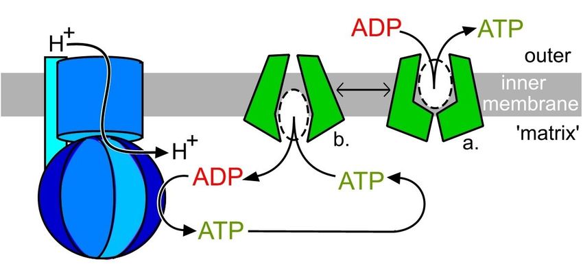
The exchange cycle of ADP and ATP. In this schematic, the ADP/ATP carrier (green) has two
interchangable states (a and b, connected by a double arrow). ADP will exchange for ATP in state a
and be released for regeneration by ATP synthase (blue) after the switch to state b.
The key player in managing the recycling of ATP is the mitochondrial ADP/ATP exchange carrier (also
known as ATP/ADP translocase), which is a pore or channel formed by six curved trans-membrane
(TM) α-helices (view-1). If we orient the molecule so that the outside of the membrane is at the top,
you can clearly see how the pore is relatively wide at that end, but much tighter towards the base
(view-2). This minimises the risk of protons leaking back across the membrane.
Three trans-membrane α-helices take an interesting turn
Stick diagram of a proline residue (Pro) in a protein chain. Atom names are indicated. Greyed out
sticks and atom names belong to adjacent residues. The arrow shows how the Pro side-chain curves
back and bonds to the backbone nitrogen atom.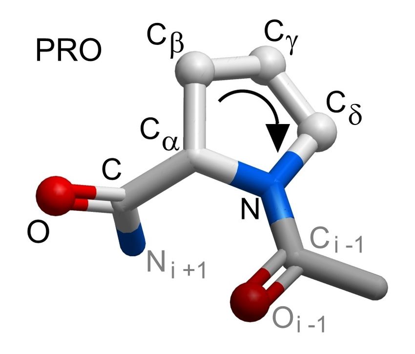
The first, third, and fifth TM α-helices that surround the pore have a distinctive kink or elbow that is
caused by proline residues (view-3). Proline residues are imino rather than amino acids and the
nitrogen atom in the backbone is bonded to a side-chain carbon atom. This unusual chemical structure
has two important consequences; first, proline residues are rather rigid in terms of the allowed
conformations of their main-chain, and second, there is no amide hydrogen atom that can be involved
in hydrogen bonds. This means that proline residues cannot be a part of a regular α-helix and in fact
introduce a disruption (kink) if they are located in helical regions (as they do here).

Three repeating units form the closed pore
At the narrowest constriction at the base of the carrier pore, a set of smaller α-helices connect each pair
of TM α-helices and lie adjacent to the inner surface of the membrane (view-4). These smaller α-
helices are called the matrix helices as they lie in the interior or “matrix” side of the mitochondrial
inner membrane. The pore structure therefore consists of a sequence of three structurally similar
repeated units of three α-helices each. Therefore, each repeating unit consists of an elbowed TM α-
helix followed by a horizontal matrix α-helix and finally a curved TM α-helix. The presence of three
structural repeats in the ADP/ATP carrier was originally postulated by Matti Saraste who observed
three repeats in the protein sequence (ref.1). This does not necessarily imply that there is rotational
symmetry around the central axis of the actual structure. However, an approximate three-fold rotational
symmetry was observed in the carrier structure, first in low resolution electron microscopy
reconstructions of the carrier and later very clearly in the crystal structure of the carrier (view-5) (ref.2).
The structural repeats in the carrier structure are shown in view-5 both superimposed and then
presented alongside each other. The repeats, which are approximately 100 amino acid residues long,
have less than 25% sequence identity, but can be superimposed with an RMSD of less than 2Å. As the
ADP and ATP substrates of the channel do not have any symmetry, it seems likely that the repeats in
the structure must relate to the mechanism of transport. There are repeated sequence motifs built into
the structural repeats. This fact suggests that interactions of key side-chains function to close the pore
on one side while opening on the other. This switching must depend on the presence of the bound
substrate – does the structure of the pore suggest how this might occur?

The crystallised protein contains an inhibitor instead of ADP
The only form of the mitotchondrial ADP/ATP carrier so far crystallised (PDB entry 1okc) has its upper
section occupied by a plant poison which acts by inhibiting the carrier’s activity. This is the glycoside
inhibitor carboxyatractyloside (view-6). You can see that the presence of this inhibitor leaves little
space in the pore interior. Perhaps this explains why neither ADP, which ought to bind to this outer part
of the pore, nor the other substrate ATP are found in the crystal structure. The effectiveness of the
glycoside compound as a poison depends on its binding tightly to the carrier from the outside, thereby
locking out the substrate ADP and trapping the carrier in a non-functional state. Since the
carboxyatractyloside is different in structure from ADP, it is difficult to use it to model the binding of
substrate to the protein. The crystal structure also provides no clues about how the pore opens to allow
ADP to pass through the membrane and be exchanged for ATP in return. In the exchange cycle
schematic shown above, state a with the carrier channel closed at the matrix side resembles the crystal
structure most.

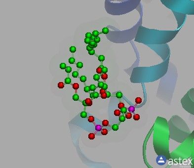
Cardiolipin - a lipid molecule with a key role in mitochondrial
function
The structure in PDB entry 1okc does, however, throw light on the binding of a key component of the
mitochondrial membrane, namely the unique lipid cardiolipin. This compound gets its name from its
abundance in cardiac muscle, which has a high content of mitochondria. The ADP/ATP carrier was
purified in detergent but has retained three molecules of cardiolipin from its original mitochondrial
membrane. These cardiolipins are bound, one to each structural repeat, around the outer surface of the
pore (view-7). You can find out more about the structure of this unique lipid compound and its presence
in other PDB entries by using the PDBe compound browser and searching for the three-letter code
CDL as described in this month's mini-tutorial. Mutation of a key enzyme (with the bizarre name
'tafazzin') that is involved in the synthesis of cardiolipin results in defective mitochondria. This is
evidence that cardiolipin plays a key role in the function of the mitochondria and the intimate
interaction revealed here with the ADP/ATP carrier may be part of this.

Further explorations
Use the PDBe small molecule compound browser to explore the inhibitor, lipids, and detergents bound
to the ADP/ATP carrier structure. This mini-tutorial will get you started. For many years, it was thought
that the ADP/ATP carrier could be a functional dimer so that the transport of ADP inwards could be
synchronised with the transfer of ATP outwards in a neighbouring partner carrier. Using the PDBePISA
service you can examine the PDB structure of the ADP/ATP carrier 1okc for likely biological
assemblies [PISA link]. There are tutorials to get you started with PDBePISA or you may have
followed the tutorial in the previous Quips on NGF. The PDBePISA analysis shows that there are no
stable dimeric contacts in the 1okc structure . This could be an artifact due to the poisoned state of the
protein in this crystal form, but the shape of the carrier does not seem to allow for a very close
association of two proteins. The exchange cycle schematic above shows that a functional dimer is not
required, provided that a single carrier requires either ADP or ATP to be bound before switching
conformation. The ATP exchange will be driven in the correct direction by the charge difference (and
concentration difference) between it and ADP. Many protein structures have repeated structural motifs
such as those observed in the carrier. If you suspect a structure contains repeats then you can use the
PDBeFold service to superimpose and align repeated motifs. This can be done for the repeats observed
here (view-5). If you are interested in structural repeats then a good place to start is Nick Grishin's
MALIDUP database.

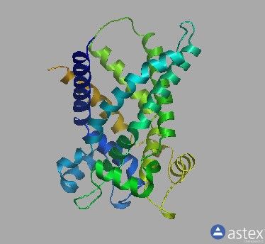
View 1
Cartoon drawing of ATP-ADP exchange pore (PDB entry 1okc).
The structure is coloured from blue at the N-terminus to red at the C-terminus.
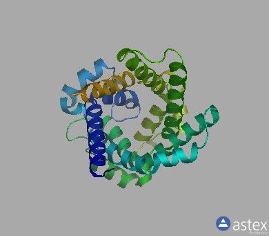
View-2
Looking down the carrier pore.
Notice the gradual narrowing of the pore from the outside towards the inside of the membrane.
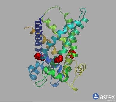
View-3
Elbowed helices in the carrier pore structure.
Elbowed helices in the carrier pore structure. As explained in the text, the unusual chemical structure of
proline residues (coloured red) causes kinks in the three long helices in the structure of the pore carrier
(PDB entry 1okc).
View-4
Trans-membrane and matrix helices.
Trans-membrane (TM) and matrix helices.
Matrix helices (red) lie adjacent to the inner surface of the mitochondrial membrane and connect the
remaining (TM) helices. Inner surface of TM helices creates a narrowing pore indicated by blue
surfaces (deeper shades of blue are deeper down the pore).
View-5
Superimposed repeats.
The three repeats (green, red, blue) are superimposable within 2Å. In the superimposition, we can see
that each consists of an elbowed helix (cyan), a matrix helix (yellow), and a curved helix (magenta).
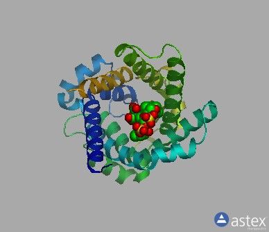
View-6
Poisoning the carrier pore.
Binding of the plant poison carboxyatractyloside to the carrier pore leaves little room for ADP to bind.
View-7
Cardiolipins bound to the carrier surface.
Cardiolipins, found in mitochondrial membranes, are seen associated with the outer surface of the
carrier.
You can also read