Guidelines for Monitoring and Management of Rectal Prolapse in Mice
←
→
Page content transcription
If your browser does not render page correctly, please read the page content below
Animal Care & Ethics Committees
Guidelines for Monitoring and
Management of Rectal Prolapse
in Mice
Purpose: This guideline has been developed to ensure the wellbeing of mice exhibiting signs of rectal
prolapse and to reduce premature killing of experimental animals due to this condition.
Background:
Rectal prolapse is a commonly encountered condition in laboratory mice. The condition may occur
spontaneously, related to specific genetic manipulations or specific experimental models as well be linked to
parasitic, viral or bacterial infections.2,4,6 Mice are particularly susceptible to rectal prolapse because they have
a very short rectum where the descending colon, enveloped in serosa, extends almost to the anus. 3 Therefore,
a rectal prolapse in mice can occur simply because of straining during parturition or defaecation.
Symptoms:
Rectal prolapse can be defined as the eversion of the rectal mucosa beyond the rectal opening3, presenting as
a small red mass at the anus. As a consequence, the prolapsed rectal tissue may bleed or become dry and
necrotic. Presence of blood staining/marking on bedding, cage walls or environmental enrichment may be
indicative of a mouse with this condition.
Assessment:
The degree of severity is assessed based on measurement of the prolapsed rectal tissue and the clinical
condition of the mouse.
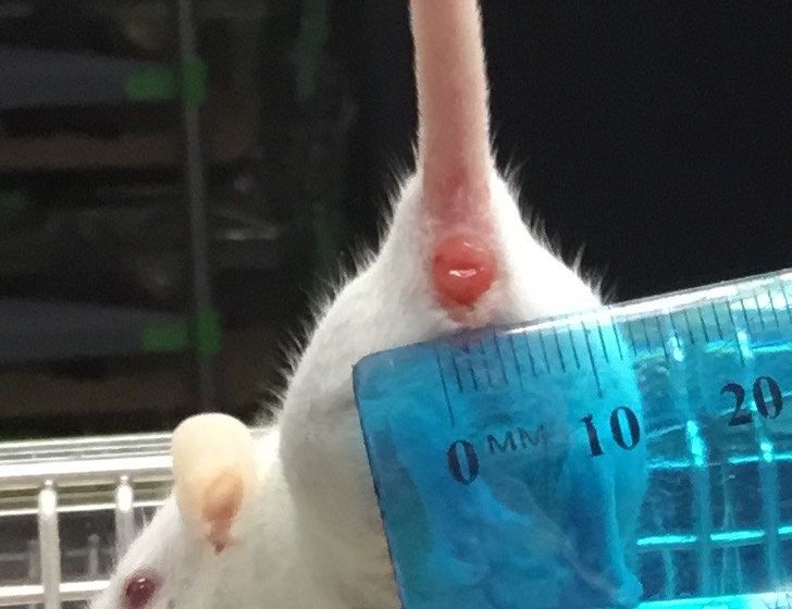
Measuring the prolapsed tissue:
A. Diameter of the rectal
prolapse: measure the
diameter of the rectal
prolapse when viewed from
the front. (Photo: UNSW,
2017a)
A B B. Protrusion of the rectal
tissue: measure the distance
the tissue is extending out of
the anus as viewed from the
side. (Photo: Manesh Kumar,
2004)
Version 2 Approved 4 July 2020 1
Expiry July 2023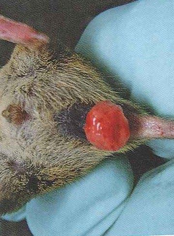
Mild Rectal Prolapse                Moderate Rectal Prolapse         Severe Rectal Prolapse

                  Photo: UNSW (2018)                    Photo: UNSW (2017)          Photo: Hankenson (2014)

            Diameter:  7 mm
            Protrusion:  4 mm

Decision Tree for Rectal Prolapse Management

                                                                                (Adapted from Hankenson, 2014)

Version 2            Approved 4 July 2020                                                                2
                     Expiry July 2023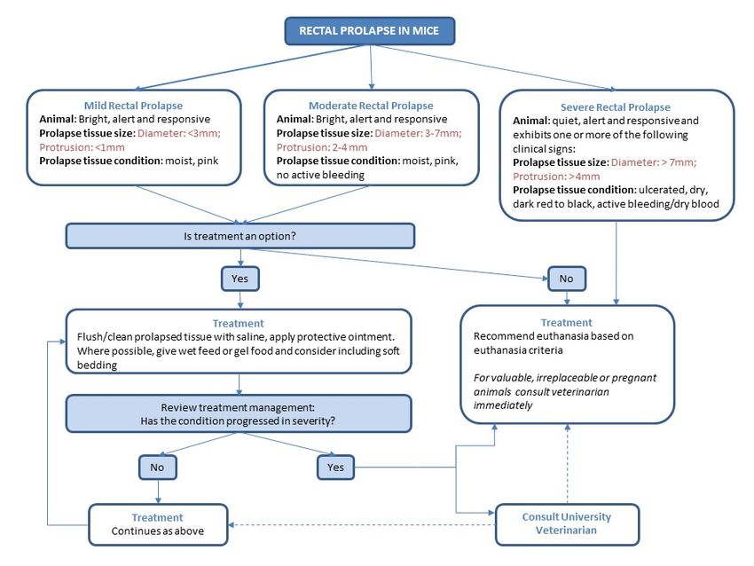
Treatment and Monitoring:
    •       It is the investigator’s responsibility to determine any potential impact of this condition and proposed
            treatment on their experimental outcome.
    •       Although a surgical procedure for reducing rectal prolapse has been described 6, surgery on small
            prolapses likely causes more damage to the mucosa than treating the site conservatively.
    •       Treatment for mild and moderate prolapses should focus on minimising damage to the prolapsed
            tissue through the following:
                    1) Flush area with sterile saline to remove any debris
                    2) Apply a thin layer of protective cream/ointment (e.g. Bepanthen™, Sudocream™,
                       Vaseline™, antibiotic ointment etc.) as determined by the University Veterinarians
                    3) Where possible, give moistened feed or commercially available gel food
                    4) Where possible, consider including soft bedding e.g. Protowels™, tissue paper to reduce
                       irritation and potential damage to prolapsed tissue
    •       It is recommended that mice are treated at least once daily (including weekends), preferably 2-3 times
            per day as needed and assessed each day to ensure that their condition is not worsening.
    •       Where possible, provision should be that animals suffering from rectal prolapse, should be allocated
            to the shortest experimental time-point.
    •       Animals are to be re-evaluated by veterinarians during the treatment period.

Euthanasia criteria:
Immediate euthanasia is required for mice with rectal prolapse that meet one or more of the following criteria:
    •       Severe rectal prolapse and/or
    •       Prolapsed tissues is ulcerated with excessive bleeding, desiccated or necrotic (dark red to black)
    •       The animal is showing any of the clinical signs pain or distress including loss of normal behaviour,
            reduced activity, ruffled fur, hunched posture, dehydration, weight loss equal to or greater than 20%,
            clinical signs of
Contact the University Veterinarians if assistance is required for critical assessment of mice with rectal
prolapse.

Comments
An increased incidence of rectal prolapses in a colony may be a symptom of underlying conditions such as
enteric diseases, underlying parasitic, viral or bacterial infections or linked to a specific phenotype. This should
be reported to the animal facility and the University Veterinary Staff for further investigation and
management.
Female breeding mice exhibiting clinical signs of rectal prolapses should not be bred, unless approved by
University Veterinarians.

Version 2               Approved 4 July 2020                                                                  3
                        Expiry July 2023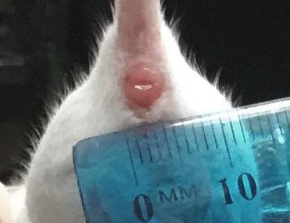
References:

    1. Beck, T.F., Shchelochkov, O.A., Yu, Z., Kim, B.J., Hernandez-Garcia, A., Zaveri, H.P., Bishop, C.,
       Overbeek, P.A., Stockton, D.W., Justice, M.J. and Scott, D.A., 2013. Novel frem1-related mouse
       phenotypes and evidence of genetic interactions with gata4 and slit3. PLoS One, 8(3).

    2. Fox, J.G. and Whary M.T., 2007. ‘Helicobacter Infections in Mice’, in Fox, J.G., Barthold, S., Davisson,
       M., Newcomer, C.E., Quimby, F.W. and Smith, A. (eds.), The Mouse in Biomedical Research, 2nd edn,
       vol. 3, Elsevier, San Diego.

    3. Georgia State University Institutional Animal Care and Use Committee, 2018. Georgia State
       University Institutional Animal Care and Use Committee (IACUC) Guidelines and Procedures Manual,
       viewed 10 June 2020,

    4. Hankenson, F.C., 2014. Critical care management for laboratory mice and rats, CRC Press, Boca Raton.

    5.       Kumar, M.M., Nagarajan, P., Venkatesan, R. and Juyal, R.C., 2004. Case Report and Short
            Communication: Rectal prolapse associated with an unusual combination of pinworms and citrobacter
            species infection in FVB mice colony. Scandinavian Journal of Laboratory Animal Sciences, 31(4),
            pp.221-223.

    6. Miller, C.L., Muthupalani, S., Shen, Z. and Fox, J.G., 2014. Isolation of Helicobacter spp. from mice with
       rectal prolapses. Comparative Medicine, 64(3), pp.171-178.

    7. Uchihashi, M., Wilding, L.A. and Nowland, M.H., 2015. Surgical Correction of Rectal Prolapse in
       Laboratory Mice (Mus musculus). Journal of the American Association for Laboratory Animal
       Science, 54(4), pp.433-438.

    8. Washington State University Institutional Animal Care and Use Committee, 2020. Standard Operating
       Procedures for Common Minor Medical Conditions of Rodents and Treatments , viewed 10 June 2020,

Version 2              Approved 4 July 2020                                                                4
                       Expiry July 2023
You can also read