HEART RATE VARIABILITY, DYSAUTONOMIA AND SPORTS RELATED CONCUSSION
←
→
Page content transcription
If your browser does not render page correctly, please read the page content below
HEART RATE VARIABILITY,
DYSAUTONOMIA AND
SPORTS RELATED
CONCUSSION
Big Sky Athletic Training Sports Medicine Conference 2022

        Robert J. Baker ATC, MD, PhD
        Program Director Primary Care Sports Medicine
        Professor Departments of Family and Community Medicine and
        Orthopedics
        Western Michigan University, Homer Stryker MD School of Medicine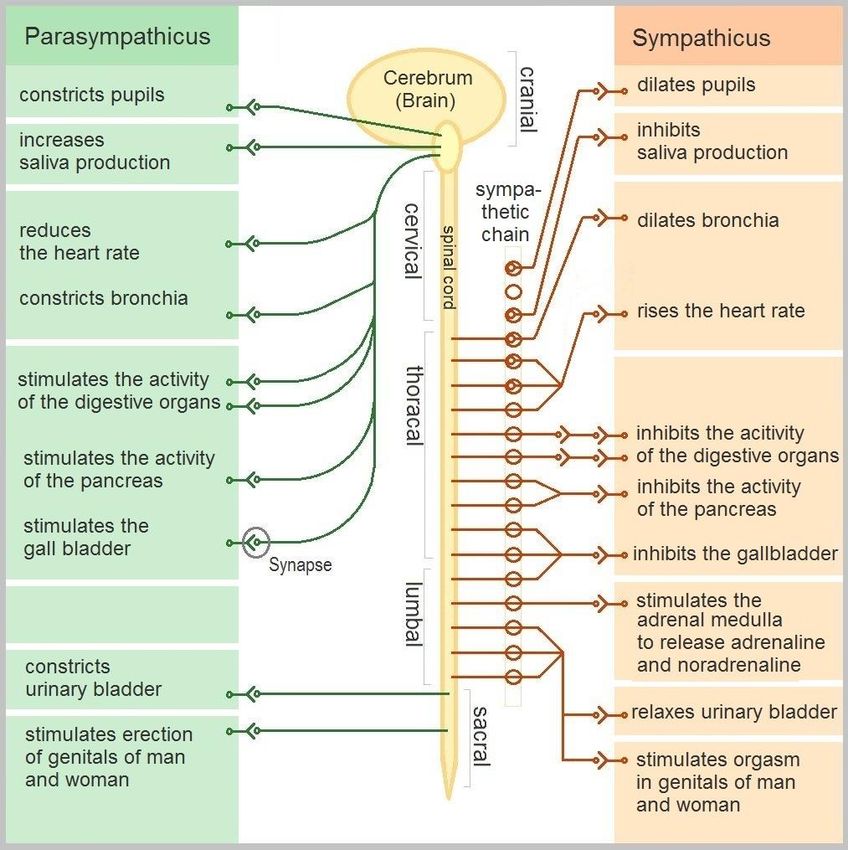
OBJECTIVES:

• To compare post-concussion recovery
  among the neurological domains of
  symptom evaluation, cognition,
  balance, ocular-motor, and autonomic
  nervous system [using the surrogate of
  heart rate variability (HRV)] and with the
  clinical determination that an athlete
  has recovered.
• To describe the neurological effects of
  concussion within these domains and
  any association among the domains
  affected.
• To describe longitudinal patterns of
  recovery from concussion within these
  domains.
ILLUSTRATIVE CASE
• 15 year old male Concussion 3 months ago
  with soccer.
• Most symptoms resolved in 1-2 weeks with PCP
• Persistent symptoms of fatigue, low energy,
  shortness of breath with running. Previous Half
  Marathon runner now with symptoms at 2-3
  miles.
• Symptoms progress and presented at the ED
• Follow up with Cardiology: EKG- ICRBB (rapid
  HR 93), Normal Echo, POTS
• Labs: CBC Normal, TSH normal, Free T4 normal,
  CMP normal, Vitamin D 52, Ferritin 191
• General: Slight male, no acute distress.
                                                                                   PHYSICAL EXAM
• Heart: Normal s1, s2. No murmur, click, rub, or gallop was heard.
• Peripheral pulses are 2+ and full throughout with no radial-femoral delay.
  Extremities are warm and well-perfused with no cyanosis, clubbing or edema
  noted.
• Pulmonary/Chest: Effort normal and breath sounds normal. No respiratory
  distress. He has no wheezes. He has no rales.
• Neurological: He is alert and oriented to person, place, and time. He
  has normal strength. He displays normal reflexes. No cranial nerve deficit. He
  exhibits normal muscle tone. He displays a negative Romberg
  sign. Coordination and gait normal.
  Immediate memory: recalled the 5 words easily
  Delayed recall score: 4/5
  Concentration: digit backwards score is 4/5. Correct month in reverse order
  Balance: normal balance tests
  Coordination: normal finger to nose test

• Psychiatric: He has a normal mood and affect. His speech is
  normal. Judgment and thought content normal. Cognition and memory are
  normal. He exhibits normal recent memory and normal remote memory.
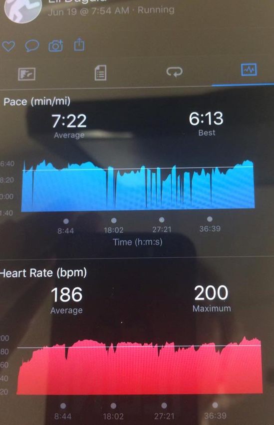
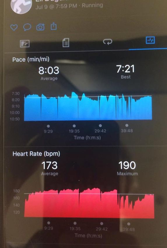
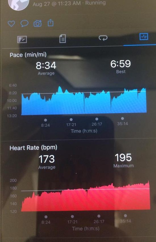
PATIENT PROVIDED HEART RATE AND PACE DATA
SAMPLE HEART RATE TRAINING
                                                         ZONE CALCULATIONS
     UPMC 5-Stage Exertion Protocol for Concussion
     • Stage 1: 30%-40% maximal exertion 113-126 Light aerobic conditioning 0.3
       (HRR) + RHR = 0.3 (124) +76
     • Stage 2: 40%-60% maximal exertion 126-150 Light to moderate aerobic
       conditioning 0.4 (HRR) + RHR = 0.4 (124) + 76
     • Stage 3: 60%-80% maximal exertion 150-175 Moderately aggressive
       aerobic exercise 0.6 (HRR) + RHR = 0.6 (124) + 76
     • Stage 4: 80% maximal exertion 175 80% exertion avoiding contact 0.8
       (HRR) + RHR = 0.8(124) + 76
     • Stage 5: 100% 200 Full exertion for sports with contact
Miranda, N. A., Boris, J. R., Kouvel, K. M., & Stiles, L. (2018). Activity and exercise intolerance after concussion: Identification and management of
postural orthostatic tachycardia syndrome. Journal of Neurologic Physical Therapy, 42(3), 163–171. https://doi.org/10.1097 /NPT.0000000000000231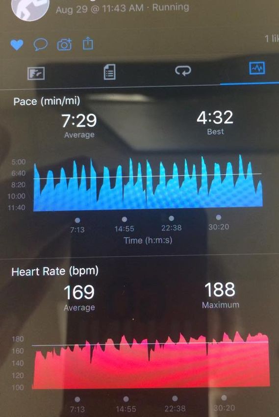
PATIENT PROVIDED HEART RATE AND PACE DATA
CLINICAL QUESTIONS

• Role of Dysautonomia
  Prolonged Concussion
  Symptoms?
• Role of POTS and Post
  Concussion Symptoms?
• Can We Measure of HRV with
  Mobile Device?
Role of Dysautonomia Prolonged Concussion
Symptoms?

            Harmon KG, Clugston JR, Dec K, et al. American Medical Society for Sports Medicine position
            statement on concussion in sport. Br J Sports Med. 2019;53(4):213-225. doi:10.1136/bjsports-2018-100338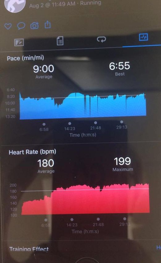
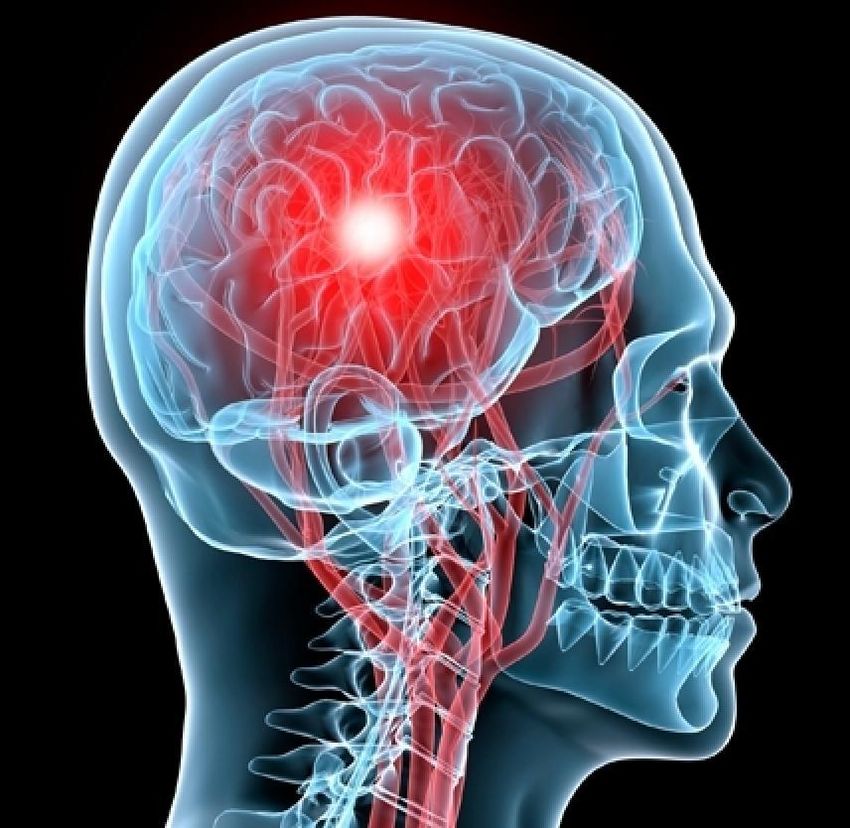
THEORY OF PROLONGED SYMPTOMS
Physiologic -> Autonomic nervous
system (ANS) dysfunction
(dysautonomia)
1. Mechanical changes & neuro-
metabolic -> alterations cerebral
circulation
2. Metabolic and physiologic
changes outside the brain
       Higher heart rates at rest
       Elevated HR cognitive and
       physiological stress
Severe TBI & SRC associated with
greater sympathetic nervous activity                         Hovda DA, Lee SM, Smith ML, et al. The neurochemical and metabolic cascade
                                                             following brain injury: moving from animal models to man. J Neurotrauma.
& lower parasympathetic activity                             1995;12(5):903-906.

              Leddy, J. J., Sandhu, H., Sodhi, V., Baker, J. G., & Willer, B. (2012).
              Rehabilitation of concussion and post-concussion syndrome. Sports Health,
              4(2), 147–154. https://doi.org /10.1177/1941738111433673
THEORY OF PROLONGED SYMPTOMS
Autonomic dysregulation
TBI severity
Improves during TBI recovery
Altered endocrine or
Neuropeptide milieu after TBI
Autoregulation: cerebral blood
flow changes in systemic
blood pressure
Symptoms often reappear or
worsen with physical and/or
mental exertion
Secondary insults:
hypotension, intracranial
hypertension, and
dehydration
Leddy, J. J., Sandhu, H., Sodhi, V., Baker, J. G., & Willer, B. (2012). Rehabilitation of concussion and post-concussion
syndrome. Sports Health, 4(2), 147–154. https://doi.org /10.1177/1941738111433673THEORY OF PROLONGED SYMPTOMS
Physiologic
Altered ANS balance ->
pulmonary ventilation
altered during exertion
The primary ANS control center is
located in the brainstem
Twisting mechanism
(glancing blow)
Alter central ANS regulation
cardiorespiratory/ventilation
fMRI ANS diffusely distributed
beyond the brainstem
Leddy, J. J., Sandhu, H., Sodhi, V., Baker, J. G., & Willer, B. (2012). Rehabilitation of concussion and post-
concussion syndrome. Sports Health, 4(2), 147–154. https://doi.org /10.1177/1941738111433673ROLE OF EVALUATION OF DYSAUTONOMIA IN
• ANS dysfunction after a mild TBI -> changes in heart
CONCUSSION?
rate variability
• Autonomic disfunction in the post-acute stage after
a concussion
• long term effects of mild TBI on the autonomic
nervous system unknown?
• systemic effects of ANS dysfunction in mild TBI?
• Persistence of autonomic dysfunction after
symptomatic resolution of concussions -> return-to-
play protocols?
Esterov, D., Greenwald, BD. (2017). Autonomic dysfunction after mild
traumatic brain injury. Brain Sci. 7 (100); doi:10.3390/brainsci7080100ROLE OF POTS AND POST CONCUSSION
SYMPTOMS?
Pediatric: 11.4% of individuals Activation of the sympathetic
diagnosed with POTS report onset nervous system
of symptoms within 3 months of
sustaining a concussion lightheadedness
shortness of breath
chest pain
tachycardia
palpitations standing/with
exertion
exercise intolerance
Miranda, N. A., Boris, J. R., Kouvel, K. M., & Stiles, L. (2018). Activity and exercise intolerance after concussion:
Identification and management of postural orthostatic tachycardia syndrome. Journal of Neurologic Physical
Therapy, 42(3), 163–171. https://doi.org/10.1097 /NPT.0000000000000231HEART RATE AND HEART RATE VARIABILITY AT
REST AND DURING EXERCISE IN BOYS WHO
SUFFERED A SEVERE TRAUMATIC BRAIN INJURY
AND TYPICALLY-DEVELOPED CONTROLS
• Heart rate (HR) and heart rate variability (HRV) at rest and during exercise in children with post-severe traumatic brain injury
• Children post-TBI demonstrated higher mean HR values at rest (TBI 91.87.0 beats per minute vs 72.07.1 beats per minute in
controls, pHEART RATE VARIABILITY IN NEUROREHABILITATION
PATIENTS WITH SEVERE ACQUIRED BRAIN INJURY
• Patients with traumatic brain injury
(TBI) by means of reduced heart
rate variability (HRV). It was
hypothesized that patient groups
with other ABI etiology (mainly
stroke, subarachnoid hemorrhage
and anoxia) would also present
reduced HRV
• HRV appeared identical across ABI
etiology
• HRV was considerably reduced in
an heterogenic ABI patient group
admitted for neurorehabilitation
Vistisen, S. T., Hansen, T. K., Jensen, J., Nielsen, J. F., & Fleischer, J. (2014). Heart rate variability in neurorehabilitation
patients with severe acquired brain injury. Brain Injury, 28(2), 196–202. https://doi.org/10.3109 /02699052.2013.860477MULTIMODAL ASSESSMENT OF SPORT-
RELATED CONCUSSION
• Determine which assessments best identify athletes with sport-related
concussion (SRC) from healthy controls in the acute/early subacute phase
(within 10 days of SRC) of injury.
• Prospective, cohort, specialty concussion clinic
• Multimodal evaluation that are most robust at discriminating SRC
acute/early subacute phase: 1. Symptom report 2. Symptoms on
vestibular/oculomotor assessment
Sherry NS, Fazio-Sumrok V, Sufrinko A, Collins MW, Kontos AP. Multimodal Assessment of Sport-Related Concussion.
Clin J Sport Med. 2021;31(3):244-249. doi:10.1097/JSM.0000000000000740HEART RATE VARIABILITY OF RECENTLY
CONCUSSED ATHLETES AT REST AND EXERCISE
• Neuroautonomic cardiovascular
regulation (HRV) in recently concussed
athletes at rest and in response to low-
moderate steady-state exercise, using
heart rate variability
• No difference at rest was detected
between the concussed athletes and
their matched controls
• Exercise tests: concussed group
demonstrated a significant decrease in
the mean RR interval, and low- and high-
frequency power (P 0.05)
• Low-moderate steady-state exercise
elicits a neuroautonomic cardiovascular
dysfunction in concussed athletes
(exercise induced uncoupling between Gall, B., Parkhouse, W., & Goodman, D. (2004). Heart rate
the autonomic and cardiovascular variability of recently concussed athletes at rest and exercise.
systems). Medicine & Science in Sports & Exercise, 36(8), 1269–1274.
https://doi.org/10.1249/01.MSS.0000135787.73757.4DHEART RATE VARIABILITY OF ATHLETES ACROSS
CONCUSSION RECOVERY MILESTONES: A
PRELIMINARY STUDY.
• Heart rate variability (HRV) in athletes
with concussion across three phases of
recovery
• Prospective matched control group: 1.
HRV 2. Symptoms Questionnaire
• 3 phases of recovery (1) symptomatic;
(2). asymptomatic; and (3) one-week
after return-to-play (RTP)
• Athletes with concussion displayed
autonomic dysfunction in some
measures of HRV that persisted beyond
RTP and were related to a previous
history of concussion
Senthinathan, A., Mainwaring, L. M., & Hutchison, M. (2017). Heart rate
variability of athletes across concussion recovery milestones: A
preliminary study. Clinical Journal of Sport Medicine, 27(3), 288–295.
https://doi.org/10.1097 /JSM.0000000000000337Autonomic Function Following Concussion in Youth
Athletes: An Exploration of Heart Rate Variability
Using 24-hour Recording Methodology
• Heart rate variability (HRV) is a non-invasive
physiological indicator of the autonomic
nervous system, capturing the reciprocal
interplay between the sympathetic and
parasympathetic nervous systems
• 24 h recording methodology
• (1) evaluate the physiological effects of a
concussion in youth athletes
• (2) describe the trajectory of physiological
change resolution of self-reported post-
concussion symptoms.
• The raw beat-to-beat time intervals
captured can be transformed to derive
time domain and frequency domain
measures
Paniccia M, Taha T, Keightley M, et al. Autonomic Function Following
Concussion in Youth Athletes: An Exploration of Heart Rate Variability
Using 24-hour Recording Methodology. J Vis Exp. 2018;(139):58203.
Published 2018 Sep 21. doi:10.3791/58203POST CONCUSSIVE DISORDERS (PCD)
1. Physiological post-PCD: characterized by concussion
symptoms from alterations in cerebral blood flow
secondary to autonomic nervous system dysfunction
2. Vestibulo-ocular PCD: characterized by symptoms
secondary to dysfunction of the vestibular and oculomotor
systems
3. Cervicogenic PCD: characterized by muscle trauma and
inflammation secondary to cervical spine somatosensory
system.
4. Clinical depression
5. Post traumatic mood disorders
6. Migraine headaches
Leddy, J. J., Sandhu, H., Sodhi, V., Baker, J. G., & Willer, B.
(2012). Rehabilitation of concussion and post-concussion
syndrome. Sports Health, 4(2), 147–154. https://doi.org
/10.1177/1941738111433673
Esterov, D., Greenwald, BD. (2017). Autonomic dysfunction after
mild traumatic brain injury. Brain Sci. 7 (100);
doi:10.3390/brainsci7080100CAN WE MEASURE OF HRV WITH MOBILE
DEVICE?
Paniccia M, Taha T, Keightley M, et al. Autonomic Function Following Concussion in Youth Athletes: An Exploration
of Heart Rate Variability Using 24-hour Recording Methodology. J Vis Exp. 2018;(139):58203. Published 2018 Sep 21.
doi:10.3791/58203VALIDITY OF THE ELITE HRV SMARTPHONE
APPLICATION FOR EXAMINING HEART RATE
VARIABILITY IN A FIELD-BASED SETTING
• Relationship and validity between a
vagal-related HRV index, rMSSD, when
derived from a smartphone application
accessible with most operating systems
against a frequently used computer
software program, Kubios HRV 2.2
• While differences exist between the two
sources of HRV analysis however, further
research is warranted, as this smartphone
HRV application may offer a reliable
platform when assessing parasympathetic
modulation
Perrotta, A. S., Jeklin, A. T., Hives, B. A., Meanwell, L. E., & Warburton, D. E. R. (2017). Validity of the elite HRV
smartphone application for examining heart rate variability in a field-based setting. The Journal of Strength
and Conditioning Research, 31(8), 2296–2302. https://doi.org /10.1519/JSC.0000000000001841FACE COOLING EXPOSES CARDIAC
PARASYMPATHETIC AND SYMPATHETIC
DYSFUNCTION IN RECENTLY CONCUSSED COLLEGE
• Concussed college ATHLETES
athletes (CA) have
attenuated
parasympathetic and
sympathetic responses to
face cooling (FC)
• These data indicate that
symptomatic concussed
patients have impaired
cardiac parasympathetic
and sympathetic
activation
Johnson, B. D., O’Leary, M. C., McBryde, M., Sackett, J. R., Schlader, Z. J., & Leddy, J. J. (2018). Face
cooling exposes cardiac parasympathetic and sympathetic dysfunction in recently concussed college
athletes. Physiological Reports, 6(9), e13694. https://doi.org/10.14814/phy2.13694MOBILE EVALUATION OF HEART RATE
VARIABILITY USING THE DIVER’S REFLEX
Significantly decreased compared to at rest
• Validates prior research with larger
sample sizes and proposes a model for
establishing baseline HRV reactivity in
healthy participants
• RMSSD was elevated at 1 and 2 min
(+47.4 ms, p < .0001; +16.5 ms, p = .014)
following face cooling and fell to
baseline at 3 min (+4.6 ms, p = .52)
• LF/HF ratio decreased following face
cooling at 2 and 3 min (change from rest
%: 2 min, −33%, p = .007; 3 min, −50%, p
= < .0001)
• The Elite HRV platform can detect an
elevation in RMSSD in the first minute
following face cooling with a return to
baseline in the second and third
minutes. It can also detect a consistent
decrease in LF/HF following face cooling
Seltzer, H., Pellman, M., Warchock, R., Billian, J., & Baker, R. (2021). Mobile evaluation of heart rate variability
using the Diver’s Reflex. NeuroRegulation, 8(2), 96–103. https://doi.org/10.15540/nr.8.2.96MOBILE EVALUATION OF HEART RATE
VARIABILITY USING THE DIVER’S REFLEX
Significantly increased compared to at rest
Seltzer, H., Pellman, M., Warchock, R., Billian, J., & Baker, R. (2021). Mobile evaluation of heart rate variability
using the Diver’s Reflex. NeuroRegulation, 8(2), 96–103. https://doi.org/10.15540/nr.8.2.96AUTONOMIC DYSFUNCTION SUMMARY
• Ongoing central and systemic physiologic
regulatory dysfunction has been proposed
as a mechanism for persistent systems in
patients with PCS
• Concussion can result in transient or
more persistent autonomic
dysregulation and ultimately POTS and
hyperadrenergic states
• Fatigue, dizziness, tachycardia,
headaches, nausea, exercise intolerance
Leddy Neurorehabilitation 2007
Miranda J Neurol Phys Ther 2018AUTONOMIC DYSFUNCTION SUMMARY FUTURE
DIRECTIONS
• Understanding ANS dysfunction at rest
following concussion recovery and post
recovery
• Current literature is limited by small
sample sizes, lack of female or pediatric
participants, methodological
heterogeneity and lack of follow-up
• While there is some evidence to suggest
changes during physical activity
following concussion, methodological
limitations
• Understanding the effect of concussion
on ANS will contribute to the
development of more comprehensive
concussion management strategies
Blake, T. A., McKay, C. D., Meeuwisse, W. H., & Emery, C. A. (2016). The impact of concussion on cardiac autonomic
function: A systematic review. Brain Injury, 30(2), 132–145. https://doi.org/10.3109/02699052.2015.1093659REFERENCES
• Patricios JS, et al. Br J Sports Med 2018;52:635–641. doi:10.1136/bjsports-2018-099079
• Hovda DA, Lee SM, Smith ML, et al. The neurochemical and metabolic cascade following brain injury: moving from animal models to man. J
Neurotrauma. 1995;12(5):903-906.
• Leddy, J. J., Sandhu, H., Sodhi, V., Baker, J. G., & Willer, B. (2012). Rehabilitation of concussion and post-concussion syndrome. Sports Health,
4(2), 147–154. https://doi.org /10.1177/1941738111433673
• Miranda, N. A., Boris, J. R., Kouvel, K. M., & Stiles, L. (2018). Activity and exercise intolerance after concussion: Identification and management
of postural orthostatic tachycardia syndrome. Journal of Neurologic Physical Therapy, 42(3), 163–171. https://doi.org/10.1097
/NPT.0000000000000231
• Esterov, D., Greenwald, BD. (2017). Autonomic dysfunction after mild traumatic brain injury. Brain Sci. 7 (100); doi:10.3390/brainsci7080100
• Shaffer, F., & Ginsberg, J. P. (2017). An overview of heart rate variability metrics and norms. Frontiers in Public Health, 5, 258.
https://doi.org/10.3389/fpubh.2017.00258
• Senthinathan, A., Mainwaring, L. M., & Hutchison, M. (2017). Heart rate variability of athletes across concussion recovery milestones: A
preliminary study. Clinical Journal of Sport Medicine, 27(3), 288–295. https://doi.org/10.1097 /JSM.0000000000000337
• Gall, B., Parkhouse, W., & Goodman, D. (2004). Heart rate variability of recently concussed athletes at rest and exercise. Medicine & Science
in Sports & Exercise, 36(8), 1269–1274. https://doi.org/10.1249/01.MSS.0000135787.73757.4D
• Paniccia M, Taha T, Keightley M, et al. Autonomic Function Following Concussion in Youth Athletes: An Exploration of Heart Rate Variability
Using 24-hour Recording Methodology. J Vis Exp. 2018;(139):58203. Published 2018 Sep 21. doi:10.3791/58203
• Leddy, J., Baker, J. G., Haider, M. N., Hinds, A., & Willer, B. (2017). A physiological approach to prolonged recovery from sport-related
concussion. Journal of Athletic Training, 52(3), 299–308. https://doi.org/10.4085/1062-6050-51.11.08REFERENCES
• Sherry NS, Fazio-Sumrok V, Sufrinko A, Collins MW, Kontos AP. Multimodal Assessment of Sport-Related Concussion. Clin J Sport Med.
2021;31(3):244-249. doi:10.1097/JSM.0000000000000740
• Johnson, B. D., O’Leary, M. C., McBryde, M., Sackett, J. R., Schlader, Z. J., & Leddy, J. J. (2018). Face cooling exposes cardiac
parasympathetic and sympathetic dysfunction in recently concussed college athletes. Physiological Reports, 6(9), e13694.
https://doi.org/10.14814/phy2.13694
• Perrotta, A. S., Jeklin, A. T., Hives, B. A., Meanwell, L. E., & Warburton, D. E. R. (2017). Validity of the elite HRV smartphone application for
examining heart rate variability in a field-based setting. The Journal of Strength and Conditioning Research, 31(8), 2296–2302. https://doi.org
/10.1519/JSC.0000000000001841
• Blake, T. A., McKay, C. D., Meeuwisse, W. H., & Emery, C. A. (2016). The impact of concussion on cardiac autonomic function: A systematic
review. Brain Injury, 30(2), 132–145. https://doi.org/10.3109/02699052.2015.1093659
• Vistisen, S. T., Hansen, T. K., Jensen, J., Nielsen, J. F., & Fleischer, J. (2014). Heart rate variability in neurorehabilitation patients with severe
acquired brain injury. Brain Injury, 28(2), 196–202. https://doi.org/10.3109 /02699052.2013.860477
• Katz-Leurer, M., Rotem, H., Keren, O., & Meyer, S. (2010). Heart rate and heart rate variability at rest and during exercise in boys who suffered
a severe traumatic brain injury and typically-developed controls. Brain Injury, 24(2), 110–114. https://doi.org/10.3109/02699050903508234
• Seltzer, H., Pellman, M., Warchock, R., Billian, J., & Baker, R. (2021). Mobile evaluation of heart rate variability using the Diver’s Reflex.
NeuroRegulation, 8(2), 96–103. https://doi.org/10.15540/nr.8.2.96
• Harmon KG, Clugston JR, Dec K, et al. American Medical Society for Sports Medicine position statement on concussion in sport. Br J Sports
Med. 2019;53(4):213-225. doi:10.1136/bjsports-2018-100338
• Hovda DA, Lee SM, Smith ML, et al. The neurochemical and metabolic cascade following brain injury: moving from animal models to man. J
Neurotrauma. 1995;12(5):903-906.You can also read