Heatstroke induced late onset neurological deficits in mice caused by white matter demyelination, Purkinje cell degeneration, and synaptic ...
←
→
Page content transcription
If your browser does not render page correctly, please read the page content below
www.nature.com/scientificreports
OPEN Heatstroke‑induced late‑onset
neurological deficits in mice caused
by white matter demyelination,
Purkinje cell degeneration,
and synaptic impairment
in the cerebellum
Kazuyuki Miyamoto1,2,3*, Motoyasu Nakamura1,2, Hirokazu Ohtaki2,4, Keisuke Suzuki1,2,
Hiroki Yamaga1,2, Kaoru Yanagisawa1,2, Atsuo Maeda1, Masaharu Yagi1, Munetaka Hayashi1,
Kazuho Honda2 & Kenji Dohi1,2
Global warming increases heatstroke incidence. After heatstroke, patients exhibit neurological
symptoms, suggesting cerebellar damage. However, the potential long-term adverse outcomes are
poorly understood. We studied the cerebellum after heatstroke in mouse heatstroke models. In this
study, motor coordination disorder significantly appeared 3 weeks after heatstroke and gradually
improved to some extent. Although white matter demyelination was detected at 1 and 3 weeks after
heatstroke in the cerebellum, it was not found in the corpus callosum. The Purkinje cell numbers
significantly decreased at 1, 3, and 9 weeks after heatstroke. The intensity of synaptophysin and
postsynaptic density-95 temporarily appeared to attenuate at 3 weeks after heatstroke; however,
both appeared to intensify at 9 weeks after heatstroke. Motor coordination loss occurred a few weeks
after heatstroke and recovered to some extent. Late-onset motor impairment was suggested to be
caused by cerebellar dysfunctions morphologically assessed by myelin staining of cerebellar white
matter and immunostaining of Purkinje cells with pre- and postsynaptic markers. Purkinje cell number
did not recover for 9 weeks; other factors, including motor coordination, partially recovered, probably
by synaptic reconstruction, residual Purkinje cells, and other cerebellar white matter remyelination.
These phenomena were associated with late-onset neurological deficits and recovery after heatstroke.
Heatstroke, a systemic disease caused by exposure to high ambient temperature (AT) and relative humidity
(RH)1, is estimated to increase owing to the recent global w
arming2,3. Since patients with heatstroke often develop
multiple organ damage in critical care, they are primarily treated with whole-body cooling, hemodialysis, and
plasma exchange4. Although the acute effects after a heatstroke are well-recognized, potential long-term adverse
outcomes are poorly understood. Notably, damage to the central nervous system (CNS) after a heatstroke has
not been fully elucidated, despite being known to decrease the level of consciousness and function, which results
in a condition similar to neuronal damage following a high fever caused by viral or bacterial infections and
inflammation5. Approximately 25% of patients with heatstroke exhibit convalescent or long-term neurological
deficits, including motor dysfunction and cognitive impairment.
Moreover, more than 70% of patients with neurological symptoms exhibit long-term cerebellar damage,
most commonly presenting with adverse long-term neurological outcomes6. The cerebellum plays an important
1
Department of Emergency, Critical Care and Disaster Medicine, Showa University School of Medicine, 1‑5‑8
Hatanodai, Shinagawa‑ku, Tokyo 142‑8555, Japan. 2Department of Anatomy, Showa University School of
Medicine, 1‑5‑8 Hatanodai, Shinagawa‑ku, Tokyo 142‑8555, Japan. 3Department of Emergency and Disaster
Medicine, Showa University Northern Yokohama Hospital, 35‑1 Chigasaki‑chuo Tsuzuki‑ku, Yokohama 224‑8503,
Japan. 4Department of Functional Neurobiology, Tokyo University of Pharmacy and Life Science, School of
Pharmacy, 1432‑1 Horinouchi, Hachioji, Tokyo 192‑0392, Japan. *email: k-miyamoto@med.showa-u.ac.jp
Scientific Reports | (2022) 12:10598 | https://doi.org/10.1038/s41598-022-14849-9 1
Vol.:(0123456789)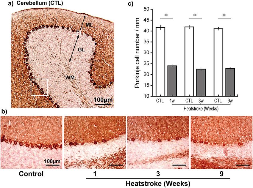
www.nature.com/scientificreports/
role in motor coordination/learning, sensory integration, and coordinate transformation. Cerebellar damage
results in increased postural sway, hypermetric postural responses to perturbations and optokinetic stimuli, and
postural responses that are poorly coordinated with volitional movement, which induces deficits in balance and
walking7. After a heatstroke, patients who exhibit certain neurological symptoms—disorientation, wobbling
(Supplementary Video S1), and vertigo—are bedridden, thereby preventing patients from returning to normal
life, suggesting cerebellar damage. In addition to these neurological symptoms, several reports have revealed
cerebellar abnormalities on head magnetic resonance imaging (MRI) in patients with heatstroke (Supplemen-
tary Fig. S1)8,9. Moreover, several interesting reports showed that neurological deficits appeared several weeks
after heatstroke rather than immediately a fterward10,11, and some cases improved with t ime9, while others are
permanent. However, it is not well understood how these late-onset neurological deficits post heatstroke appear
and, in some cases, improve over time. Therefore, we focused on the influence of heatstroke on the cerebellum
using a mouse heatstroke model.
Few studies have examined the CNS using a heatstroke model. Most CNS studies performed on animals
after heatstroke have evaluated heat-induced cerebral ischemic injury12, hypothalamic injury13, and alcohol
influence on the central amygdaloid n ucleus14. These studies did not consider RH, and other heatstroke studies
that did not examine the CNS also disregarded the RH impact and created desert-like conditions15–18. How-
ever, summer in temperate or tropical zones has high AT and RH. Even if AT remains the same, a higher RH
accounts for a higher wet-bulb globe temperature (WBGT), suggesting a higher risk of heatstroke. Therefore,
we recently established a mouse heatstroke model that accounts for AT and RH while monitoring WBGT. Our
animal model presents low mortality (< 20%, 4 days after heatstroke) and is considered suitable for evaluating
long-term adverse outcomes19. In this study, we examined late-onset motor coordination and cerebellar damage
for 9 weeks in mouse heatstroke models.
Results
Motor coordination disorder appeared 3 weeks after heatstroke. Before heatstroke, the mean
rotarod running time of mice in the control (CTL) and heatstroke (HS) groups was 85.6 ± 5.1 and 84.7 ± 5.2 s,
respectively. Notably, 2 out of 36 mice in the HS group died within 1 day after heatstroke, and the remaining
mice survived for 9 weeks. One week after heatstroke, the running time of the rotarod test in the HS group was
81.8 ± 6.8 s, whereas the running time in the CTL group was 93.1 ± 7.0 s. The running time of the rotarod test in
the HS group 3 weeks after heatstroke further decreased to 68.8 ± 6.2 s and was significantly lower than that in
the CTL group (92.7 ± 7.7 s, P < 0.05). The running time of the HS group gradually increased over time, whereas
no differences were observed in the CTL group (Fig. 1a). To eliminate a feeding disorder after heatstroke, both
groups of mice were weighed during the experiment, but these results were not statistically significant through-
out the experiment (Fig. 1b).
Demyelination detected at 1 and 3 weeks after heatstroke in the cerebellar white mat‑
ter. The cerebellar white matter in the CTL mice was dense and homogeneous to fast blue. However, the
white matter in the HS mice was heterogeneous and sponge-like. Semi-quantification of myelin showed that the
myelin percentage was significantly decreased at 1 (CTL, 70.4% ± 2.8%; HS, 59.7% ± 2.8%; P < 0.05) and 3 weeks
(CTL, 69.3% ± 1.6%; HS, 63.8% ± 1.3%; P < 0.05) after heatstroke (Fig. 2), suggesting demyelination. However,
this decrease was not statistically significant among the groups at 9 weeks, suggesting recovery.
In contrast, we compared the myelin quantity in the cerebellum with that in the corpus callosum for the same
sections to determine region specificity. Interestingly, we found no significant differences between the CTL and
HS groups during the experiments.
Purkinje cell number significantly decreased after heatstroke. The Purkinje cells in the CTL mice
aligned clearly in an equidistant manner (Fig. 3a). However, after heatstroke, the calbindin-positive Purkinje
cells showed unequal distances and decreased numbers (Fig. 3b). The Purkinje cell number in the CTL versus HS
groups was as follows: 1 week (41.7 ± 1.2 vs. 24.0 ± 0.4 cells/mm, P < 0.05), 3 weeks (41.9 ± 0.9 vs. 22.5 ± 0.5 cells/
mm, P < 0.05), and 9 weeks (41.1 ± 0.7 vs. 22.9 ± 0.4 cells/mm, P < 0.05) after heatstroke (Fig. 3c).
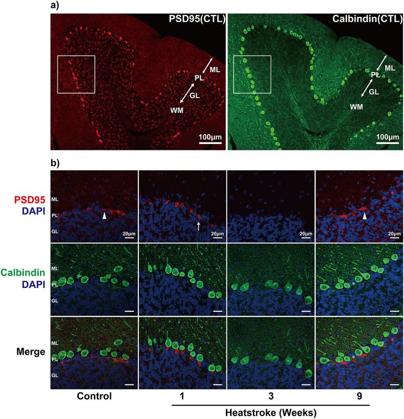
Expression of postsynaptic density‑95 (PSD95) appeared to have temporarily attenuated at
3 weeks after heatstroke. We examined Purkinje cell synaptic plasticity based on PSD95 expression.
The distribution of PSD95-positive cells was strongly localized to the axon plexus. The PSD95-positive cells
in the CTL group had a wide morphology, such as plates, but had low density. The PSD95-positive cells at
1 week after heatstroke were localized to the axon hillock of the Purkinje cells (looked similar to pen points).
After 3 weeks, the immunoreactions appeared to have attenuated, although the calbindin-positive Purkinje cells
were stained in the region. The positive reactions reappeared more heterogeneously at 9 weeks after heatstroke.
Moreover, PSD95 immunoreactivity was observed again around the axon hillock of some residual Purkinje
cells and appeared to have stronger intensities than that observed in the CTL group (Fig. 4). The co-staining of
PSD95 with synaptophysin revealed that the intensity of synaptophysin immunoreactivity around the Purkinje
cells appeared to have been the weakest at 3 weeks after heatstroke; however, both intensified at 9 weeks after
heatstroke (Fig. 5).
Discussion
It is estimated that the recent global warming will cause an increase in patients with heatstroke in the near future.
However, there is poor understanding of the potential long-term adverse outcomes on the CNS, although patients
often exhibit neurological d eficits8,20.
Scientific Reports | (2022) 12:10598 | https://doi.org/10.1038/s41598-022-14849-9 2
Vol:.(1234567890)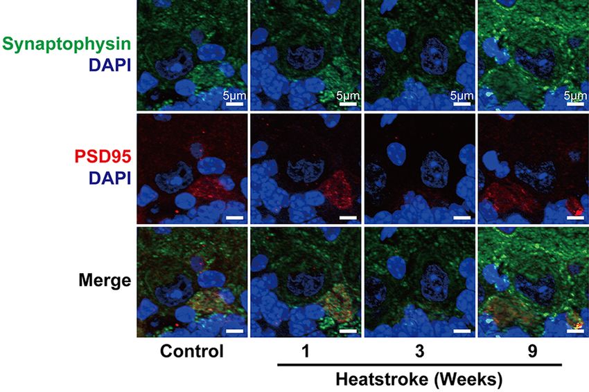
www.nature.com/scientificreports/
Figure 1. Motor coordination and body weight for 9 weeks after heatstroke. Running time of the rotarod
test (a) and body weight (b, BW) in the control (CTL) and heatstroke (HS) animals. The running time of the
rotarod test in the HS animals 3 weeks after heatstroke significantly decreased compared with that in CTL
animals. BW in the HS and CTL groups did not change significantly over the experiments. Data are expressed as
mean ± standard deviation (SD) (*P < 0.05, Tukey–Kramer test). HS heatstroke, CTL control, BW body weight.
In this study, we evaluated the neurological deficits using the rotarod test and brain histological evaluations (in
the cerebellum) for 9 weeks using a mouse heatstroke model because disappearing and swelling of the Purkinje
cells have been reported in autopsied brain tissues of patients with heatstroke21. Observation of brain diffusion-
weighted MRI revealed hyperintensity in the bilateral cerebellar medullas in patients with h eatstroke22. These
reports indicate that cerebellar protection is a key therapeutic target in patients with heatstroke.
In our study, the running time of rotarod test was significantly shorter at 3 weeks after heatstroke and gradu-
ally returned to baseline at 9 weeks after heatstroke. These results suggest motor function/coordination impair-
ment in our heatstroke model. In patients with multiple sclerosis, cerebellar ataxia is commonly observed, and
diffuse white matter demyelination has been r eported23. Therefore, we examined the morphology of the brain
by Klüver–Barrera (KB) staining, but we could not recognize apparent cerebral infarction or hemorrhage. How-
ever, the cerebellum showed decreased intensity with KB staining and was recognized as sponge-like in the HS
group. Semi-quantification of the KB-stained region revealed that the cerebellar white matter was significantly
demyelinated between 1 and 3 weeks after heatstroke in the HS group. However, the intensity did not significantly
differ from that of the CTL group at 9 weeks after heatstroke, suggesting remyelination. In contrast, demyelina-
tion could not be recognized in the corpus callosum during the experiments, and demyelination was cerebellar
specific and might not have recovered until 9 weeks.
The calbindin-positive Purkinje cells in the CTL mice were aligned equidistantly on the Purkinje cell layer. The
cell numbers had significantly decreased at 1 week after heatstroke and did not recover during the experimental
periods, suggesting that the Purkinje cells degenerated. These results, including the rotarod test results, suggested
that demyelination after heatstroke-induced cerebellar ataxia resulted in impaired motor coordination. These
results resemble the outcomes observed in human patients with h eatstroke8,20–22.
Scientific Reports | (2022) 12:10598 | https://doi.org/10.1038/s41598-022-14849-9 3
Vol.:(0123456789)www.nature.com/scientificreports/
Figure 2. Cerebellum myelin sheaths in heatstroke animals temporarily decreased with Klüver–Barrera
staining after heatstroke. The amount of myelination was semi-quantified in the white matter (WM) of the
cerebellum and corpus callosum (CC) after heatstroke. Semi-quantification of the myelin staining revealed
that the myelinated percentages were significantly decreased at 1 and 3 weeks after heatstroke, and it recovered
9 weeks later. No differences were observed between the HS and CTL groups in the CC. Data are expressed as
mean ± SD (*P < 0.05, Student’s t test). SD standard deviation, CTL control.
Figure 3. Purkinje cell number significantly decreased after heatstroke. To assess Purkinje cells, calbindin was
immunostained in the brain and the calbindin-positive Purkinje cell numbers were counted. (a) Representative
image of calbindin-immunoreactions in the cerebellum. Calbindin-immunoreactions stained the soma of
Purkinje cells, the dendritic fibers that were widely stained in the molecular layer (ML), and the white matter.
(b) Higher magnification images of the Purkinje cell layer in the control (CTL; left) group from 1 to 9 weeks
after heatstroke. Purkinje cells in the CTL animals were aligned in an equidistant manner. However, the number
of cells decreased after heatstroke. (c) The Purkinje cell numbers of the HS group were significantly decreased
during the experimental periods after heatstroke (1, 3, and 9 weeks, n = 9, respectively) compared with the CTL
group (n = 9). Data are expressed as means ± SD (*P < 0.05, Student’s t test). ML molecular layer, PL Purkinje cell
layer, GL granular cell layer, WM white matter, SD standard deviation.
Scientific Reports | (2022) 12:10598 | https://doi.org/10.1038/s41598-022-14849-9 4
Vol:.(1234567890)www.nature.com/scientificreports/
Figure 4. Immunostaining of postsynaptic density 95 in Purkinje cells after heatstroke. (a) Representative
images of anti-postsynaptic density 95 (PSD95) (red), a postsynaptic marker, and calbindin (green)
immunoreactions in the cerebellum in control mice. The PSD95 immunoreactions were observed in the
Purkinje cells. Blue is due to 4′,6-diamidino-2-phenylindole nucleic staining. (b) The higher magnified
images indicated that the PSD95-immunoreactions were located in the axon hillock of each Purkinje cell. The
immunoreactions in the control group were widely recognized, such as plates, with relatively lower density in
the Purkinje cells (arrowhead). The PSD95 immunoreactions 1 week after heatstroke (arrow) were gathered and
concentrated in the axon hillock of the Purkinje cells like pen points. The immunoreactions appeared to have
attenuated at 3 weeks and reappeared more heterogeneously at 9 weeks. ML molecular layer, PL Purkinje cell
layer, GL granular cell layer, WM white matter, CTL control.
However, myelin recovered 9 weeks after heatstroke along with motor coordination, although the number
of Purkinje cells did not recover during the experimental periods. To explain this discrepancy, we performed
immunostaining of PSD95 and synaptophysin to examine synaptic plasticity and observed the Purkinje layer
at a higher magnification. PSD95 was highly concentrated and tightly bound to the PSD of type 1 glutamater-
gic synapses, suggesting its critical role in protein assembly, synaptic development, and neural plasticity23–25.
Synaptophysin is localized to the presynaptic vesicle membrane and contributes to membrane transport26. Its
reduction has been considered to correlate with synaptic and neural impairments in Alzheimer’s disease and
traumatic brain injury27–30. The PSD95 immunoreactions were recognized in the same direction of the axon
plexus surrounding the axon hillock of each Purkinje cell as previously r eported31 and were widely distributed,
similar to plates or sheets, along with Purkinje cells. The degenerated Purkinje cells could not recover, and the
cerebellar neuronal connections were temporally disrupted after heatstroke. However, the synapse and myelin
staining results, including motor coordination, suggested that the surviving and residual Purkinje cells and the
other cerebellar neurons were remyelinated and reconstructed new synaptic connections, probably inducing the
recovery of partial motor coordination. It was suggested that these phenomena were associated with late-onset
neurological deficits and recovery after heatstroke.
Scientific Reports | (2022) 12:10598 | https://doi.org/10.1038/s41598-022-14849-9 5
Vol.:(0123456789)www.nature.com/scientificreports/
Figure 5. Expression of postsynaptic density-95 and synaptophysin around Purkinje cells. To estimate
synaptic connections, pre- (synaptophysin, green) and postsynaptic (PSD95, red) markers were co-stained
in the cerebellum and were observed with higher magnification images. Both postsynaptic density 95 and
synaptophysin immunoreactions in the Purkinje cell layer appeared to have attenuated 3 weeks after heatstroke
and intensified again after 9 weeks.
The cerebellum exhibits diversity and d ynamism32, and cerebellar dysfunction improved to some extent with
time in our mouse heatstroke model, although there is a case that showed a permanent neurological deficit in
human heatstroke. Synaptic impairment might not be improved if the Purkinje cell damages were severe after
heatstroke. Our results indicated there is a time before demyelination or synaptic impairment. Therefore, some
therapeutic interventions during this period may contribute to improving neurological deficit after heatstroke.
In this study, we neither analyzed skeletal muscle impairment nor determined the spinal cord impairment and
the peripheral nerve fiber innervation, because of a circulatory failure due to massive hemorrhagic shock and
ischemia-induced motor neuron necrosis in the spinal cord’s ventral h orn33. We did not systematically determine
the presence of diffuse cerebral ischemia. Moreover, we did not perform electrophysiological examination and
quantification of immunofluorescence staining (PSD95 and synaptophysin) to assess synaptic function after
heatstroke. Further analysis is needed to clarify the heatstroke pathogeny and progression.
Conclusions
In the present study, we determined the motor coordination loss within a few weeks after the onset of heatstroke.
Motor impairment was suggested to be caused by cerebellar dysfunctions that were morphologically assessed
by myelin staining of cerebellar white matter and immunostaining of Purkinje cells with pre- and postsynaptic
markers. However, although the Purkinje cell number did not recover for 9 weeks, other factors, including motor
coordination, were partially recovered, probably by synaptic reconstruction, residual Purkinje cell, and other
cerebellar white matter remyelination. It was possible that these phenomena were associated with late-onset
neurological deficits and recovery after heatstroke. Further research is required to clarify the mechanism of
cerebellar dysfunction in heatstroke to establish new therapeutic strategies.
Methods
Male C57/BL6J mice (age 10 weeks) were used in this study. All animals were purchased from SLC Japan Inc.
(Shizuoka, Japan). The mice were allowed free access to food and water and were maintained on a 12-h light/dark
cycle at room temperature (24 ± 2 °C) with constant humidity (40 ± 15%). All experimental procedures involving
animals and clinical data were approved and overseen by the Institutional Animal Care and Use Committee of
Showa University (#09022, 02003), which adhered to the ARRIVE guidelines. All methods were performed in
accordance with the relevant guidelines and regulations.
Video of wobbling after human heatstroke and head MRI in patients with heatstroke were obtained from
the participants. All experimental protocols were approved and overseen by the Clinical Trial Review Board
of Showa University (F2019C83), which adhered to the CIOMS Ethical Guidelines for Biomedical Research.
Informed consent was obtained from all participants and/or their legal guardians. Research has been performed
in accordance with the principles of the Declaration of Helsinki.
Scientific Reports | (2022) 12:10598 | https://doi.org/10.1038/s41598-022-14849-9 6
Vol:.(1234567890)www.nature.com/scientificreports/
Protocol for heatstroke. The high AT and RH, resembling temperate/tropical summers, heatstroke model
was used, which was based on our previous study19. A semi-enclosed heatstroke chamber (200 × 340 × 300 mm)
made of acrylic was created by vertically stacking the animal cages in a greenhouse-like construction. An ultra-
sonic humidifier (USB-68, Sanwa, Japan) and a digital thermo-hygrometer (AD-5696; CA&D Company, Japan)
were used for the humidification and monitoring of AT, RH, and WBGT. The heatstroke chamber was placed in
an incubator (Bio-chamber, BCP-120F; TITEC, Japan) and preheated to the desired experimental temperature
for ≥ 3 h. The humidifier was started 3 h before heatstroke to create a hot and humid environment. Meanwhile,
the mice were allowed 3 h of water restriction, and the mildly dehydrated mice were placed in the heatstroke
chamber and exposed to high AT (41 °C) and RH (> 99.0%) for 60 min. Subsequently, they were returned to the
animal cage, where they could access food and water. Mice that were not heat-exposed were used as controls
(CTL group).
Behavioral study (rotarod test). The rotarod test was performed according to a previous report34. A
rotarod treadmill (Muromachi Kikai, Japan) consisted of a plastic rod (diameter, 3 cm; length, 10 cm) flanked
by four large round plates (diameter, 57 cm). The rod rotates at a constant speed of 4 rpm at the beginning and
continuously accelerates to a speed of 40 rpm for 5 min. The time each mouse spent on the rod was measured.
Mice were trained with rotarod test once a week for five times before heatstroke. The behavior test in a mouse
was performed twice within an interval of 5 min each and was expressed as the average of the trials. The animals
were divided into two groups (HS and CTL) according to the scores of the last training (n = 36 in each group).
The HS group mice were subjected to heat for 1 h. The CTL group (without HE) mice were prepared as a control.
The HS and CTL groups were subjected to behavioral tests at 1, 3, 5, 7, and 9 weeks after heatstroke (Supplemen-
tary Fig. S2a).
Tissue preparation. Under sodium pentobarbital (50 mg/kg, i.p.) anesthesia, the mice (HS, n = 9, per time
course in each group) at 1, 3, and 9 weeks after heatstroke were transcardially perfused with 0.9% sodium chlo-
ride, followed by 10% neutralized formalin. The brain was removed and divided into two parts along the lon-
gitudinal cerebral fissure. Paraffin-embedded specimens of the right hemisphere of the brain were prepared.
Subsequently, eight sagittal sections were sliced at a thickness of 5 μm at every 200 μm interval from the cerebral
longitudinal fissure for histological examination, as described below. Age-matched CTL group mice were pre-
pared to eliminate the influence of senescence (Supplementary Fig. S2b).
Semi‑quantification of cerebellar white matter demyelination. Cerebellar white matter demyeli-
nation was examined with the KB method without Nissl staining33,35. Eight sagittal sections of the cerebellar
hemispheres were prepared at 200 µm intervals at the thickness 5 µm, stained with KB. These eight sections
were tile scanned of the entire cerebellar hemisphere at 400 × magnification in each mouse using 9 animals per
time course. The Luxol fast blue intensity, which stains myelin (fast blue) in the white matter, was then semi-
quantified to evaluate demyelination. These areas were manually traced with blinded investigators and converted
to black and white; black areas were semi-quantified using Scion Image for Windows (Scion Corporation, USA).
The pixels of the black myelinated regions were divided by the pixels from the total traced areas and expressed as
percentages. Eight serials of sagittal sections were averaged for each animal. These procedures were performed
by two investigators (H.Y. and K.Y.) who were blinded to the experimental groups.
Immunochemical staining and counting of Purkinje cells. Another serial series of eight sagittal sec-
tions were immunostained with antibodies against calbindin D-28k (calbindin) and used for counting Purkinje
cells36. After removing the paraffin, using a series of xylene/alcohol solutions, the sections were incubated in
10 mM sodium citrate buffer (pH = 5.0) for 25 min to retrieve heat-induced antigen and immersed in 0.3%
hydrogen peroxide/methanol for 30 min to block the endogenous peroxidase reaction. The sections were then
incubated with a mouse Ig blocking reagent (M.O.M.; Vector, USA), followed by 5% goat serum wash to dimin-
ish the mouse endogenous immunoglobulin and non-specific reactions. The sections were then incubated over-
night with a monoclonal mouse anti-calbindin antibody (1:2000; Swant, Switzerland) and incubated for 90 min
with a biotinylated goat anti-mouse IgG the next day (1:200, DAKO, CA, USA). The immunoreactions were
visualized by incubating with an avidin–biotin complex solution (Vector, USA) and diaminobenzidine (Sigma,
USA). The calbindin-immunopositive Purkinje cells were determined and counted using the CellSens Standard
software (Olympus, Japan). The number of Purkinje cells in the molecular layer of the cerebellum was counted
twice manually in all lobules of the same section, and the average was calculated. Cell counting was repeated in
the eight sagittal sections. This was also performed by an investigator (H.Y. and K.Y.) who was blinded to the
experimental groups.
Immunostaining of synaptic markers. Post- and presynaptic markers were co-stained in the cerebellum
at 1, 3, and 9 weeks after heatstroke to estimate the synapse around Purkinje cells using four of the nine mice.
After heat-induced antigen retrieval and blocking according to the protocol as mentioned above, the sections
were incubated with either monoclonal rabbit anti-calbindin antibody (1:100, Cell Signaling, USA) or polyclonal
rabbit anti-synaptophysin antibody (1:200, Proteintech, USA) with monoclonal mouse anti-postsynaptic density
95 (PSD95) antibody (1:400, BD, USA) overnight at 4 °C. After washing, the sections were incubated with Alexa
488-conjugated goat anti-rabbit IgG antibody (1:400; Thermo Fisher Scientific, USA) and Alexa 546-conjugated
goat anti-mouse IgG antibody (1:400; Invitrogen, USA). Subsequently, cell nuclei were stained with 4,6-diami-
dine-2-phenylindole dihydrochloride (1:10,000; Roche, Germany) and incubated in 1.0 mM C uSO4 in 50 mM
Scientific Reports | (2022) 12:10598 | https://doi.org/10.1038/s41598-022-14849-9 7
Vol.:(0123456789)www.nature.com/scientificreports/
ammonium acetate buffer (pH = 5.0) to diminish autofluorescence37,38. Fluorescence was detected using an Axio
Imager optical sectioning microscope with ApoTome II (Carl Zeiss, Germany). For control staining, the same
steps were performed except incubation with primary antibodies.
Statistical analysis. Data are reported as mean ± standard error of the mean. Student’s t test was performed
for comparisons between the two groups. A repeated measures analysis of variance and Tukey–Kramer test were
performed for multiple comparisons. Statistical significance was set at a P-value of < 0.05.
Ethics declarations. All experimental procedures involving animals were approved and overseen by the
Institutional Animal Care and Use Committee of Showa University, which adhered to the ARRIVE guidelines.
All human research protocols were approved and overseen by the Clinical Trial Review Board of Showa Univer-
sity, which adhered to the CIOMS Ethical Guidelines for Biomedical Research. Informed consent was obtained
from all participants and/or their legal guardians. The research has been performed in accordance with the
Declaration of Helsinki.
Approval for animal experiments. All experimental procedures involving animals and clinical data were
approved and overseen by the Institutional Animal Care and Use Committee of Showa University (#09022,
02003), which adhered to the ARRIVE guidelines.
Approval for human experiments. All human research protocols were approved and overseen by the
Clinical Trial Review Board of Showa University (F2019C83), which adhered to the CIOMS Ethical Guidelines
for Biomedical Research. Informed consent was obtained from all participants and/or their legal guardians.
Research has been performed in accordance with the principles of the Declaration of Helsinki.
Data availability
The datasets used and analysed during the current study available from the corresponding author on reasonable
request.
Received: 9 February 2022; Accepted: 13 June 2022
References
1. Epstein, Y. & Yanovich, R. Heatstroke. N. Engl. J. Med. 380, 2449–2459. https://doi.org/10.1056/NEJMra1810762 (2019).
2. Meehl, G. A. & Tebaldi, C. More intense, more frequent, and longer lasting heat waves in the 21st century. Science 305, 994–997.
https://doi.org/10.1126/science.1098704 (2004).
3. Sherwood, S. C. & Huber, M. An adaptability limit to climate change due to heat stress. Proc. Natl Acad. Sci. U.S.A. 107, 9552–9555.
https://doi.org/10.1073/pnas.0913352107 (2010).
4. Hifumi, T., Kondo, Y., Shimizu, K. & Miyake, Y. Heat stroke. J. Intensive Care 6, 30. https://doi.org/10.1186/s40560-018-0298-4
(2018).
5. Harden, L. M., Kent, S., Pittman, Q. J. & Roth, J. Fever and sickness behavior: Friend or foe?. Brain Behav. Immun. 50, 322–333.
https://doi.org/10.1016/j.bbi.2015.07.012 (2015).
6. Lawton, E. M., Pearce, H. & Gabb, G. M. Review article: Environmental heatstroke and long-term clinical neurological outcomes:
A literature review of case reports and case series 2000–2016. Emerg. Med. Australas. 31, 163–173. https://doi.org/10.1111/1742-
6723.12990 (2019).
7. Marsden, J. F. Cerebellar ataxia. Handb. Clin. Neurol. 159, 261–281. https://doi.org/10.1016/B978-0-444-63916-5.00017-3 (2018).
8. Li, J. et al. Diffusion tensor imaging of the cerebellum in patients after heat stroke. Acta Neurol. Belg. 115, 147–150. https://doi.
org/10.1007/s13760-014-0343-6 (2015).
9. De Cori, S. et al. Clinical recovery despite cortical cerebral and cerebellar damage in heat stroke. Neuroradiol. J. 23, 35–37. https://
doi.org/10.1177/197140091002300105 (2010).
10. Jung, I., Choi, S. Y., Kim, H. J. & Kim, J. S. Delayed vestibulopathy after heat exposure. J. Neurol. 264, 49–53. https://doi.org/10.
1007/s00415-016-8322-x (2017).
11. Lo, Y. C., Yen, D. H., Guo, W. Y. & Yang, C. C. Diffuse cerebral cortex, cerebellar cortex and basal ganglia injury: A rare MR imaging
manifestation of heat stroke. Neuroradiol. J. 20, 37–40. https://doi.org/10.1177/197140090702000105 (2007).
12. Lin, M. T. Heatstroke-induced cerebral ischemia and neuronal damage. Involvement of cytokines and monoamines. Ann. N. Y.
Acad. Sci. 813, 572–580. https://doi.org/10.1111/j.1749-6632.1997.tb51748.x (1997).
13. Lee, K. L., Niu, K. C., Lin, M. T. & Niu, C. S. Attenuating brain inflammation, ischemia, and oxidative damage by hyperbaric oxygen
in diabetic rats after heat stroke. J. Formos. Med. Assoc. 112, 454–462. https://doi.org/10.1016/j.jfma.2012.02.017 (2013).
14. Kibayashi, K., Nakao, K. & Shojo, H. Hyperthermia combined with ethanol administration induces c-fos expression in the central
amygdaloid nucleus of the mouse brain. A possible mechanism of heatstroke under the influence of ethanol intake. Int. J. Legal
Med. 123, 371–379. https://doi.org/10.1007/s00414-008-0278-7 (2009).
15. Shen, K. H., Lin, C. H., Chang, H. K., Chen, W. C. & Chen, S. H. Premarin can act via estrogen receptors to rescue mice from
heatstroke-induced lethality. Shock 30, 668–674. https://doi.org/10.1097/SHK.0b013e31817538cb (2008).
16. Bouchama, A. et al. Inflammatory, hemostatic, and clinical changes in a baboon experimental model for heatstroke. J. Appl. Physiol.
98, 697–705. https://doi.org/10.1152/japplphysiol.00461.2004 (2005).
17. Leon, L. R., DuBose, D. A. & Mason, C. W. Heat stress induces a biphasic thermoregulatory response in mice. Am. J. Physiol. Regul.
Integr. Comp. Physiol. 288, R197–R204. https://doi.org/10.1152/ajpregu.00046.2004 (2005).
18. Chen, C. M. et al. Activated protein C therapy in a rat heat stroke model. Crit. Care Med. 34, 1960–1966. https://doi.org/10.1097/
01.CCM.0000224231.01533.B1 (2006).
19. Miyamoto, K. et al. A novel mouse model of heatstroke accounting for ambient temperature and relative humidity. J. Intensive
Care 9, 35. https://doi.org/10.1186/s40560-021-00546-8 (2021).
20. Albukrek, D., Bakon, M., Moran, D. S., Faibel, M. & Epstein, Y. Heat-stroke-induced cerebellar atrophy: Clinical course CT and
MRI findings. Neuroradiology 39, 195–197. https://doi.org/10.1007/s002340050392 (1997).
21. Malamud, N., Haymaker, W. & Custer, R. P. Heat stroke; A clinico-pathologic study of 125 fatal cases. Mil. Surg. 99, 397–449 (1946).
Scientific Reports | (2022) 12:10598 | https://doi.org/10.1038/s41598-022-14849-9 8
Vol:.(1234567890)www.nature.com/scientificreports/
22. Hirayama, I., Inokuchi, R., Ueda, Y. & Doi, K. Heat stroke lesions in the globus pallidus. Intern. Med. 59, 1015–1016. https://doi.
org/10.2169/internalmedicine.3317-19 (2020).
23. Redondo, J. et al. Purkinje cell pathology and loss in multiple sclerosis cerebellum. Brain Pathol. 25, 692–700. https://doi.org/10.
1111/bpa.12230 (2015).
24. Kim, E., Cho, K. O., Rothschild, A. & Sheng, M. Heteromultimerization and NMDA receptor-clustering activity of Chapsyn-110,
a member of the PSD-95 family of proteins. Neuron 17, 103–113. https://doi.org/10.1016/s0896-6273(00)80284-6 (1996).
25. Sheng, M. & Hoogenraad, C. C. The postsynaptic architecture of excitatory synapses: A more quantitative view. Annu. Rev. Biochem.
76, 823–847. https://doi.org/10.1146/annurev.biochem.76.060805.160029 (2007).
26. Kwon, S. E. & Chapman, E. R. Synaptophysin regulates the kinetics of synaptic vesicle endocytosis in central neurons. Neuron 70,
847–854. https://doi.org/10.1016/j.neuron.2011.04.001 (2011).
27. Reiss, A. B., Arain, H. A., Stecker, M. M., Siegart, N. M. & Kasselman, L. J. Amyloid toxicity in Alzheimer’s disease. Rev. Neurosci.
29, 613–627. https://doi.org/10.1515/revneuro-2017-0063 (2018).
28. Lecca, D. et al. (–)-Phenserine and the prevention of pre-programmed cell death and neuroinflammation in mild traumatic brain
injury and Alzheimer’s disease challenged mice. Neurobiol. Dis. 130, 104528. https://doi.org/10.1016/j.nbd.2019.104528 (2019).
29. Aggarwal, S. et al. Relationship of cerebral blood flow and cerebral swelling to outcome in patients with acute fulminant hepatic
failure. Transplant. Proc. 23, 1978–1979 (1991).
30. Manek, R. et al. Protein biomarkers and neuroproteomics characterization of microvesicles/exosomes from human cerebrospinal
fluid following traumatic brain injury. Mol. Neurobiol. 55, 6112–6128. https://doi.org/10.1007/s12035-017-0821-y (2018).
31. Hunt, C. A., Schenker, L. J. & Kennedy, M. B. PSD-95 is associated with the postsynaptic density and not with the presynaptic
membrane at forebrain synapses. J. Neurosci. 16, 1380–1388. https://doi.org/10.1523/JNEUROSCI.16-04-01380.1996 (1996).
32. De Zeeuw, C. I., Lisberger, S. G. & Raymond, J. L. Diversity and dynamism in the cerebellum. Nat. Neurosci. 24, 160–167. https://
doi.org/10.1038/s41593-020-00754-9 (2021).
33. Kudo, Y. et al. Neuronal damage in rat brain and spinal cord after cardiac arrest and massive hemorrhagic shock. Crit. Care Med.
34, 2820–2826. https://doi.org/10.1097/01.CCM.0000242522.48734.64 (2006).
34. Matsuura, S., Shuvaev, A. N., Iizuka, A., Nakamura, K. & Hirai, H. Mesenchymal stem cells ameliorate cerebellar pathology in a
mouse model of spinocerebellar ataxia type 1. Cerebellum 13, 323–330. https://doi.org/10.1007/s12311-013-0536-1 (2014).
35. Wakita, H. et al. Axonal damage and demyelination in the white matter after chronic cerebral hypoperfusion in the rat. Brain Res.
924, 63–70. https://doi.org/10.1016/s0006-8993(01)03223-1 (2002).
36. Girard, F., Venail, J., Schwaller, B. & Celio, M. R. The EF-hand Ca(2+)-binding protein super-family: A genome-wide analysis of
gene expression patterns in the adult mouse brain. Neuroscience 294, 116–155. https://doi.org/10.1016/j.neuroscience.2015.02.
018 (2015).
37. Schnell, S. A., Staines, W. A. & Wessendorf, M. W. Reduction of lipofuscin-like autofluorescence in fluorescently labeled tissue. J.
Histochem. Cytochem. 47, 719–730. https://doi.org/10.1177/002215549904700601 (1999).
38. Hiraiwa, A. & Milner, E. C. Locus-specific vector/primer systems for rapid cloning of allelic variants. Gene 71, 193–199. https://
doi.org/10.1016/0378-1119(88)90091-1 (1988).
Acknowledgements
We would like to thank Editage (http://www.editage.com) for their English language editing.
Author contributions
K.M. designed and performed this research. M.N. also performed this research and contributed equally to this
article. H.O. contributed to the immunostaining of tissues. K.H. advised the histopathological findings as a clini-
cal pathologist. K.D. supervised the contents. All authors reviewed the manuscript.
Funding
This work was supported by JSPS KAKENHI Grant Numbers 19K09442 (K.M.), 19K22779 (K.D.), and 21H03036
(K.D.).
Competing interests
The authors declare no competing interests.
Additional information
Supplementary Information The online version contains supplementary material available at https://doi.org/
10.1038/s41598-022-14849-9.
Correspondence and requests for materials should be addressed to K.M.
Reprints and permissions information is available at www.nature.com/reprints.
Publisher’s note Springer Nature remains neutral with regard to jurisdictional claims in published maps and
institutional affiliations.
Open Access This article is licensed under a Creative Commons Attribution 4.0 International
License, which permits use, sharing, adaptation, distribution and reproduction in any medium or
format, as long as you give appropriate credit to the original author(s) and the source, provide a link to the
Creative Commons licence, and indicate if changes were made. The images or other third party material in this
article are included in the article’s Creative Commons licence, unless indicated otherwise in a credit line to the
material. If material is not included in the article’s Creative Commons licence and your intended use is not
permitted by statutory regulation or exceeds the permitted use, you will need to obtain permission directly from
the copyright holder. To view a copy of this licence, visit http://creativecommons.org/licenses/by/4.0/.
© The Author(s) 2022
Scientific Reports | (2022) 12:10598 | https://doi.org/10.1038/s41598-022-14849-9 9
Vol.:(0123456789)You can also read