HIGH-SENSITIVITY AND SELECTIVITY DETECTION OF PERMANGANATE IONS BASED ON PIG LIVER-BASED CARBON QUANTUM DOTS
←
→
Page content transcription
If your browser does not render page correctly, please read the page content below
Tu et al.: High-sensitivity and selectivity detection of permanganate ions based on pig liver-based carbon quantum dots
- 7249 -
HIGH-SENSITIVITY AND SELECTIVITY DETECTION OF
PERMANGANATE IONS BASED ON PIG LIVER-BASED CARBON
QUANTUM DOTS
TU, Y. J.1,2,3 –TIAN, Y. H.4 –YANG, Y. L.1*
1
Faculty of Life Science and Technology, Kunming University of Science and Technology,
Yunnan Province 650500, China
2
Faculty of Environmental Science and Engineering, Kunming University of Science and
Technology, Yunnan Province 650500, China
3
Department of Chemical Science and Technology, Kunming University, Yunnan Province
650214, China
4
School of Metallurgy and Energy Engineering, Kunming University of Science and
Technology, Kunming 650500 Yunnan Province, China
*Corresponding author
e-mail: yilyil8@163.com
(Received 26th Dec 2018; accepted 3rd May 2019)
Abstract. In this paper, novel fluorescent carbon quantum dots (CQDs) were successfully prepared from
pig liver by a simple one-step hydrothermal method. The prepared CQDs were characterized using TEM,
XRD, XPS, FT-IR, UV-Vis, and fluorescence spectroscopy. It was found that the CQDs were uniformly
distributed in size and exhibited good excitation dependence and excellent photostability. Fluorescent
probe was used for detection of permanganate. This paper also studied the fluorescent staining
performance of pig liver-based CQDs on four live/dead bacteria to evaluate the low toxicity and
application prospects of the CQDs. Under the optimum experimental conditions, the probe showed a good
linear range (R2 = 0.9967) and a low detection limit (0.06 μM) in the concentration range of 0.1-50 μM.
The pig liver based-CQDs were proved to be a low toxicity fluorescent dye. These attractive properties
indicated that the new CQDs could adapt to various complex pH environments, and thus had extensive
prospect and promising application for detection of permanganate in complex environmental samples.
Keywords: carbon quantum dots, pig liver, bacterial staining, in vivo bio-imaging, MnO4-
Introduction
Manganese is an essential trace element of human body. Too much or too little
manganese in the body can cause adverse consequences (Teo and Chen, 2001).
Manganese deficiency affects the body’s reproductive function, lipid metabolism (da
Silva et al., 2006), glucose metabolism (Doroschuk et al., 2004) and immune function
(Rask et al., 2015), thus causing mental retardation, dysmotility and balance disorders
(Gunter et al., 2010), and skeletal dysfunction (Liang et al., 2006). It can also deform
the next generation. The excessive amount of manganese in the body can lead to mental
symptoms (Zeng et al., 2017). Therefore, the analysis of manganese in food and
environment samples is of important practical significance, and it is highly necessary to
monitor the level of manganese in body fluids or environmental samples.
Permanganate is usually oxidable, hazardous explosion chemicals, precursor
chemicals, and easy to decompose by heating because they all have the same anions-
permanganate ions. Permanganate ion has strong oxidation in acidic solution (acidic
APPLIED ECOLOGY AND ENVIRONMENTAL RESEARCH 17(4): 7249-7263.
http://www.aloki.hu ● ISSN 1589 1623 (Print) ● ISSN 1785 0037 (Online)
DOI: http://dx.doi.org/10.15666/aeer/1704_72497263
2019, ALÖKI Kft., Budapest, Hungary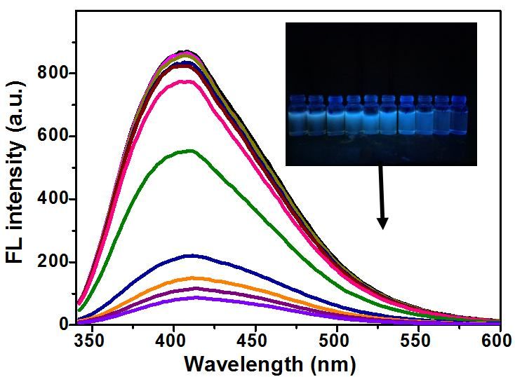
Tu et al.: High-sensitivity and selectivity detection of permanganate ions based on pig liver-based carbon quantum dots
- 7250 -
KMnO4 (potassium permanganate)). It also has certain oxidation in alkaline solution,
which is purple red in solution. The oxidation of potassium permanganate can be found
in both acidity and alkalinity, but it is more obvious in acidity. In recent years, many
methods of manganese detection have been developed, including catalytic kinetic
spectrophotometric for the determination of trace manganese, but there are few methods
for the determination of permanganate ions. A new fluorescence method for the
determination of trace manganese was established (Xu et al., 2004), where the
concentration range of MnO4 - is 1.0~2.4 mg/L, the maximum excitation wavelength is
278 nm, the maximum emission wavelength is 616 nm, and the fluorescence intensity
and concentration show a good linear relationship by transforming the manganese into
MnO4-. The results obtained in this method were satisfactory.
Carbon quantum dots (CQDs) are one of the most popular carbon nanomaterials after
fullerene, carbon nanotubes and graphene. They were first discovered in 2004 (Wang
and Hu, 2014) when electrophoresis was used to purify single-walled carbon nanotubes.
They have attracted great attention due to their excellent optical properties, such as
water solubility, chemical inertia, low toxicity, easy functionalization (Li et al., 2012)
and high fluorescence intensity, photobleaching resistance and adjustable luminescence
color. They have been widely used in detection of metal ions (Lu et al., 2012), anions
(Zhao et al., 2011), small organic molecules (Li et al., 2013) and biological molecules
(Deng et al., 2014). The method of detecting metal ions or other environmental
pollutants using CQDs as a fluorescent probe has received extensive attention due to its
excellent sensitivity and selectivity (Clarkson and Magos, 2006). Zhang and Chen
(2014) used folic acid as carbon source and nitrogen source to trace mercury ions by
nitrogen-doped carbon dots, and the detection limit was 0.23 mol/L.
In this study, fluorescent carbon quantum dots (CQDs) were successfully prepared
by simple one-step hydrothermal method using pig liver as a carbon source. Since pig
liver is a complex that contains a number of organics and biomolecules including fat,
proteins, vitamin B, vitamin C, vitamin E, carbohydrates, cholesterol and minerals, it
can be useful for doping of multiple hetero atoms in the CQDs without addition of any
additives. In the experiment, we found that MnO4- could quench the fluorescence of
CQDs, and the quenching degree was related to the concentration of MnO4 -. To our
knowledge, there is no report used CQDs fluorescent probe from pig liver for detection
of MnO4- yet, which provides a new method for sensitive detection of MnO4 -. In
addition, this method has been successfully applied to detecting MnO4 - in
environmental water and soil samples.
Experiment
Instruments and reagents
Pig liver was purchased from a local supermarket in Kunming (Yunnan, China); all
analytical reagents, including potassium permanganate (KMnO4), citric acid, disodium
hydrogen phosphate (Na2HPO4), FeCl3•6H2O and FeCl2 •4H2O, sodium nitrite, sodium
nitrate, KI, NaCl, NaF, copper sulfate, aluminum sulfate, calcium chloride, MnSO4,
BaCl2, MgCl2 and sodium sulfide (NaS) were purchased from Shanghai Aladdin
Biochemical Technology Co., Ltd. (Shanghai, China); and all solutions were prepared
from Milli-Q system ultrapure water (18.2 MΩ.cm, 25 °C).
G9800A fluorospectrophotometer (Agilent Technologies, USA) was used to
determine the fluorescence spectrum and its intensity; UV-2600 spectrophotometer
APPLIED ECOLOGY AND ENVIRONMENTAL RESEARCH 17(4): 7249-7263.
http://www.aloki.hu ● ISSN 1589 1623 (Print) ● ISSN 1785 0037 (Online)
DOI: http://dx.doi.org/10.15666/aeer/1704_72497263
2019, ALÖKI Kft., Budapest, Hungary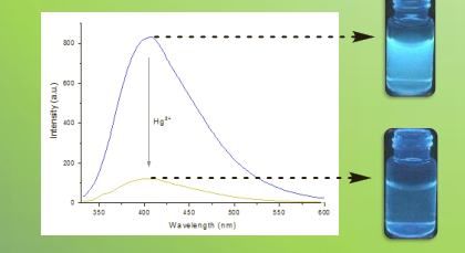
Tu et al.: High-sensitivity and selectivity detection of permanganate ions based on pig liver-based carbon quantum dots
- 7251 -
(Shimadzu, Japan) was used to determine UV spectra; Tecnai G2 F30S-Twin high
resolution field emission transmission electron microscope (FEI, Netherlands) was used
to detect the particle size and morphology of fluorescent quantum dots; TENSOR 27
Fourier transform infrared spectrometer (Bruker, Germany) was used to determine the
infrared spectrum and the structure of the material; D8-advance X-ray powder
diffractometer (Bruker, Germany) was used to determine the crystal structure
morphology; Thermo Scientific K-Alpha X-ray photoelectron spectrometer (Thermo
Fisher Scientific Inc. U.S.A.) was used to analyze the elemental composition ratio and
chemical oxidation state; XH-B vortex instrument (Shanghai Hanuo Instrument Co.,
Ltd., China) was used for vortex mixing; and 80-2 high speed centrifuge (Shanghai
Surgical Instrument Factory, China) was used for centrifugal filtration.
Preparation of pig liver-based CQDs
The pig liver was dried naturally at room temperature and grinded into powders in a
mortar. The CQDs of pig liver were prepared by simple hydrothermal method using pig
liver powder as carbon source. Five grams of pig liver powder was dispersed in 150 mL
deionized water. Then, the mixture was transferred into a high-pressure autoclave
(200 mL) lined with polytetrafluoroethylene (PTFE), heated at 180 °C for 10 h, cooled
naturally to room temperature, and centrifuged at 13,000 rpm for 20 min to obtain
bright yellow solution, and filtered to remove insoluble substances. In order to obtain
pure pig liver-based CQDs, the solution was filtered by 0.22 μm membrane. Finally, the
obtained pig liver CQDs were stored at 4 °C for further use. Compared with other
synthetic methods, the hydrothermal synthesis of pig liver CQDs is simpler and more
feasible. In addition, pig liver as raw material for synthesis is cheap and easily available,
and this method is greener and more environmentally friendly.
Fluorescence quenching test for permanganate
Typically, 50 μL of prepared pig liver-based CQDs solution and 1 mL of pH 7.0
citric acid-disodium hydrogen phosphate buffer solution mixed and diluted to 3.0 mL
with deionized water were placed in a 10 mL glass centrifuge tube. Then, 2mL
permanganate solution or sample with a different concentration was added to the
centrifuge tube and the vortex was mixed with 30 s. After 3 min, the fluorescence
intensity was measured at excitation wavelength 278 nm and emission wavelength
616 nm on the fluorescence spectrophotometer, and the fluorescence intensity was
recorded. The slit width of excitation and emission was 5 nm. In order to evaluate the
effect of coexisting interfering substances on the fluorescence intensity of pig liver-
based CQDs and the selectivity of permanganate and other substances or ions (including
FeCl3•6H2O and FeCl2•4H2O, sodium nitrite, sodium nitrate, KI, NaCl, NaF, copper
sulfate, aluminum sulfate, calcium chloride, MnSO4, BaCl2, MgCl2 and sodium sulfide),
the fluorescence quenching test was carried out under the same experimental conditions.
The schematic diagram of detection of MnO4 - with pig liver-based CQDs was shown in
Figure 1.
APPLIED ECOLOGY AND ENVIRONMENTAL RESEARCH 17(4): 7249-7263.
http://www.aloki.hu ● ISSN 1589 1623 (Print) ● ISSN 1785 0037 (Online)
DOI: http://dx.doi.org/10.15666/aeer/1704_72497263
2019, ALÖKI Kft., Budapest, Hungary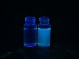
Tu et al.: High-sensitivity and selectivity detection of permanganate ions based on pig liver-based carbon quantum dots
- 7252 -
MnO4-
M
MnO4-
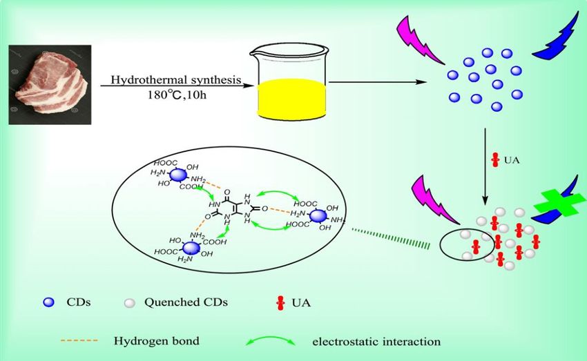
Figure 1. Processing diagram for the synthesis and application of CQDs
Collection and pretreatment of environmental samples
In this study, water samples were collected directly from our laboratory, Laoyuhe
Lake and the domestic wastewater of the community next to a school. All water samples
were filtered by simple centrifugal filtration to remove solid particles and suspended
solids and then by 0.22 μm microporous membrane, and stored at 4 °C. The
permanganate ion was added to the samples at a concentration of 0.01 mg/mL for
spiked fluorescence analysis.
Soil samples were collected from the road outside the school after rain and dried
under suitable environmental conditions (final moisture content 0.32%). One milliliter
of methanol solution containing different concentrations of permanganate ions was
added to 200 mg soil, mixed for 1 h on a 50 rpm shaker, and then allowed to stand
overnight at room temperature (RT). On the second morning, the mixture was again
vortexed and centrifuged for 2 min at 5,000 rpm and the supernatant was used for
fluorescence detection.
Results and discussion
Characterization of the CQDs
The transmission electron microscopy (TEM) technique was used to explore the
morphology and particle size distributions of CQDs. As shown in Figure 2, the CQDs
present high dispersity, uniform spherical shapes, and a size distribution within the
range of 3–10 nm and an average diameter of about 5.0 nm. The surface chemistry of
CQDs was studied using Fourier transform infrared (FT-IR) spectroscopy. As shown in
Figure 3, the strong absorption peak at 1469 cm-1 reveals the existence of -COOH
stretching vibrations; the peak at 1652 cm-1 reveals the existence of O = C-NH
stretching vibrations; and the absorption peak at 3190 cm-1 displays O-H stretching
vibrations. As shown in Figure 4, the full scan XPS spectra present distinct peaks at
APPLIED ECOLOGY AND ENVIRONMENTAL RESEARCH 17(4): 7249-7263.
http://www.aloki.hu ● ISSN 1589 1623 (Print) ● ISSN 1785 0037 (Online)
DOI: http://dx.doi.org/10.15666/aeer/1704_72497263
2019, ALÖKI Kft., Budapest, HungaryTu et al.: High-sensitivity and selectivity detection of permanganate ions based on pig liver-based carbon quantum dots
- 7253 -
287.3 and 532.7 eV, which are attributed to C1s and O1s, respectively. The result of
XPS diagram was consistent with that of FT-IR analysis. Therefore, there should be
hydrophilic groups on the surface of pig liver-based CQDs, such as -COOH, -OH, etc.,
which have good water solubility and broad application prospects.
Figure 2. TEM image (inset: HR-TEM image) of the synthesized CQDs
Transmittance(%)
COOH O-H
O=C-NH
0 500 1000 1500 2000 2500 3000 3500 4000 4500
Wavenumber(cm-1)
Figure 3. FT-IR spectrum of pig liver-based CQDs
APPLIED ECOLOGY AND ENVIRONMENTAL RESEARCH 17(4): 7249-7263.
http://www.aloki.hu ● ISSN 1589 1623 (Print) ● ISSN 1785 0037 (Online)
DOI: http://dx.doi.org/10.15666/aeer/1704_72497263
2019, ALÖKI Kft., Budapest, HungaryTu et al.: High-sensitivity and selectivity detection of permanganate ions based on pig liver-based carbon quantum dots
- 7254 -
O1s
C1s
Intensity(a.u.)
0 200 400 600 800 1000 1200 1400
Binding energy(eV)
Figure 4. XPS spectra of pig liver-based CQDs
In order to study the optical properties of pig liver-based CQDS, the pig liver-based
CQDs were characterized by UV-vis absorption spectrum and fluorescence emission
spectrum. The UV-vis absorption spectrum (UV-vis) of pig liver-based CQDs is shown
in Figure 5. The prepared pig liver-based CQDs had a weak absorption peak around
258 nm and an excitation wavelength of 278 nm at the maximum emission wavelength
of 616 nm, as shown in Figure 6, a fluorescence spectrum (FS) and an excitation-
dependent emission spectrum of pig liver-based CQDs. The results showed that when
the excitation wavelength was changed from 300 to 360 nm in increments of 10 nm, the
emission peak shifted toward the long wavelength direction.
2.5 MnO4-
猪肝基CQDs
2.0
Absorbance
1.5
1.0
258nm
0.5
0.0
200 300 400 500 600 700 800
Wavelength(nm)
Figure 5. UV-Vis absorption spectra of the synthesized pig liver-based CQDs. (Inset:
photographs of pig liver-based CQDs under daylight (left) and UV (365 nm) irradiation (right))
APPLIED ECOLOGY AND ENVIRONMENTAL RESEARCH 17(4): 7249-7263.
http://www.aloki.hu ● ISSN 1589 1623 (Print) ● ISSN 1785 0037 (Online)
DOI: http://dx.doi.org/10.15666/aeer/1704_72497263
2019, ALÖKI Kft., Budapest, HungaryTu et al.: High-sensitivity and selectivity detection of permanganate ions based on pig liver-based carbon quantum dots
- 7255 -
1000
300 nm
310 nm
800
FL intensity (a.u.)
320 nm
330 nm
600 340 nm
350 nm
400 360 nm
200
0
350 400 450 500 550 600
Wavelength (nm)
Figure 6. Fluorescence emission spectra of pig liver-based CQDs at different excitation
wavelengths
Light stability test of pig liver-based CQDs
In order to study the stability of pig liver-based CQDs in detecting permanganate,
the fluorescence intensity of pig liver-based CQDs at different storage time was studied.
As shown in Figure 7A, the fluorescence intensity of the pig liver based-CQDs is
substantially constant. In addition, the reaction time increased while the fluorescence
intensity of the experimental system remained constant, indicating that the storage time
had no effect on the fluorescence intensity of pig liver-based CQDs. The effects of ionic
strength and pH on the fluorescence intensity of pig liver-based CQDs were also
studied, as shown in Figure 7B and C. It is observed from the figures that with the
gradual increase of the concentration of NaCl solution, the fluorescence intensity only
slightly fluctuated without significant change; the pH of the citric acid-dibasic sodium
phosphate buffer changed from 2.0 to 8.0; and the fluorescence intensity of pig liver-
based CQDs remain unchanged, this demonstrates that pig liver-based CQDs can be
used as fluorescent nanoprobe in any environmental conditions.
APPLIED ECOLOGY AND ENVIRONMENTAL RESEARCH 17(4): 7249-7263.
http://www.aloki.hu ● ISSN 1589 1623 (Print) ● ISSN 1785 0037 (Online)
DOI: http://dx.doi.org/10.15666/aeer/1704_72497263
2019, ALÖKI Kft., Budapest, HungaryTu et al.: High-sensitivity and selectivity detection of permanganate ions based on pig liver-based carbon quantum dots
- 7256 -
D
10000
Pig liver-CQDs
8000 Pig liver-CQDs+MnO4-
6000
Counts
4000
2000
0
0 20 40 60 80 100
Time/ns
Figure 7. Effects of various conditions on the fluorescence intensity of pig liver-based CQDs:
(A) storage time; (B) NaCl concentration; (C) pH; (D) the Fluorescence lifetime decay curve
APPLIED ECOLOGY AND ENVIRONMENTAL RESEARCH 17(4): 7249-7263.
http://www.aloki.hu ● ISSN 1589 1623 (Print) ● ISSN 1785 0037 (Online)
DOI: http://dx.doi.org/10.15666/aeer/1704_72497263
2019, ALÖKI Kft., Budapest, HungaryTu et al.: High-sensitivity and selectivity detection of permanganate ions based on pig liver-based carbon quantum dots
- 7257 -
Selectivity
The selectivity of pig liver based-CQDs was investigated by monitoring the
fluorescence intensity of different possible interfering substances such as MnO4 -, Fe3+,
Fe2+, NO2, NO3, I-, Cl-, F-, Na+, K+, Cu2+, Al3+, Ca2+, Mn2+, Ba2+, Mg2+ and S2- in the
presence of different concentrations or ions. As shown in Figure 8, only permanganate
ion can effectively quench the fluorescence intensity of CQDs in pig liver, while the
quenching effect of other possible interfering substances is negligible. The results show
that there may be some quenching relationship between pig liver-based CQDs and
permanganate ions, which is worthy of further study. Therefore, there is a high
selectivity between permanganate ions and pig liver-based CQDs. This demonstrates
that pig liver-based CQDs can be used to detect permanganate ions.
1000
800
600
F
400
200
0
- - -
k - 3+ 2+ 2- 3- + +
2 3 + 2 +2 +
2 2+ + + 2-
BlanMnO 4 Fe Fe NO NO I F Cl Na K Cu Al Ca Mn Ba Mg S
Figure 8. Evaluation of method selectivity against possible interferents
Optimization of experimental conditions
PH affects not only the fluorescence intensity of pig liver-based CQDs, but also the
fluorescence quenching effect of permanganate ions on pig liver-based CQDs.
Therefore, the effect of pH from 2.2 to 8.0 on the experimental system was investigated.
As shown in Figure 9A, the fluorescence intensity of pig liver-based CQDs is related to
pH. In the range of pH2.0-7.0, the fluorescence intensity increased gradually and the
fluorescence quenching efficiency reached its maximum, but after pH 7.0, the
fluorescence intensity decreased gradually. The results show that the fluorescence
quenching response of the system is the largest at pH 7.0, according to which pH 70 is
the optimum pH value of citric acid-disodium hydrogen phosphate buffer.
In order to study the stability of pig liver-based CQDs in detecting permanganate
ions, the fluorescence intensity of pig liver-based CQDs in the presence of
permanganate ions at the same concentration under different reaction time was studied.
As shown in Figure 9B, the fluorescence intensity of pig liver based-CQDs decreased
sharply within 0-1 min and gradually within 1-10 min, then tended to be stable, and
reached the lowest fluorescence intensity in 2 min. Therefore, the reaction time of 2 min
was chosen as the optimal fluorescence quenching response time.
APPLIED ECOLOGY AND ENVIRONMENTAL RESEARCH 17(4): 7249-7263.
http://www.aloki.hu ● ISSN 1589 1623 (Print) ● ISSN 1785 0037 (Online)
DOI: http://dx.doi.org/10.15666/aeer/1704_72497263
2019, ALÖKI Kft., Budapest, HungaryTu et al.: High-sensitivity and selectivity detection of permanganate ions based on pig liver-based carbon quantum dots
- 7258 -
Figure 9. Effect of pH (A) and (B) reaction time on the fluorescence intensity of the pig liver-
based CQDs
Method performance study
Under the optimal experimental conditions, the relationship between the fluorescence
quenching intensity of pig liver-based CQDs and the concentration of MnO4 - was
studied, and the quantitative analysis method for fluorescence quenching of MnO4- was
established. As shown in Figure 10, the fluorescence intensity of pig liver-based CQDs
decreases gradually with the addition of MnO4 - of different concentrations, and shows
regular changes. As shown in Figure 11, the ratio of fluorescence intensity of the
system was linear with the concentration of MnO4 - in the range of 0-50 μM both before
and after addition of pig liver-based CQDs. The regression equation is F/F0 = 0.0151C
– 0.0112 (R2 = 0.9967), where F and F0 are the fluorescence intensity of pig liver-based
CQDs in the presence or absence of MnO4- respectively and C is the concentration of
MnO4-. The detection limit of MnO4 - was 0.06 μM and the signal-noise ratio was
3(S/N).
APPLIED ECOLOGY AND ENVIRONMENTAL RESEARCH 17(4): 7249-7263.
http://www.aloki.hu ● ISSN 1589 1623 (Print) ● ISSN 1785 0037 (Online)
DOI: http://dx.doi.org/10.15666/aeer/1704_72497263
2019, ALÖKI Kft., Budapest, HungaryTu et al.: High-sensitivity and selectivity detection of permanganate ions based on pig liver-based carbon quantum dots
- 7259 -
Figure 10. Fluorescence response of pig liver-based CQDs upon addition of different
concentrations of MnO4- (concentration gradient of pig liver based-CQDs under ultraviolet
light)
0.8
0.7 F/F0=0.0151C-0.0112
0.6 R2=0.9967
0.5
0.4
F/F0
0.3
0.2
0.1
0.0
-0.1
0 10 20 30 40 50
-
Concentration of MnO4 (μM)
Figure 11. Linear correlation of F0/F values versus the concentration of MnO4- over the range
from 0.1 to 50 μM
Possible fluorescence quenching mechanism
In recent years, many sensors have been established based on the fluorescence
variation of carbon quantum dots. These sensors were primarily analytes that quenched
or increased the fluorescence of the carbon dots (Mariappan et al., 2017). Theoretically,
this fluorescence phenomenon is related to different mechanisms. For fluorescence
APPLIED ECOLOGY AND ENVIRONMENTAL RESEARCH 17(4): 7249-7263.
http://www.aloki.hu ● ISSN 1589 1623 (Print) ● ISSN 1785 0037 (Online)
DOI: http://dx.doi.org/10.15666/aeer/1704_72497263
2019, ALÖKI Kft., Budapest, HungaryTu et al.: High-sensitivity and selectivity detection of permanganate ions based on pig liver-based carbon quantum dots
- 7260 -
quenching, the mechanism is usually divided into dynamic quenching and static
quenching, caused by the collision between fluorescent materials and quencher and the
formation of ground state complexes respectively (Kaviyarasu et al., 2017). In case of
dynamic quenching, the Stern-Volmer equation can be used for analysis (Guo et al.,
2015) with the following formula:
F0/F = 1 + Kqτ0[Q] = 1 + KSV[Q]
where: F0 and F indicate the fluorescence intensity before and after the addition of
MnO4- respectively, Kq is the bimolecular quenching constant, τ0 is the average lifetime
of CQDs, generally 10-8 s, KSV is the Stern-Walmer quenching constant, and [Q] was
the concentration of MnO4 -. Therefore, the fluorescence quenching process may be
caused by static quenching, i.e. the formation of ground state complexes in the reaction
system (Lakowicz, 2006; Sun et al., 2015; Formica et al., 2012). The absorption
strength of CQDs increased and the peak shifted to blue after addition of MnO4 -,
indicating that the interaction between MnO4- and CQDs produced the ground state
complex. According to almost constant fluorescence lifetime, the fluorescence
quenching caused by MnO4- may be static quenching due to the formation of stable non-
fluorescent complexes (Zuo et al., 2016; Sun and Lei, 2017; Wu et al., 2011; Liu et al.,
2017, 2014). FT-IR and XPS confirmed the presence of -COOH, -OH, etc. on the
surface of CQDs, indicating good water solubility and good application prospect of the
synthesized CQDs.
Analysis of MnO4- in real samples
The above mentioned environmental water and soil samples were pretreated, and
then MnO4 - in environmental water and soil samples was detected by prepared pig liver-
based CQDs. The environmental water samples and soil samples were spiked and
recovered using different concentrations of MnO4 - and pretreated according to the same
procedure. As shown in Table 1, the recovery of MnO4 - in water samples is
94.2~102.5% and the relative standard deviations (RSD) of all samples are less than
5.8%. As shown in Table 2, the recovery of MnO4 - in soil samples is 90.5 to 99.5% and
the relative standard deviations (RSD) of all samples are below 4.7%. Therefore, this
method has the potential for detecting MnO4 - in environmental water samples and soil
samples.
Table 1. Analytical results of MnO4- in the water samples (n = 3)
Spiked Found Recovery RSD
Samples
(μg/mL) (μg/mL) (%) (%, n = 3)
0 ND - -
Tap water 5 4.71 94.2 4.3
20 19.3 96.5 3.6
0 ND - -
Lake water 5 4.92 98.4 5.8
20 20.5 102.5 2.5
0 ND - -
Waste water 5 5.10 102.0 5.3
20 20.05 100.3 4.7
ND = not detected
APPLIED ECOLOGY AND ENVIRONMENTAL RESEARCH 17(4): 7249-7263.
http://www.aloki.hu ● ISSN 1589 1623 (Print) ● ISSN 1785 0037 (Online)
DOI: http://dx.doi.org/10.15666/aeer/1704_72497263
2019, ALÖKI Kft., Budapest, HungaryTu et al.: High-sensitivity and selectivity detection of permanganate ions based on pig liver-based carbon quantum dots
- 7261 -
Table 2. Analytical results of MnO4- in the soil samples (n = 3)
Spiked Found Recovery RSD
Samples
(μg/mL) (μg/mL) (%) (%, n = 3)
0 ND - -
Sample 1 5 4.97 99.5 4.5
20 18.1 90.5 2.8
0 ND - -
Sample 2 10 9.58 95.8 3.5
20 18.34 91.7 3.1
ND = not detected
Conclusion
In summary, novel fluorescent CQDs were prepared from pig liver by hydrothermal
synthesis method and the fluorescence intensity of CQDs could be quenched by MnO4 -,
so the CQDs were used as nanosensors for the sensitive and selective detection of
MnO4- in environmental samples. For instance, morphology, particle size, crystal form,
chemical element composition and optical properties of the prepared pig liver-based
CQDs were characterized by TEM, FT-IR, XPS, UV-Vis and FS. The results showed
that it had good stability and fluorescence characteristics. In addition, a good linear
relationship was achieved between the fluorescence response of CQDs and the
concentration of MnO4 - in the range from 0.1 to 50 μM. The detection limit was
0.06 μM under optimized conditions. The developed method has been successfully
applied to the determination of MnO4 - in water samples and soils of environmental
samples. This method is green, environmental friendly, simple and easy to operate, and
shows great potential for application.
REFERENCES
[1] Bhunia, S. K., Saha, A., Maity, A. R., Ray, S. C., Jana, N. R. (2013): Carbon
nanoparticle-based fluorescent bioimaging probes. – Sci. Rep. 3: 1473.
[2] Clarkson, T. W., Magos, L. (2006): The toxicology of mercury and its chemical
compounds. – Crit. Rev. Toxicol. 36(8): 609-662.
[3] da Silva, E. G. P., do N. Santos, A. C., Costa, A. C. S., da N. Fortunato, D. M., José, N.
M., Korn, M. G. A., dos Santos, W. N. L., Ferreira, S. L. C. (2006): Determination of
manganese and zinc in powdered chocolate samples by slurry sampling using sequential
multi-element flame atomic absorption spectrometry. – Microchemical Journal 82(2):
159-162.
[4] Deng, X. Y., Li, J. Y., Tan, K. J. (2014): Rapid screening and confirmation of pesticide
residues in potato by high-resolution benchtop Q exactive LC-MS. – Chinese J. Anal.
Chem. 42(4): 579-584.
[5] Doroschuk, V. O., Lelyushok, S. O., Ishchenko, V. B., Kulichenko, S. A. (2004): Flame
atomic absorption determination of manganese (II) in natural water after cloud point
extraction. – Talanta 64(4): 853-856.
[6] Formica, M., Fusi, V., Giorgi, L., Micheloni, M. (2012): New fluorescent chemosensors
for metal ions in solution. – Coord. Chem. Rev. 256 170-192.
[7] Gunter, T. E., Miller, L. M., Gavin, C. E., Eliseev, R., Salter, J., Buntinas, L.,
Alexandrov, A., Hammond, S., Gunter, K. K. (2010): Determination of the oxidation
states of manganese in brain, liver, and heart mitochondria. – Journal of Neurochemistry
88(2): 266-280.
APPLIED ECOLOGY AND ENVIRONMENTAL RESEARCH 17(4): 7249-7263.
http://www.aloki.hu ● ISSN 1589 1623 (Print) ● ISSN 1785 0037 (Online)
DOI: http://dx.doi.org/10.15666/aeer/1704_72497263
2019, ALÖKI Kft., Budapest, HungaryTu et al.: High-sensitivity and selectivity detection of permanganate ions based on pig liver-based carbon quantum dots
- 7262 -
[8] Guo, Y., Zhang, L., Zhang, S., Yang, Y., Chen, X., Zhang, M. (2015): Fluorescent carbon
nanoparticles for the fluorescent detection of metal ions. – Biosens. Bioelectron. 63 61-
71.
[9] Hua, X.-W., Bao, Y.-W., Wang, H.-Y., Chen, Z., Wu, F.-G. (2017): Bacteria-derived
fluorescent carbon dots for microbial live/dead differentiation. – Nanoscale 9: 2150-2161.
[10] Jones, K. H., Senft, J. A. (1985): An improved method to determine cell viability by
simultaneous staining with fluorescein diacetate-propidium iodide. – J. Histochem.
Cytochem. 33: 77-79.
[11] Kaviyarasu, K., Kanimozhi, K., Matinise, N., Maria, C. Magdalane, Mola, G. T.,
Kennedy, J., Maaza, M. (2017): Antiproliferative effects on human lung cell lines A549
activity of cadmium selenide nanoparticles extracted from cytotoxic effects: Investigation
of bio-electronic application. – Mater. Sci. Eng. C. 76 1012-1025.
[12] Lakowicz, J. R. (2206): Principles of Fluorescence Spectroscopy. 3rd. Ed. – Springer,
Singapore.
[13] Li, H., Kong, W. Q., Liu, J., Liu, N. Y., Huang, H., Liu, Y., Kang, Z. H. (2015):
Fluorescent N-doped carbon dots for both cellular imaging and highly-sensitive catechol
detection. – Carbon 91: 66-75.
[14] Li, H. T., Kang, Z. H, Liu, Y. et al. (2012): Carbon nanodots: synthesis, properties and
applications. – J. Mater. Chem. 22(46): 24230-24253.
[15] Li, J. Z., Wang, N. Y., Tran, T. T. et al. (2013): Electrogenerated chemiluminescence
detection of trace level pentachlorophenol using carbon quantum dots. – Analyst 138(7):
2038-2043.
[16] Li, Z., Yu, H. J., Bian, T., Zhao, Y. F., Zhou, C., Shang, L., Liu, Y. H., Wu, L. Z., Tung,
C. H., Zhang, T. R. . (2015): Highly luminescent nitrogen-doped carbon quantum dots as
effective fluorescent probes for mercuric and iodide ions. – Mater. Chem. C 3: 1922-
1928.
[17] Liang, P., Sang, H., Sun, Z. (2006): Cloud point extraction and graphite furnace atomic
absorption spectrometry determination of manganese (II) and iron(III) in water samples. –
Journal of Colloid & Interface Science 304(2): 486-490.
[18] Lim, S. Y., Shen, W., Gao, Z. Q. (2015): – Chem. Soc. Rev. 44: 362.
[19] Liu, Y., Zhao, Y., Zhang, Y. (2014): One-step green synthesized fluorescent carbon
nanodots from bamboo leaves for copper(II) ion detection. – Sens. Actuators B 196: 647-
652.
[20] Liu, Y., Duan, W., Song, W., Liu, J., Ren, C., Wu, J., Liu, D., Chen, H. (2017): Red
emission B, N, S-co-doped carbon dots for colorimetric and fluorescent dual mode
detection of Fe3+ ions in complex biological fluids and living cells. – ACS Appl. Mat.
Interfaces 9(14): 12663-12672.
[21] Lu, W., Qin, X., Liu, S. et al. (2012): Economical, green synthesis of fluorescent carbon
nanoparticles and their use as probes for sensitive and selective detection of mercury(II)
ions. – Analytical Chemistry 84(12): 5351-5357.
[22] Mariappan, A., Kaviyarasu, K., Neyvasagam, K., Ayeshamariam, A., Pandi, P.,
Palanichamy, R. R., Gopinathan, C., Mola, G. T., Maaza, M. (2017): – Surf. Interface. 6
247-255.
[23] Ming, H., Ma, Z., Liu, Y., Pan, K. M., Yu, H., Wang, F., Kang, Z. H. (2012): Large scale
electrochemical synthesis of high quality carbon nanodots and their photocatalytic
property. – Dalton Trans. 41: 9526-9531.
[24] Nocker, A., Cheung, C. Y., Camper, A. K. (2006): Comparison of propidium monoazide
with ethidium monoazide for differentiation of live vs. dead bacteria by selective removal
of DNA from dead cells. – J. Microbiol. Methods 67(2): 310-320.
[25] Rask, J. H., Miner, B. A. Buseck, P. R. (2015): Determination of manganese oxidation
states in solids by electron energy-loss spectroscopy. – Ultramicroscopy 21(4): 321-326.
[26] Sun, J., Yang, S. W., Wang, Z. Y., Shen, H., Xu, T., Sun, L. T., Li, H., Chen, W. W.,
Jiang, X. Y., Ding, G. Q., Kang, Z. H., Xie, X. M., Jiang, M. H. (2015): Ultra-high
APPLIED ECOLOGY AND ENVIRONMENTAL RESEARCH 17(4): 7249-7263.
http://www.aloki.hu ● ISSN 1589 1623 (Print) ● ISSN 1785 0037 (Online)
DOI: http://dx.doi.org/10.15666/aeer/1704_72497263
2019, ALÖKI Kft., Budapest, HungaryTu et al.: High-sensitivity and selectivity detection of permanganate ions based on pig liver-based carbon quantum dots
- 7263 -
quantum yield of graphene quantum dots: aromatic-nitrogen doping and
photoluminescence mechanism. – Part. Part. Syst. Charact. 32: 434-440.
[27] Sun, X., Lei, Y. (2017): Fluorescent carbon dots and their sensing applications. – Trends
in Analytical Chemistry 89 163-180.
[28] Sun, X., Wang, Y., Lei, Y. (2015): Fluorescence based explosive detection: from
mechanisms to sensory materials. – Chem. Soc. Rev. 44 8019-8061.
[29] Teo, K. C., Chen, J. (2001): Determination of manganese in water samples by flame
atomic absorption spectrometry after cloud point extraction. – Analyst 126(4): 534.
[30] Wang, Y. F., Hu, A. G. (2014): Carbon quantum dots: synthesis, properties and
applications. – J. Mater. Chem. C 2(34): 6921-6939.
[31] Wu, J., Liu, W., Ge, J., Zhang, H., Wang, P. (2011): New sensing mechanisms for design
of fluorescent chemosensors emerging in recent years. – Chem. Soc. Rev. 40: 3483-3495.
[32] Xu, X. Y., Robert, R., Gu, Y. L. et al. (2004): Electrophoretic analysis and purification of
fluorescent single-walled carbon nanotube fragments. – J. am. Chem. Soc. 126(40):
12736-12737.
[33] Yu, H. J., Shi, R., Zhao, Y. F., Waterhouse, G. I. N., Wu, L. Z., Tung, C. H., Zhang, T. R.
(2016): Nitrogen-doped porous carbon nanosheets templated from g-C3 N4 as metal-free
electrocatalysts for efficient oxygen reduction reaction. – Adv. Mater. 28(25): 9454-9477.
[34] Zeng, C., Qin, P., Lan, L., Wei, H., Wu, W. (2017): Chemical vapor generation coupled
with atomic fluorescence spectrometry for the determination of manganese in food
samples. – Microchemical Journal 131: 31-35.
[35] Zhang, R., Chen, W. (2014): Nitrogen-doped carbon quantum dots: facile synthesis and
application as a “turn-off” fluorescent probe for detection of Hg2+ ions. – Biosensors and
Bioelectronics 55: 83-90.
[36] Zhao, H. X., Liu, L. Q., Liu, Z. D. et al. (2011): Highly selective detection of phosphate
in very complicated matrixes with an off-on fluorescent probe of europium-adjusted
carbon dots. – Chem. Commun. 47(9): 2604-2606.
[37] Zuo, P., Lu, X., Sun, Z., Guo, Y., He, H. (2016): A review on syntheses, properties,
characterization and bioanalytical applications of fluorescent carbon dots. – Microchim.
Acta 183 519-542.
APPLIED ECOLOGY AND ENVIRONMENTAL RESEARCH 17(4): 7249-7263.
http://www.aloki.hu ● ISSN 1589 1623 (Print) ● ISSN 1785 0037 (Online)
DOI: http://dx.doi.org/10.15666/aeer/1704_72497263
2019, ALÖKI Kft., Budapest, HungaryYou can also read