Hypokalaemic rhabdomyolysis as initial presentation of primary aldosteronism - Hypokalaemic rhabdomyolysis as initial presentation ...
←
→
Page content transcription
If your browser does not render page correctly, please read the page content below
J R Coll Physicians Edinb 2021; 51: 149–52 | doi: 10.4997/JRCPE.2021.211                                              CASE REPORT

Clinical
Hypokalaemic rhabdomyolysis as initial
presentation of primary aldosteronism
Aye Chan Maung1, Ann Kwee Kerwen2, Lee Phong Ching3

                   Rhabdomyolysis is a clinical syndrome characterised by the destruction of            Correspondence to:
                   skeletal muscle with subsequent leakage of intracellular muscle contents             Aye Chan Maung
                                                                                                        Department of
 Abstract into          the systemic circulation. It is commonly caused by trauma, strenuous
                   physical activities, medications, illicit drug use, toxins, infections and           Endocrinology
                   electrolyte abnormalities. It can manifest as myalgia, muscle weakness,              Singapore General Hospital
                   or myoglobinuria with or without acute kidney injury. Severe hypokalaemia            Academia, Level 3
leading to rhabdomyolysis is a rare initial presentation of primary aldosteronism, with limited         20 College Road
case reports in existing medical literature. Here, we report a case of primary hyperaldosteronism       Singapore 169856
presenting with rhabdomyolysis due to profound hypokalaemia.
                                                                                                        Email:
Keywords: rhabdomyolysis, hypokalaemia, primary aldosteronism                                           chanmaung.aye@mohh.
                                                                                                        com.sg
Financial and Competing Interests: No conflict of interests declared.

Informed Consent: Written informed consent for the paper to be published (including
images, case history and data) was obtained from the patient/guardian for publication of
this paper, including accompanying images.

Introduction                                                         hypertension which was diagnosed two years earlier. She was
                                                                     on amlodipine 5 mg once daily, without the use of diuretics
Rhabdomyolysis is a clinical syndrome characterised by acute         or over the counter medications.
damage of the skeletal muscle leading to the release of
potentially toxic muscle cell constituents such as creatine          At presentation, her blood pressure (BP) was 150/75 mmHg,
kinase (CK) and myoglobin into the systemic circulation. The         heart rate 88 beats/minute and SpO2 99% on room air.
common causes of rhabdomyolysis include intense exercise,            Her height was 160 cm and weight 62 kg, giving her a body
trauma, drugs (e.g. statins), infections, electrolyte disorders      mass index of 24.2 kg/m2. On neurological exam, proximal
(e.g. hypokalaemia, hypophosphataemia) and very rarely,              weakness (Medical Research Council grading 3/5) was
genetic defects in muscle enzymes or channelopathies.                detected in upper and lower limbs bilaterally. There were no
This can result in acute kidney injury due to renal tubular          features of Cushing’s syndrome. She was clinically euthyroid
blockage by the released myoglobin. Profound hypokalaemia            without a goitre or signs of thyroid eye disease. Systemic
can trigger rhabdomyolysis, which can present with muscle            examination was unremarkable.
cramps, weakness and fatigability. Rhabdomyolysis due
to hypokalaemia is a rare initial presentation of primary            Initial investigations performed at the Emergency Department
hyperaldosteronism (PA). Patients with PA usually present            revealed profound hypokalaemia (potassium 2.1 mmol/L),
with hypertension and may have biochemical abnormalities             hypomagnesaemia (magnesium 0.49 mmol/L) and metabolic
such as hypokalaemia secondary to kaliuresis or metabolic            alkalosis (bicarbonate 35.1 mmol/L). Her CK and urine
alkalosis. The diagnosis is confirmed with evidence of               myoglobin were significantly elevated (Table 1). She was
autonomous aldosterone production with suppressed renin              diagnosed with rhabdomyolysis based on the findings of
concentration or activity.                                           elevated serum CK and urine myoglobin levels, together
                                                                     with proximal muscle weakness. She denied any acute
Case presentation                                                    infections, trauma or intoxication. She was not taking any
                                                                     statin or fibrate. There were no signs of autoimmune disease
A 42-year-old Filipino lady presented with progressive proximal      or dermatomyositis.
upper and lower limb weakness for two weeks. She had
difficulty in climbing stairs and standing up from a squatting       Her serum potassium in 2017 was normal (4.1 mmol/L).
position. The weakness gradually progressed, resulting in            There was no history of vomiting or diarrhoea to suggest
difficulty ambulating on flat ground. She had a history of           gastrointestinal loss of potassium. She was not given

1
 Senior Resident, 2Associate Consultant, 3Consultant, Department of Endocrinology, Singapore General Hospital, Singapore

               50TH ANNIVERSARY YEAR    JUNE 2021 VOLUME 51 ISSUE 2 JOURNAL OF THE ROYAL COLLEGE OF PHYSICIANS OF EDINBURGH   149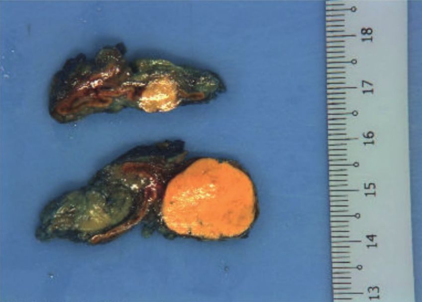
AC Maung, AK Kerwen, LP Ching

Table 1 Initial investigations performed on admission
                                                                         potassium was also significantly elevated at 86 (10–44
 Blood test                   Results       Reference range              mmol/day). Hypokalaemia with renal potassium wasting,
 Urea                         4.4           2.7–6.9 mmol/L               metabolic alkalosis and history of hypertension suggest the
 Sodium                       140           136–146 mmol/L               possibility of mineralocorticoid excess, and hence screening
                                                                         for PA was carried out.
 Potassium                    2.1           3.6–5.0 mmol/L
 Chloride                     87            100–107 mmol/L
                                                                         Her plasma aldosterone concentration (PAC) was 424.6
 Bicarbonate                  35.1          19–29 mmol/L                 (97.3–834 pmol/L) and direct renin concentration was 1.3
 Glucose                      5.8           3.9–11.0 mmol/L              (upright: 1.8–59.4 ng/mL, supine: 1.1–20.2 ng/mL), with
 Creatinine                   83            37–75 μmol/L                 an aldosterone renin ratio (ARR) of 11.8. The corresponding
 Magnesium                    0.49          0.74–0.97 mmol/L             serum potassium was 3.3 mmol/L. ARR of greater than or
                                                                         equal to 3.8 is suggestive of a positive case detection for
 Creatine kinase              1,579         44–201 U/L
                                                                         PA.1 ARR is expected to be higher if it would have been sent
 Urine myoglobin              179
Hypokalaemic rhabdomyolysis
Figure 2 Macroscopic appearance of two left adrenal nodules
showing well-circumscribed and encapsulated with a typical golden and heart failure.10 Moreover, PA also significantly increased
yellow appearance the risk of atherogenic conditions such as diabetes mellitus
and metabolic syndrome.10
Given our patient’s young age, and the presence of unilateral
aldosterone-producing adenoma (APA), laparoscopic
adrenalectomy would be the treatment of choice as it offers
the possibility of a cure. It is the preferred surgical approach
and is associated with a shorter recovery time, decreased
length of stay and fewer postoperative complications
compared to open approach.1 Alternatively, medical therapy
would involve a lifetime of medications and monitoring.
Recent studies have shown that adrenalectomy is superior
to medical therapy in relation to cardiovascular and renal
outcomes. In a large prospective study, medically treated PA
patients had a higher incidence of atrial fibrillation compared
with adrenalectomised PA patients during the 12-year follow-
up period.11 Another study showed that arterial stiffness was
Discussion reduced following adrenalectomy, but not after one year of
Rhabdomyolysis presenting with severe hypokalaemia spironolactone treatment for PA.12 Patients with unilateral APA
as the first manifestation of PA is extremely rare. Severe had reduced progression to end-stage renal failure and lower
muscle weakness or rhabdomyolysis usually occurs only if mortality than hypertensive controls after adrenalectomy.
serum potassium is below 2.5 mmol/L To the best of our Mineralocorticoid receptor antagonist did not significantly
knowledge, there are 16 cases of rhabdomyolysis caused alter those outcomes in a population database study of
by hypokalaemia from PA reported in the literature thus far.2 PA patients from Taiwan.13 In relation to the resolution
Hypokalaemia is present in only 9–37% of patients with PA.1 of hypertension, surgical but not medical treatment was
PA associated with hypokalaemia-induced rhabdomyolysis is significantly associated with amelioration of hypertension
more common in Asians, as seen in our patient.3 among those with APA in a Japanese epidemiological study.14
Female gender, fewer than or equal to two anti-hypertensive
Multiple epidemiological studies have shown that PA has medications, duration of hypertension less than or equal to
a prevalence of >5% (possibly even >10%) in hypertensive six years and a body mass index less than or equal to 25
patients, both in general and specialty settings.4 PA occurs kg/m2 were identified as the best predictors of complete
in ~5% of the adult hypertensive patients in Singapore.5 The resolution of hypertension post-adrenalectomy.15 Our patient
prevalence of PA is increased in relation to the severity of fulfilled all this criteria and she achieved normalisation of BP
the hypertension.6 PA was present in only 2% of patients with after adrenalectomy.
mild hypertension (AC Maung, AK Kerwen, LP Ching
References
1 Funder JW, Carey RM, Mantero F et al. The management 9 Milliez P, Girerd X, Plouin P-F et al. Evidence for an increased
of primary aldosteronism: case detection, diagnosis, and rate of cardiovascular events in patients with primary
treatment: an endocrine society clinical practice guideline. J aldosteronism. J Am Coll Cardiol 2005; 45: 1243–8.
Clin Endocrinol Metab 2016; 101: 1889–916. 10 Monticone S, D’Ascenzo F, Moretti C et al. Cardiovascular
2 Martínez JJA, Oliveira CL, Meneses AL et al. Rhabdomyolysis events and target organ damage in primary aldosteronism
due to primary hyperaldosteronism. Endocrinol Nutr 2009; 56: compared with essential hypertension: a systematic review
431–4. and meta-analysis. Lancet Diabetes Endocrinol 2018; 6:
3 Ma JT, Wang C, Lam KS et al. Fifty cases of primary 41–50.
hyperaldosteronism in Hong Kong Chinese with a high 11 Rossi GP, Maiolino G, Flego A et al. Adrenalectomy lowers
frequency of periodic paralysis. Evaluation of techniques for incident atrial fibrillation in primary aldosteronism patients at
tumour localisation. Q J Med 1986; 61: 1021–37. long term. Hypertension 2018; 71: 585–91.
4 Rossi GP, Bernini G, Caliumi C et al. A prospective study of the 12 Strauch B, Petrák O, Zelinka T et al. Adrenalectomy improves
prevalence of primary aldosteronism in 1,125 hypertensive arterial stiffness in primary aldosteronism. Am J Hypertens
patients. J Am Coll Cardiol 2006; 48: 2293–300. 2008; 21: 1086–92.
5 Loh KC, Koay ES, Khaw MC et al. Prevalence of primary 13 Chen Y-Y, Lin Y-HH, Huang W-C et al. Adrenalectomy improves
aldosteronism among Asian hypertensive patients in the long-term risk of end-stage renal disease and mortality of
Singapore. J Clin Endocrinol Metab 2000; 85: 2854–9. primary aldosteronism. J Endocr Soc 2019; 3: 1110–26.
6 Monticone S, Burrello J, Tizzani D et al. Prevalence and clinical 14 Miyake Y, Tanaka K, Nishikawa T et al. Prognosis of
manifestations of primary aldosteronism encountered in primary aldosteronism in Japan: results from a nationwide
Primary Care Practice. J Am Coll Cardiol 2017; 69: 1811–20. epidemiological study. Endocr J 2014; 61: 35–40.
7 Mosso L, Carvajal C, González A et al. Primary aldosteronism 15 Zarnegar R, Young WF, Lee J et al. The aldosteronoma
and hypertensive disease. Hypertension 2003; 42: 161–5. resolution score: predicting complete resolution of
8 Rossi GP. Does primary aldosteronism exist in normotensive hypertension after adrenalectomy for aldosteronoma. Ann Surg
and mildly hypertensive patients, and should we look for it? 2008; 247: 511–8.
Hypertens Res 2011; 34: 43–6.
152 JOURNAL OF THE ROYAL COLLEGE OF PHYSICIANS OF EDINBURGH VOLUME 51 ISSUE 2 JUNE 2021 50TH ANNIVERSARY YEARYou can also read