In silico analysis of Naegleria fowleri cathepsin B paralogs: important drug targets
←
→
Page content transcription
If your browser does not render page correctly, please read the page content below
European Review for Medical and Pharmacological Sciences                         2021; 25: 3162-3172

In silico analysis of Naegleria fowleri cathepsin
B paralogs: important drug targets
M. AURONGZEB1,3, S.A. HAQ2, Y. RASHID2, S.H. AHMED NAQVI3, S.I. HUSSAIN4,
A.U. REHMAN4, I. KALEEM6, S. BASHIR7
1
 Jamil ur Rehman Center for Genome Research, Dr. Punjwani Center for Molecular Medicine and
 Drug Research, International Center for Chemical and Biological Sciences, University of Karachi,
 Karachi, Pakistan
2
 Department of Biochemistry, University of Karachi, Karachi, Pakistan
3
  Institute of Biotechnology and Genetic Engineering, University of Sindh, Jamshoro, Pakistan
4
  Department of Zoology, Federal Urdu University of Arts, Science & Technology, Gulshan-e-Iqbal,
 Karachi, Pakistan
5
  Department of Biochemistry, Abdul Wali khan University Mardan, Mardan, Khyber Pakhtunkhwa,
 Pakistan
6
  Department of Bioinformatics and Bioscience, COMSATS University (CUI), Islamabad, Pakistan
7
 Neuroscience Center, King Fahad Specialist Hospital, Dammam, Saudi Arabia

Muhammad Aurongzeb and Sadia Mohammed contributed equally

Abstract. – Naegleria fowleri is a deadly hu-          information can contribute to the discovery of
man pathogen that causes primary amoebic me-           novel and selective treatments that are effective
ningoencephalitis (PAM). In this study, in silico      against N. fowleri.
investigations of two important N. fowleri cathep-
sin B paralogs, i.e., copies of genes resulting        Key Words:
from a gene duplication event, were carried out            Primary amoebic meningoencephalitis, Naegle-
using comparative modeling and molecular dy-           ria fowleri, Cysteine proteases, Homology modeling,
namics (MD) simulations. Comparative models            All-atom molecular dynamic simulations.
of both paralogs showed significant architectur-
al similarity with their template, i.e., rat cathep-
sin B. However, in N. fowleri cathepsin B (UniProt
ID: X5D761) and putative cathepsin B (UniProt ID:      Abbreviations
M1HE19) enzymes, eleven and fifteen residues
in the occluding loop regions were deleted, re-        PAM: Primary amoebic meningoencephalitis; VMD:
spectively, suggesting that these enzymes have         Visual Molecular Dynamics; MD simulation: Molecu-
a short occluding loop. Thus, it is concluded that     lar Dynamics simulation; CatB-M1: Cathepsin B Mod-
N. fowleri cathepsin B and putative cathepsin B        el-1; CatB-M2: Cathepsin B Model-2; PBL: Proseg-
enzymes lack exopeptidase activity but possess         ment-Binding Loop; ECM: Extracellular Matrix.
enhanced endopeptidase activity and an affini-
ty for macromolecular inhibitors. MD simulations
further confirmed that prosegments (macromo-
lecular inhibitors) bond more tightly with both                         Introduction
enzymes than with wild-type cathepsin B. Addi-
tionally, a mutation was identified at an import-
ant N-glycosylation site; this mutation is believed       Primary amoebic meningoencephalitis (PAM)
to affect cathepsin B targeting inside the cell and    is a destructive brain disease caused by the
make cathepsin B available in the extracellular        thermotolerant protozoa Naegleria fowleri. This
environment. Due to this important N-glycosyla-        free-living amoeba is found in recreational wa-
tion site mutation, these enzymes are secreted         ters, mainly ponds, lakes, rivers, hot springs, and
in the extracellular environment via an alterna-
tive, still unknown, posttranslational processing
                                                       swimming pools. It has also been found in water
strategy. The present study is the first to predict    reservoirs and water storage tanks in houses1,2.
the three-dimensional folds of N. fowleri cathep-      Due to the high mortality rate, PAM is a serious
sin B paralogous enzymes, including a detailed         medical concern worldwide3. In Pakistan, Ka-
description of the active site architecture and in-    rachi is the most affected city by PAM, and by
formation about propeptide binding mode. This          October 2019, 146 cases have been reported.

3162                  Corresponding Author: Yasmeen Rashid, MD; e-mail: yasmeen.rashid@uok.edu.pk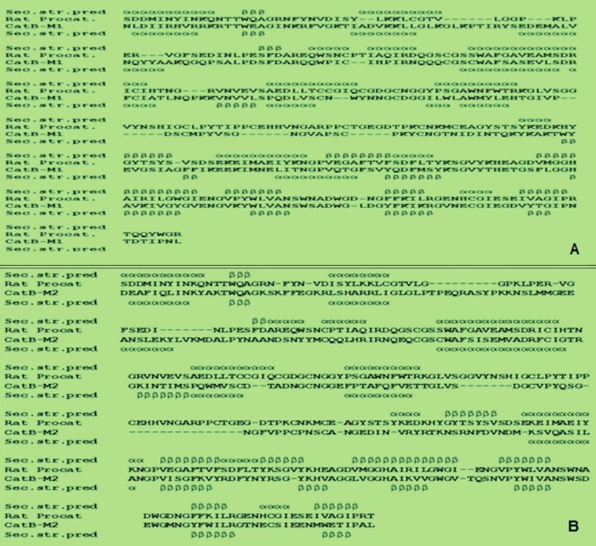
In silico analysis of Naegleria fowleri cathepsin B paralogs: important drug targets

   N. fowleri multiplies rapidly in high tempera-     of structural homologs, PSI-BLAST was used
tures, usually in summer. Infection occurs when       against Protein Data Bank (PDB). Depending on
amoebae adhere to the nasal mucosa, travel along      the high percentage similarities and the low num-
the olfactory nerve, and cross the cribriform         ber of gaps, full-length structural homologs were
plate to enter into the brain, where they cause       selected as templates for X5D761 and M1HE19
PAM4-6. Certain cytolytic molecules, including        cysteine proteases, whereas cathepsin B-like pro-
hydrolases, neuraminidases, phospholipases, and       tease (UniProt ID: X5D911) could not be aligned
phospholipolytic enzymes, are responsible for         well with any of the structures present in PDB.
the pathogenicity of N. fowleri. These mole-          Multiple sequence alignment was carried out
cules damage the cells and nerves of the host,        using homologous sequences from different spe-
often leading to death7. Current treatments for       cies using ClustalX software18 to determine the
PAM rarely ensure survival. The primary drug          consensus patterns in the cathepsin B cysteine
of choice for treating PAM is amphotericin B;         protease family. The pattern was used to optimize
however, this treatment requires a high dosage,       the pairwise sequence alignment of target and
which can lead to renal toxicity8. The identifi-      template.
cation and structural characterization of factors
that influence the pathogenicity of N. fowleri are    Secondary Structure Prediction
crucial and can support the development of more          PSIPRED program was used for secondary
effective therapeutic interventions.                  structure prediction of X5D761 and M1HE19.
   The important role of cysteine proteinases in      The information obtained was used to build struc-
protozoan pathogenesis, including tissue invasion     ture-based pairwise sequence alignment between
and the intracellular survival of several patho-      the target and template protein sequences.
genic parasites, has been reported by many stud-
ies9-12. N. fowleri genome contains three paralogs    Comparative Modeling
of cysteine cathepsin B proteases. Paralogous            Comparative models of both N. fowleri cathep-
genes (or paralogs) are a type of homologous          sin B paralogs (UniProt ID: X5D761; UniProt ID:
genes resulting from a gene duplication event         M1HE19) were built using the same template, i.e.,
within the same organism and may have the             the crystal structure of rat procathepsin B (PDB
same or different function after the event13. N.      ID: 1MIR). All steps of homology modeling and
fowleri cathepsin B proteases may be involved in      refinement were carried out using the program
the proteolytic degradation of collagen, fibronec-    MODELLER v9.20. Evaluation of homology
tin, immunoglobulins, albumin, and hemoglobin.        models was done using the stand-alone programs
This suggests that these proteases play an im-        PROCHECK19 and ProSA20. Structural superpo-
portant role in the pathogenesis of N. fowleri by     sition was performed using options available in
facilitating host tissue attachment, evading host     the SUPERPOSE script file of the MODELLER 21.
immunity, and easing nutrient uptake14. Cathep-
sin B proteases have been found in N. fowleri         Protein Modeling and Structural Analysis
lysate and excretory-secretory protein fractions,        Analysis of three-dimensional protein struc-
demonstrating their crucial role in the pathogene-    tures is a more mature field of study than se-
sis of N. fowleri15-17.                               quence analysis. DS Visualizer and Visual Mo-
   The present study aims to elucidate three-di-      lecular Dynamics (VMD)22 software programs
mensional structures of N. fowleri cathepsins B       were used for the molecular analysis of these
enzymes, delineate their active site architectures,   enzymes.
and observe enzyme propeptide binding modes
via in silico investigations.                         All-Atom Molecular Dynamics Simulation
                                                         Molecular Dynamics (MD) simulations can
                                                      help shed light on protein-protein and pro-
          Materials and Methods                       tein-ligand interactions at the molecular level 23.
                                                      In the present study, all-atom MD simulations
Sequence Analyses and Template Search                 and reasonable analysis procedures were con-
   Amino acid sequences of N. fowleri cyste-          ducted using the Amber software package, ver-
ine proteases (UniProt ID: X5D761; UniProt ID:        sion 1824. Initially, two systems were prepared,
M1HE19; UniProt ID: X5D911) were obtained             exemplified as Model-1 and Model-2. The LEaP
from the UniProt database. For the identification     module was used to add hydrogen atoms to

                                                                                                   3163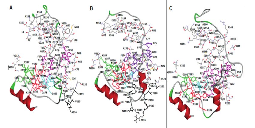
M. Aurongzeb, S.A. Haq, Y. Rashid, S.H. Ahmed Naqvi, S.I. Hussain, A.U. Rehman, et al

both systems. Counterions (Na+ and Cl-) were         Homology Modeling of N. fowleri
added to maintain system neutrality. All sys-        Cathepsin B and Putative Cathepsin B
tems were solvated in a truncated octahedral         Proteases
box of TIP3P water model with 10 Å buffer.              Homology models of N. fowleri cathepsin B
The Particle Mesh Ewald (PME) method 25 was          (CatB-M1) and putative cathepsin B (CatB-M2)
used to treat long-range electrostatic interac-      were constructed using optimized pairwise se-
tions, and ff99IDPs force field was used for all     quence alignments (Figures 1A and 1B). Both
simulations26,27. All the bonds involving hydro-     models were found to be of good quality us-
gen atoms were constrained using the SHAKE           ing different evaluation procedures. Overall, the
algorithm 28. The PMEMD of CUDA version              three-dimensional folds of N. fowleri CatB-M1
was used to accelerate all the MD simulations29.     (Figures 2A and 2B) and CatB-M2 (Figures 2C
The steepest descent method was used to min-         and 2D) were comparable to the crystal structure
imize the solvated systems for 20,000 steps,         of rat procathepsin B. Similar to the rat proca-
then 400 ps heating, and 200 ps equilibration in     thepsin B fold, both the N. fowleri cathepsin B
the NVT ensemble. The production ran under           models were found to be comprised of two dis-
the NPT ensemble at 298 K with a time step of        tinct domains: a five-stranded beta-sheet domain
2 ps in Berendsen thermostat and barostat. The       and an alpha-helical domain with three alpha-he-
CPPTRAJ package was used to analyze the              lices and a small beta-sheet structure.
trajectories in Amber 18. Finally, all analyses         A proregion lacking any globular structure
were conducted using the CPPTRAJ module              tends to wrap around cathepsin B while inter-
implemented in Amber, i.e., root-mean-square         acting with three key areas, including a proseg-
deviation (RMSD); root-mean-square fluctua-          ment-binding loop (PBL), a substrate-binding
tion (RMSF); the CA distance between the             cleft, and an occluding loop crevice. The proseg-
bound peptide and CatB protein; the radius of        ment and mature cathepsin B interactions were
gyradius (Rg); hydrogen bonding population           analyzed in both N. fowleri cathepsin B models
among peptides, respectively.                        and in the rat procathepsin B by selecting an
                                                     area of 5 Å around their respective prosegments
                                                     (Figure 3). The center of PBL that includes Y183
                    Results                          and Y188 forms a depression on the surface of
                                                     cathepsin B. This prosegment interacts with PBL
   Based on the importance of cysteine cathepsins    via a hairpin element that directs the β-strand and
to N. fowleri pathogenesis and PAM progression,      α-helix of the proregion on the other side of PBL,
we aimed to perform protein sequence analyses        thereby positioning the W24 side chain in the
and identify three-dimensional architectures of      center of the depression. The side chain of W24
N. fowleri cathepsin B using standard bioinfor-      forms an H-bond with the carbonyl group of K189
matics tools. UniProt database search provided       residue of PBL. All the interactions between N.
three N. fowleri cathepsin B protein sequences,      fowleri prosegment and PBL were well conserved
including cathepsin B (UniProt ID: X5D761),          in both models suggesting that the interactions
putative cathepsin B (UniProt ID: M1HE19), and       of PBL with prosegment are generally similar
cathepsin B-like protein (Uniprot ID: X5D911).       in both N. fowleri cathepsin B models and in the
The crystal structure of rat procathepsin B (PDB     crystal structure the template, rat procathepsin B.
ID: 1MIR) was found to be a common optimal           Residues of the substrate-binding cleft, includ-
template for both N. fowleri cathepsin B and pu-     ing G27, C29, G73, G74, W30, Y75, H110, F174,
tative cathepsin B, with sequence similarities of    V176, L181, M196, G197, G198, H199, A200, and
54% and 51%, respectively. However, no suitable      W221, bind with the prosegment (Figure 3). All
template was found for the N. fowleri cathepsin      residues were strictly conserved in both N. fowl-
B-like protein (UniProt ID: X5D911) in a PDB.        eri cathepsin B models except for H110, which is
Structure-based pairwise sequence alignments         missing due to the deletion of eleven and fifteen
of cathepsin B and putative cathepsin B, along       residues in the occluding loop region of N. fowleri
with their common template of rat procathepsin       CatB-M1 and CatB-M2, respectively (Figure 4).
B (PDB ID: 1MIR), were optimized with consid-
eration of protein secondary structures and con-     Active Site Analyses
servation patterns across the cathepsin B protease     The active site triad, i.e., Cys29-His199-
family.                                              Asp219, was conserved and found to be located

3164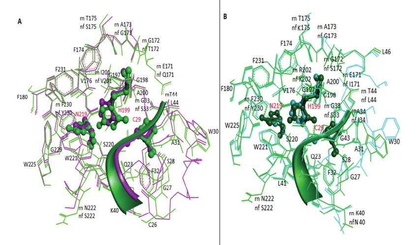
In silico analysis of Naegleria fowleri cathepsin B paralogs: important drug targets
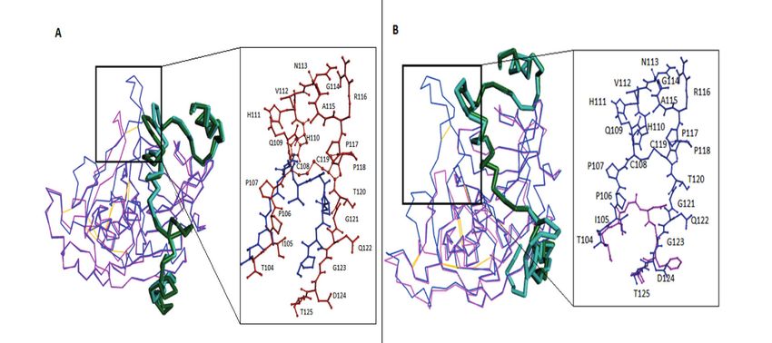
Figure 1. Optimized pairwise sequence alignments of N. fowleri cathepsin B (A) and putative cathepsin B (B) with rat
procathepsin B (PDB ID: 1MIR).
at the domain interface, like rat procathepsin B.
An area of 5 Å around the catalytic triad was
found to consist of 36 amino acid residues in
both models (Figure 5). Twenty-seven residues,
including the catalytic triad, were strictly con-
served in N. fowleri CatB-M1 compared to the
crystal structure of rat procathepsin B. Six of
these residues showed conservative substitu-
tions: I200→V, A173→G, T175→S, R200→K,
N222→S, and F230→Y. Only three noncon-
servative substitutions were observed: G23→S,
E171→Q, and G172→T. In CatB-M2, 28 resi-
dues, including the catalytic triad, were strictly
conserved compared to rat procathepsin B. Five
active site residues showed conservative substi-
tutions: A34→I, A173→G, R20→K, N222→S,
and F177→Y. Only three nonconservative sub-
stitutions were observed: T175→K, E171→I, and
G172→S.
Figure 2. Ribbon representation of three-dimensional models
of N. fowleri CatB-M1 (A, front view; B, back view) and
Active site analysis of both models revealed
CatB-M2 (C, front view; D, back view). The proregion is shown a similar abundance of active site residues in
in red and purple in CatB-M1 and CatB-M2, respectively. the N. fowleri CatB-M1 and CatB-M2 models
3165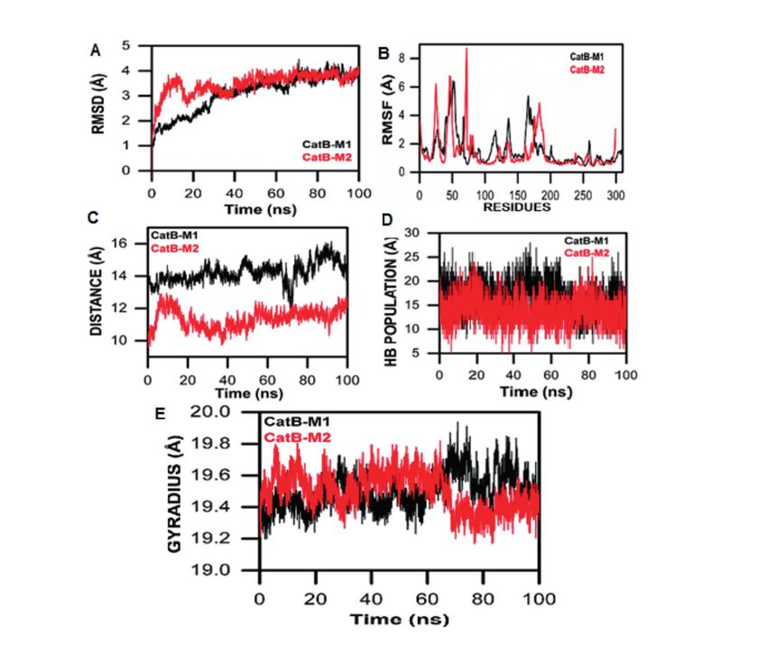
M. Aurongzeb, S.A. Haq, Y. Rashid, S.H. Ahmed Naqvi, S.I. Hussain, A.U. Rehman, et al

Figure 3. Interactions of proregion in CatB-M1 (A), 1MIR (B), and CatB-M2 (C) with the three key areas of the mature
cathepsin, including prosegment-binding loop (green color), substrate-binding cleft (purple color), and occluding loop crevice
(black color). The residues common among the three regions are shown in red, whereas the residues of the base part are shown
in cyan.

compared to rat procathepsin B. Here, we believe                  anism of action. The missing residues, which are
that the overall architectures of both N. fowleri                 important for exopeptidase activity, especially
cathepsin B models, including active sites related                H110 and H111 that were missing in both models,
to endopeptidase activity, are quite similar to that              suggest that N. fowleri cathepsin B lacks exopep-
of rat procathepsin B, suggesting a similar mech-                 tidase activity.

Figure 4. Superposition of rat procathepsin B (1MIR) with CatB-M1 (A) and CatB-M2 (B) of N. fowleri. The proregions of
the template and the model are shown in c-alpha stick presentation in dark green and cyan, respectively. Cathepsin B parts
of the template and the models are depicted in c-alpha wire presentation in blue and pink, respectively. The residues of the
occluding loop of the template and model are shown in brown and blue in CatB-M1 (A), respectively, and blue and pink in
CatB-M2 (B), respectively.

3166
In silico analysis of Naegleria fowleri cathepsin B paralogs: important drug targets

Figure 5. Analysis of the active site residues of N. fowleri cathepsin B (A) and putative cathepsin B (B) homology models
after superposition. 1MIR is shown in light (A) and dark green (B), whereas CatB-M1 active site residues are shown in pink
(A) and CatB-M2 active site residues are shown in cyan. The catalytic triad (C29, H199, and N219) is shown in a ball-and-stick
presentation.

Localization of N. fowleri Cathepsin B                            simulated in an explicit water environment for
Proteases                                                         100 ns. The deviation of backbone atoms was
   Multiple sequence alignment of human, rat,                     examined using RMSD to ensure the stability
and N. fowleri cathepsin B proteases showed                       of the simulation process. CatB-M1 showed a
strict conservation of the catalytic triad (Figure                gradual increase starting 1 Å into the MD simu-
6). Normally, it is expected that cathepsin B-con-                lation time, whereas CatB-M2 initially increased
taining N-glycosylation site, i.e., N38 (human                    drastically in deviation reaching 3.9 Å at 18 ns,
cathepsin B numbering30), is transported to the                   and then showed a consistent behavior and os-
lysosome via the vesicular MPR pathway31. How-                    cillated till 100 ns. A smaller RMSD curve indi-
ever, the nonconservative substitutions N38→R                     cates high stability and vice versa (Figure 7A).
and N38→A were observed at the N-glycosyla-                       When comparing 1MIR-M1 with 1MIR-M2, the
tion site in N. fowleri cathepsin B and putative                  RMSD results revealed the inconsistent behavior
cathepsin B, respectively (Figure 6). These sub-                  of the M1 complex throughout MD simulation,
stitutions suggest the extralysosomal distribution                whereas the 1M1R-M2 showed a consistent be-
of cathepsin B proteases via an unknown alter-                    havior that favors the stability and reliability
nate mechanism in N. fowleri. Cathepsin B prote-                  of the complex. RMSF were analyzed to deter-
ases are known to be differentially expressed and                 mine the flexibility of individual residues in the
secreted in the trophozoite stage of N. fowleri14,                CatB-M1 and CatB-M2 systems. This analysis
which suggests that they play a role in its patho-                showed that the CatB-M1 complex exhibited lo-
genesis.                                                          cal fluctuations, especially in residues 100-200,
                                                                  with residue fluctuations of up to 2.5 Å, 2.9 Å,
All-Atom Molecular Dynamics Simulation                            and 4.3 Å (Figure 7B). These residue fluctuations
   The three-dimensional folds of N. fowleri                      were reduced in the CatB-M2 complex, clear-
CatB-M1 and CatB-M2, comparable to the crys-                      ly indicating the stability and tightness of the
tal structure of rat procathepsin B, were MD                      prosegment in this complex. The RMS analysis

                                                                                                                       3167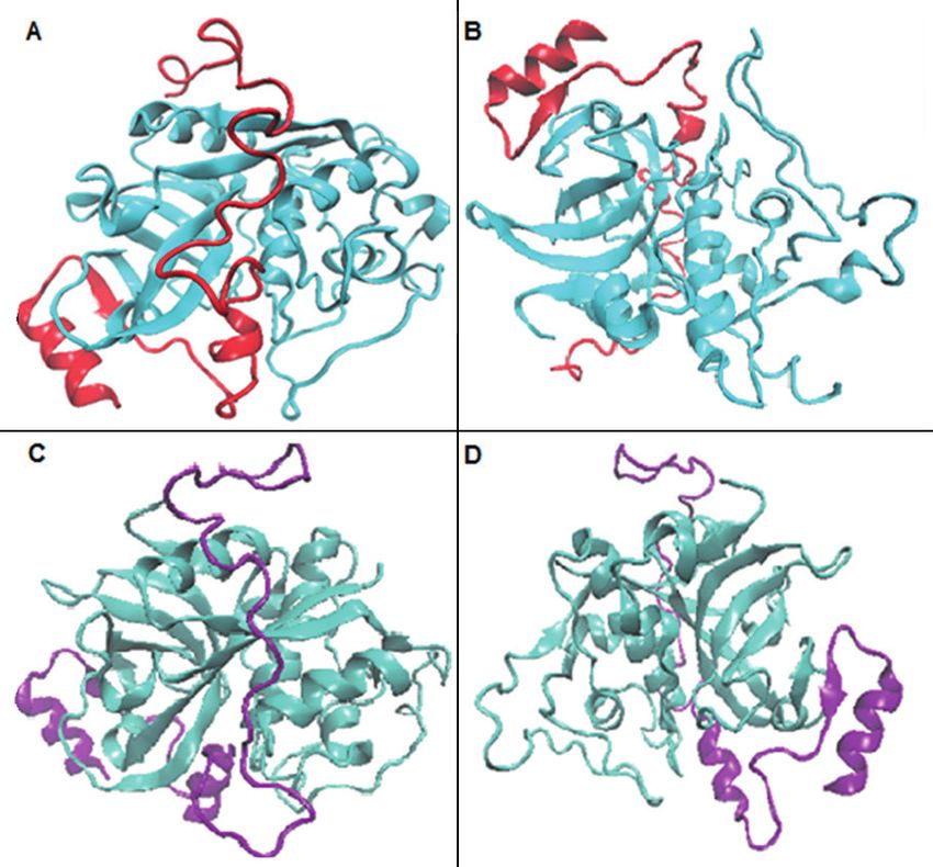
M. Aurongzeb, S.A. Haq, Y. Rashid, S.H. Ahmed Naqvi, S.I. Hussain, A.U. Rehman, et al

Figure 6. Multiple sequence alignment of human, rat, N. fowleri cathepsin B (X5D761), and putative cathepsin B (M1HE19)
proteases. The prosegment residues are shown in red, showing the important glycosylation site. The occluding loop region is
shown in a red box, whereas the catalytic triad is shown in blue.

indicates that the prosegments in both models                   tance for the CatB-M2 complex remains steady,
exhibit consistent, steady binding, particularly                i.e., around 1.5 Å. This steadiness indicates that
marked in the CatB-M2 complex, based on the                     the propeptide in CatB-M2 binds tightly and
RMSD/F results.                                                 possesses a higher affinity for binding than the
   The alpha carbon distance between the pep-                   CatB-M1 complex (Figure 7C). A hydrogen pop-
tide and mature enzyme in both models was also                  ulation analysis further revealed that the pro-
analyzed and compared. The alpha carbon dis-                    peptide showed a high affinity to bind CatB-M1

3168
In silico analysis of Naegleria fowleri cathepsin B paralogs: important drug targets

Figure 7. All-atom MD simulation studies of N. fowleri cathepsin B (X5D761) and putative cathepsin B (M1HE19) proteases.

enzyme and consistent behavior throughout the                 consistent behaviors in overall conformation,
MD simulation. In contrast, the CatB-M2 ex-                   probably due to the tight binding of the propep-
hibited dramatic behaviors, including high HB                 tide to the enzyme.
interactions between the enzyme and the pro-
peptide during the 1-10 ns and 40-70 ns MD
simulations (Figure 7D). The consequences of                                       Discussion
the backbone fluctuations and deviations in both
CatB-M1 and CatB-M2 indicated the need for                       N. fowleri is the only etiological agent of PAM
further exploration of the compactness of the                 in human beings. Although some information
overall conformation. We also analyzed the ra-                about the genome sequence and important patho-
dius of gyration (Rg), which indicated that the               genic molecules of N. fowleri is available, there
CatB-M2 was initially more compact than the                   is still no selective potential treatment for PAM32.
CatB-M1 for 60 ns to 100 ns, and the compact-                 N. fowleri invades the CNS via nasal mucosa
ness of CatB-M2 decreased. The compactness of                 by crossing the cribriform plate, where it causes
the CatB-M1 increased and decreased through-                  the characteristic meningoencephalitis. Cytolytic
out the MD simulations (Figure 7E). The analy-                molecules are responsible for the pathogenicity
sis revealed that the CatB-M2 complex showed                  of N. fowleri; these molecules are known to cause

                                                                                                                 3169
M. Aurongzeb, S.A. Haq, Y. Rashid, S.H. Ahmed Naqvi, S.I. Hussain, A.U. Rehman, et al nerve and cell destruction in the host, often re- but increases endopeptidase activity. It also in- sulting in death7. Cysteine proteases, especially creases the ability of macromolecular inhibitors cysteine cathepsins secreted by N. fowleri, can to bind to the active site43. MD simulation studies destroy host tissues and host immunity1. In addi- confirmed tight binding of the prosegment, its tion to their primary function of cellular protein natural inhibitor, with both mature cathepsin B degradation and catabolism, cysteine cathepsins models. This suggests that the prosegment plays are involved in numerous important physiologi- a protective role against uncontrolled proteolytic cal processes, including ECM turnover, immune degradation within the pathogen. Additionally, invasion, digestion, and parasite invasion and nonconservative substitutions at important N-gly- escape, and certain pathologies, such as rheu- cosylation sites in N. fowleri cathepsin B and matoid arthritis, atherosclerosis, leishmaniasis, putative cathepsin B suggest their extracellular amebiasis, and malaria31,33,34. Since it is a lyso- secretion and role in tissue damage to the host. somal enzyme, cathepsin B is normally believed This article is the first to present the tertiary ar- to be transported to the lysosomes via man- chitectures of N. fowleri cathepsin B proteases, nose-6-phosphate receptor pathway35. However, along with information about their active sites this distribution of cathepsin B shifts towards the and the mechanics of cathepsin B propeptide. cell periphery in tumor cells suggesting the ex- We believe that this information will support the tracellular secretion of cathepsin B proteases36-40. development of novel lead compounds against the Cathepsin B proteases are known to be differ- deadly protozoa and could lead to more effective entially expressed and secreted in the trophozoite treatments for PAM. stage of N. fowleri, which suggests that they play a significant role in its pathogenesis. N. fowl- eri cathepsin B enzymes play a pivotal role in Conclusions the progression of PAM41. Therefore, the present study involved in silico protein sequence analyses Cysteine proteinases, such as cathepsin B, play and sought to determine the three-dimensional a vital role in protozoan pathogenesis, including structures of N. fowleri cathepsin B and putative tissue invasion and intracellular pathogenic sur- cathepsin B proteases. Detailed analyses revealed vival. In silico studies of two paralogous N. fowl- that the overall folds of N. fowleri cathepsin B eri cathepsin B enzymes showed a large deletion and putative cathepsin B are quite similar to of eleven and fifteen residues, respectively, com- that of rat procathepsin B, suggesting a similar pared with rat cathepsin B (the template), in their mechanism of action. Three segments of mature occluding loop regions, suggesting little or no ex- cathepsin B, a part of an occluding loop (108- opeptidase activity but increased endopeptidase 122), a part of the PBL, and a loop (221-226) at activity and increased affinity between the active the base, interact together to form a crevice. The site and the inhibitory propeptide. MD simula- latter two regions were conserved in both models. tion studies of N. fowleri cathepsin B enzymes However, some residues of the occluding loop confirmed the tight binding of prosegments with involved in forming the crevice were not found in their active sites. Additionally, mutation of typ- both models. Notably, the H110 and H111 residues ical N-glycosylation site indicates extracellular (human cathepsin B numbering30) are crucial secretion of N. fowleri cathepsin B enzymes parts of the occluding loop and are responsible via an alternative yet unknown posttranslational for the exopeptidase activity in higher eukary- processing strategy. Here, we suggest that both otes. The cathepsin B protease of Giardia lacks of these N. fowleri cathepsin B enzymes have the occluding loop, suggesting that the loop was high endopeptidase activity. When these highly either deleted or inserted later during the course active cathepsin B enzymes are inside the cell, of evolution42. they are kept inactive via tight binding of their Accordingly, eleven and fifteen residues are propeptides. Once outside the cell, the enzymes deleted in the occluding loop regions of N. fowleri achieve their fully active form by cleaving off cathepsin B and putative cathepsin B, respective- their propeptide segments and become disastrous ly. These deletions, along with the missing H110 for the host tissue. and H111 residues, suggest that the occluding loop is short for cathepsin B and even shorter in putative cathepsin B (Figure 4). Deletion of the Conflict of Interest occluding loop eliminates exopeptidase activity The Authors declare that they have no conflict of interests. 3170
In silico analysis of Naegleria fowleri cathepsin B paralogs: important drug targets

                                                                   targets. PLoS Negl Trop Dis 2018; 12: e0005639.
Ethical Approval
Ethical approval is not applicable as the present study does   12) Keene WE, Petitt MG, Allen S, McKerrow JH. The
not involve any animal or human participant.                       major neutral proteinase of Entamoeba histolyti-
                                                                   ca. J Exp Med 1986; 163: 536-549.
                                                               13) Moreira D, López-García P. Paralogous Gene. In:
                                                                   Gargaud M, Amils R, Quintanilla JC, Cleaves HJ,
Informed Consent                                                   Irvine WM, Pinti D, Viso M, eds. Encyclopedia of
Informed Consent is not applicable as the present study            Astrobiology. Berlin, Heidelberg: Springer Berlin
does not involve any human participant.                            Heidelberg, 2011; 1215.
                                                               14) Lee J, Kim JH, Sohn HJ, Yang HJ, Na BK, Chwae
                                                                   YJ, Park S, Kim K, Shin HJ. Novel cathepsin B
                     References                                    and cathepsin B-like cysteine protease of Naegle-
                                                                   ria fowleri excretory-secretory proteins and their
 1) Aldape K, Huizinga H, Bouvier J, McKerrow J.                   biochemical properties. Parasitol Res 2014; 113:
    Naegleria fowleri: characterization of a secreted              2765-2776.
    histolytic cysteine protease. Exp Parasitol 1994;          15) Shin HJ, Cho MS, Jung SY, Kim HI, Park S, Kim
    78: 230-241.                                                   HJ, Im KI. Molecular cloning and characterization
 2) Ghanchi NK, Khan E, Khan A, Muhammad W,                        of a gene encoding a 13.1 kDa antigenic protein
    Malik FR, Zafar A. Naegleria fowleri meningo-                  of Naegleria fowleri. J Eukaryot Microbiol 2001;
    encephalitis associated with public water supply,              48: 713-717.
    Pakistan, 2014. Emerg Infect Dis 2016; 22: 1835-           16) Kim JH, Yang AH, Sohn HJ, Kim D, Song KJ,
    1837.                                                          Shin HJ. Immunodominant antigens in Naegleria
 3) Tung MC, Hsu BM, Tao CW, Tung MC, Hsu BM,                      fowleri excretory--secretory proteins were poten-
    Tao CW, Lin WC, Tsai HF, Ji DD, Shen SM, Chen                  tial pathogenic factors. Parasitol Res 2009; 105:
    JS, Shih FC, Huang YL. Identification and signif-              1675-1681.
    icance of Naegleria fowleri isolated from the hot          17) Kim JH, Kim D, Shin HJ. Contact-independent
    spring which related to the first primary amebic               cell death of human microglial cells due to patho-
    meningoencephalitis (PAM) patient in Taiwan. Int               genic Naegleria fowleri trophozoites. Korean J
    J Parasitol 2013; 43: 691-696.                                 Parasitol 2008; 46: 217-221.
 4) Visvesvara GS, Moura H, Schuster FL. Pathogen-
                                                               18) Thompson JD, Gibson TJ, Plewniak F, Jean-
    ic and opportunistic free-living amoebae: acan-
                                                                   mougin F, Higgins DG. The CLUSTAL_X windows
    thamoeba spp., Balamuthia mandrillaris, Naegle-
                                                                   interface: flexible strategies for multiple sequence
    ria fowleri, and Sappinia diploidea. FEMS Immu-
                                                                   alignment aided by quality analysis tools. Nucleic
    nol Med Microbiol 2007; 50: 1-26.
                                                                   Acids Res 1997; 25: 4876-4882.
 5) Jamerson M, da Rocha-Azevedo B, Cabral GA,
                                                               19) Laskowski RA, Moss DS, Thornton JM. Main-
    Marciano-Cabral F. Pathogenic Naegleria fowleri
                                                                   chain bond lengths and bond angles in protein
    and non-pathogenic Naegleria lovaniensis exhibit
                                                                   structures. J Mol Biol 1993; 231: 1049-1067.
    differential adhesion to, and invasion of, extracel-
    lular matrix proteins. Microbiology (Reading, En-          20) Sippl MJ. Recognition of errors in three-dimen-
    gland) 2012; 158: 791-803.                                     sional structures of proteins. Proteins 1993; 17:
 6) Baig AM. Primary amoebic meningoencephalitis:                  355-362.
    neurochemotaxis and neurotropic preferences of             21) Sali A, Blundell TL. Comparative protein model-
    naegleria fowleri. ACS Chem Neurosci 2016; 7:                  ling by satisfaction of spatial restraints. J Mol Biol
    1026-1029.                                                     1993; 234: 779-815.
 7) Grace E, Asbill S, Virga K. Naegleria fowleri:             22) Humphrey W, Dalke A, Schulten K. VMD: visual
    pathogenesis, diagnosis, and treatment options.                molecular dynamics. J Mol Graph 1996; 14: 33-
    Antimicrob Agents Chemother 2015; 59: 6677-                    38.
    6681.                                                      23) Rehman AU, Khan MT, Liu H, Wadood A, Malik
 8) Stevens AR, Shulman ST, Lansen TA, Cichon MJ,                  SI, Chen HF. Exploring the pyrazinamide drug re-
    Willaert E. Primary amoebic meningoencephali-                  sistance mechanism of clinical mutants T370P
    tis: a report of two cases and antibiotic and immu-            and W403G in ribosomal protein S1 of mycobac-
    nologic studies. J Infect Dis 1981; 143: 193-199.              terium tuberculosis. J Chem Inf Model 2019; 59:
 9) Karrer KM, Peiffer SL, DiTomas ME. Two distinct                1584-97.
    gene subfamilies within the family of cysteine pro-        24) Avagliano D, Sánchez-Murcia PA, González
    tease genes. Proc Natl Acad Sci USA 1993; 90:                  L. Directional and regioselective hole injection
    3063-3067.                                                     of spiropyran photoswitches intercalated into
10) McKerrow JH, Caffrey C, Kelly B, Loke P, Sajid M.              A/T-duplex DNA. Phys Chem Chem Phys 2019;
    Proteases in parasitic diseases. Annu Rev Pathol               21: 17971-17977.
    2006; 1: 497-536.                                          25) Darden T, York D, Pedersen L. Particle mesh
11) McKerrow JH. The diverse roles of cysteine pro-                Ewald: An N‧log(N) method for Ewald sums in
    teases in parasites and their suitability as drug              large systems. J Chem Phys 1993; 98: 10089.

                                                                                                                  3171
M. Aurongzeb, S.A. Haq, Y. Rashid, S.H. Ahmed Naqvi, S.I. Hussain, A.U. Rehman, et al
26) Wang W, Ye W, Jiang C, Luo R, Chen HF. New 35) Kornfeld S. Trafficking of lysosomal enzymes.
force field on modeling intrinsically disordered FASEB J 1987; 1: 462-468.
proteins. Chem Biol Drug Des 2014; 84: 253-269. 36) Calkins CC, Sameni M, Koblinski J, Sloane BF,
27) Ye W, Ji D, Wang W, Luo R, Chen HF. Test and Moin K. Differential localization of cysteine pro-
evaluation of ff99IDPs force field for intrinsically tease inhibitors and a target cysteine protease,
disordered proteins. J Chem Inf Model 2015; 55: cathepsin B, by immuno-confocal microscopy. J
1021-1029. Histochem Cytochem 1998; 46: 745-751.
28) Ryckaert JP, Ciccotti G, Berendsen HJC. Numer- 37) Sameni M, Elliott E, Ziegler G, Fortgens PH, Den-
ical integration of the cartesian equations of mo- nison C, Sloane BF. Cathepsin B and D are local-
tion of a system with constraints: molecular dy- ized at the surface of human breast cancer cells.
namics of n-alkanes. J Comput Phys 1977; 23: Pathol Oncol Res 1995; 1: 43-53.
327-341. 38) Sloane B, Moin K, Lah T. Regulation of lyso-
29) Salomon-Ferrer R, Götz AW, Poole D, Le Grand somal endopeptidases in malignant neoplasia.
S, Walker RC. Routine microsecond molecular In: Aspects of the Biochemistry and Molecular
dynamics simulations with AMBER on GPUs. 2. Biology of Tumors. New York: Academic Press,
Explicit Solvent Particle Mesh Ewald. J Chem 1994.
Theory Comput 2013; 9: 3878-3888. 39) Erdel M, Trefz G, Spiess E, Habermaas S, Spring
30) Musil D, Zucic D, Turk D, Engh RA, Mayr I, Hu- H, Lah T, Ebert W. Localization of cathepsin B in
ber R, Popovic T, Turk V, Towatari T, Katunuma N. two human lung cancer cell lines. J Histochem
The refined 2.15 A X-ray crystal structure of hu- Cytochem 1990; 38: 1313-1321.
man liver cathepsin B: the structural basis for its 40) Krepela E, Bartek J, Skalkova D, Vicar J, Rasnick
specificity. EMBO J 1991; 10: 2321-2330. D, Taylor-Papadimitriou J, Hallowes RC. Cyto-
31) Moin K, Demchik L, Mai J, Duessing J, Peters C, chemical and biochemical evidence of cathepsin
Sloane BF. Observing proteases in living cells. B in malignant, transformed and normal breast
Adv Exp Med Biol 2000; 477: 391-401. epithelial cells. J Cell Sci 1987; 87: 145-154.
32) Liechti N, Schürch N, Bruggmann R, Wittwer M. 41) Zyserman I, Mondal D, Sarabia F, McKerrow JH,
Nanopore sequencing improves the draft genome Roush WR, Debnath A. Identification of cysteine
of the human pathogenic amoeba Naegleria fowl- protease inhibitors as new drug leads against
eri. Sci Rep 2019; 9: 16040. Naegleria fowleri. Exp Parasitol 2018; 188: 36-
33) Verma S, Dixit R, Pandey KC. Cysteine proteas- 41.
es: modes of activation and future prospects as 42) Ward W, Alvarado L, Rawlings ND, Engel JC,
pharmacological targets. Front Pharmacol 2016; Franklin C, McKerrow JH. A primitive enzyme for
7: 107. a primitive cell: the protease required for excysta-
34) Vasiljeva O, Reinheckel T, Peters C, Turk D, Turk tion of Giardia. Cell 1997; 89: 437-444.
V, Turk B. Emerging roles of cysteine cathepsins 43) Illy C, Quraishi O, Wang J, Purisima E, Vernet T,
in disease and their potential as drug targets. Mort JS. Role of the occluding loop in cathepsin
Curr Pharm Des 2007; 13: 387-403. B activity. J Biol Chem 1997; 272: 1197-1202.
3172You can also read