Influence of Endodontic Sealers on Dentin Strength in Endodontically Treated Teeth
←
→
Page content transcription
If your browser does not render page correctly, please read the page content below
University of Connecticut
                                                                OpenCommons@UConn
Master's Theses                                                                       University of Connecticut Graduate School

8-6-2019

Influence of Endodontic Sealers on Dentin
Strength in Endodontically Treated Teeth
Andrew Fossum
fossum@uchc.edu

Recommended Citation
Fossum, Andrew, "Influence of Endodontic Sealers on Dentin Strength in Endodontically Treated Teeth" (2019). Master's Theses.
1416.
https://opencommons.uconn.edu/gs_theses/1416

This work is brought to you for free and open access by the University of Connecticut Graduate School at OpenCommons@UConn. It has been
accepted for inclusion in Master's Theses by an authorized administrator of OpenCommons@UConn. For more information, please contact
opencommons@uconn.edu.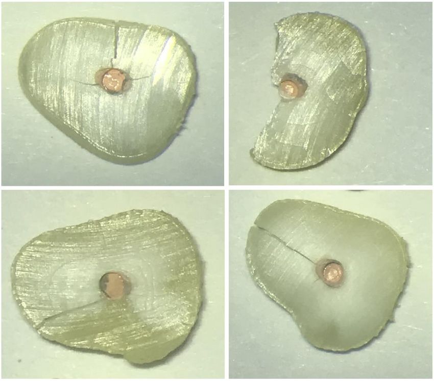
Influence of Endodontic Sealers on Dentin Strength in
             Endodontically Treated Teeth

                     Andrew C. Fossum
              D.D.S., University of Texas, 2008

                          A Thesis

            Submitted in Partial Fulfillment of the

              Requirements for the Degree of

                  Master of Dental Science

                            at the

                  University of Connecticut

                            2019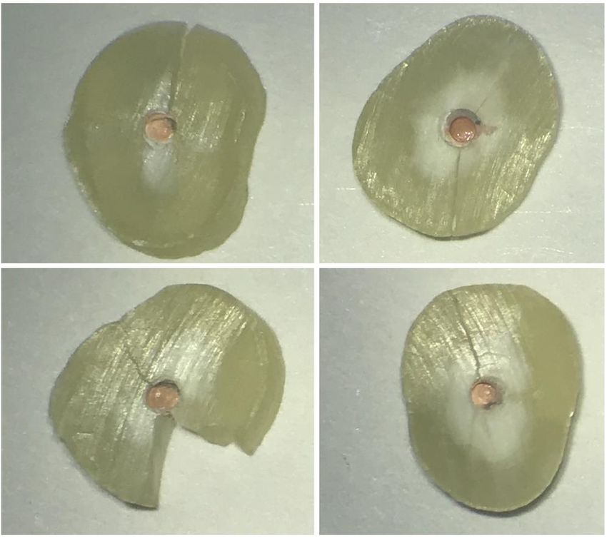
Copyright by

Andrew C. Fossum

     2019

       ii
APPROVAL PAGE

                          Master of Dental Science Thesis

Influence of Endodontic Sealers on Dentin Strength in Endodontically Treated Teeth

                                    Presented by

                             Andrew C. Fossum, D.D.S.

            Major Advisor_________________________________________

                             J. R. Kelly, D.D.S., Ph.D.

         Associate Advisor_________________________________________

                            Kamran Safavi, D.M.D., M.Ed.

         Associate Advisor_________________________________________

                           Blythe Kaufman, D.M.D., M.D.S.

                              University of Connecticut
                                         2019

                                          iii
Acknowledgments

       I would like to extend my gratefulness to my major advisor Dr. John R. Kelly.

Without his support and direction throughout this process, this project would not have

been possible. His knowledge and expertise in his field is immeasurable. Dr. Kelly

always pushed me to do the best I could and made me confident in what I was doing. I

am so appreciative of him to take on this endeavor with me and to be my major advisor.

       I also would like to extend my esteemed admiration to my program director and

associate advisor Dr. Kamran Safavi. It is futile to put into words what his mentorship

and guidance has done and will continue to do for me throughout my career and life.

Dr. Safavi is an icon not only in the field of endodontics but as a person who is caring,

generous and will always be considered part of the family.

       Additionally, I would like to express gratitude to another of my associate advisors

Dr. Blythe Kaufman. When it comes to someone who is competent and a has a

knowledge in the field of endodontics, in which it is hard to find an equal, is Dr.

Kaufman. Her dedication to provide a sound foundation of scientific knowledge will

forever support my career and understanding of endodontics.

       The authors have no financial affiliation or involvement with any commercial

organization with direct financial interest in the subject or materials discussed in this

manuscript, nor have any such arrangements existed in the past three years.

                                                                Andrew C. Fossum, D.D.S.

                                              iv
Table of Contents

Acknowledgments                                     iv

Table of Contents                                   v

List of Figures                                     vii

Abstract                                            viii

Chapter I: Introduction                             1

     A.    Endodontic Rotary Files                  1

     B.    Gutta-percha                             2

     C.    Obturation Techniques                    5

     D.    Endodontic Sealers                       8

     E.    Fractures and Cracks in Teeth            12

     F.    Hoop Stress and Thick-Walled Cylinders   15

     G.    Knowledge Gap                            17

Chapter II: Research Aim and Hypothesis             17

Chapter III: Methods and Materials                  18

     A.    Collection of Teeth                      18

     B.    Teeth Preparation                        18
                                     v
C.   Teeth Obturation            19

     D.   Load Applying               20

     E.   Statistical Analysis        23

Chapter IV: Results                   24

Chapter V: Discussion                 27

Chapter VI: Conclusion                33

Chapter VII: Appendix                 34

Chapter VIII: References              42

                                 vi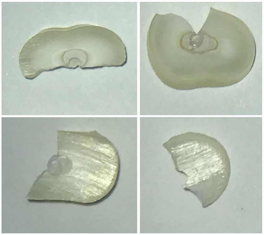
List of Figures

1.    Thick Cylinder with Both External and Internal Pressure         16
2.    MTS 858 Mini Bionix® II Biomaterials Testing System             21
3.    Stainless-steel Pistons Used with the MTS 858 Mini Bionix® II   22
4.    TestWorks® 4 Software Running During Load                       22
5.    Thick Walled Cylinder                                           23
6.    Failure Stress of Each Group                                    26
7.    Group 1 – No Sealer and Gutta-percha                            35
8.    Group 2 – AH Plus Sealer and Gutta-percha                       36
9.    Group 3 – BC Sealer and Gutta-percha                            37
10.   Group 4 – Tetranite and Gutta-percha                            38
11.   Group 5 – Tetranite®                                            39
12.   Mathcad® Software                                               40
13.   Workstation for Preparing Teeth                                 41

                                             vii
Abstract

Influence of Endodontic Sealers on Dentin Strength in Endodontically Treated Teeth
A. Fossum, K. Safavi, B. Kaufman, R. Kelly
University of Connecticut, Farmington, CT

Introduction: Gutta-percha is not compressible and tooth fracture can be initiated when
force is applied to the gutta-percha. The aim of this study was to apply a hoop stress to
roots which have been obturated with either gutta-percha and no sealer, gutta-percha
and AH Plus sealer, gutta-percha and EndoSequence Bioceramic Sealer (BC Sealer),
gutta-percha and Tetranite®, or Tetranite® and no gutta-percha until fracture occurred
and then compared the failure stresses. Methods: Teeth were divided into five groups
based on the sealer type used, no sealer used or only sealer used. The teeth were then
sectioned into 2mm thickness discs and load was applied using a piston until fracture
took place. The stress generated by the gutta-percha on the tooth wall was then
calculated using a hoop stress formula. One – way ANOVA with a 95% multiple range
test was used to compare hoop stresses at failure for all groups (SPSS, TBM). Linear
regression was used to examine failure load versus dentin wall thickness (SigmaPlot
13.0, Systa Software). Results: With respect to the amount of stress exerted by the
gutta-percha on the internal tooth wall with or without sealer types showed there were
significant differences between the Tetranite®/no gutta-percha group and all the groups
(p < 0.05). Multiple comparisons showed no significant difference between the gutta-
percha/no sealer group and gutta-percha/AH Plus sealer group (p < 0.928), gutta-
percha/no sealer group and gutta-percha/BC sealer group (p < 0.927), gutta-percha/no
sealer group and gutta-percha/Tetranite group (p < 1.000), gutta-percha/AH Plus sealer
group and gutta-percha/BC sealer group (p < 0.479), gutta-percha/AH Plus sealer group
and gutta-percha/Tetranite® group (p < 0.845) and the gutta-percha/BC sealer group
and gutta-percha/Tetranite® group (p < 0.973). Conclusions: Application of a hoop
stress provides the field of endodontics a method to test whether sealers enhance
dentin strength. Currently there is a knowledge gap in endodontics where there is no
method to test whether endodontic sealers enhance dentin strength, and this shows it is
possible. The Tetranite®/no gutta-percha group enhanced dentin strength in this study.

                                           viii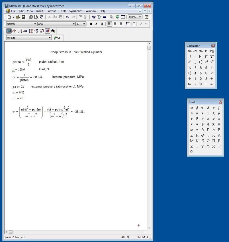
I. Introduction

       Root canal therapy is primarily completed by shaping, cleaning, and filling the

root canal space with endodontic files, irrigants, gutta-percha, and endodontic sealers

respectively. The variety of available instruments today for shaping a root canal

consists mainly of endodontic hand files and rotary file instruments. Endodontic hand

files are manually operated endodontic instruments used for cleaning and shaping of

root canals. Hand filing is very time consuming and this preparation technique can lead

to iatrogenic errors (i.e. ledging, zipping, canal transportation and apical blockage)

(Walton et al., 2002), much consideration has been directed toward root canal

preparation techniques with rotary instruments.

Endodontic Rotary Files

       Rotary endodontic instruments are primarily used to obtain most of the shaping

and are mechanically driven with a handpiece. NiTi alloy was developed by the Naval

Ordnance Laboratory (White Oak, MD, USA). It was named Nitinol; an acronym for

nickel (ni), titanium (ti) and Naval Ordnance Laboratory (Buehler et al., 1963). In the

late 1980s nickel-titanium (NiTi) rotary files were introduced to endodontics. Rotary NiTi

instruments have become popular as they can clean and shape root canals with fewer

procedural errors and more predictability than stainless steel hand files (Hargreaves et

al., 2011).

       Many designs of NiTi instruments are available. Most resemble a basic file, with

flutes along the length and a latching or attaching system to affix the file to a handpiece.

Some are available in different tapers and with noncutting tips. NiTi rotary instruments

                                             1
are used to flare either with the step-back or the crown-down methods. NiTi rotary
instrumentation has advantages as well as disadvantages compared with stainless steel
hand instrumentation. Because of their flexibility, the files have less tendency to
transport curved canals. Finger fatigue is less because the handpiece is doing much of
the work. Somewhat less time is required to prepare the canal. Debridement
effectiveness is comparable to that with hand instrumentation. There are also
disadvantages. Expense is greater if one of the special motor systems is purchased; in
addition, the files are costly. Files are prone to breakage, without warning, particularly if
overused. Overall, no difference is seen with NiTi rotary instruments for either quality of
debridement or prognosis; there are no substantive data on either. (Walton et al., 2002)
In the past decade, several proprietary processing procedures for nickel titanium
(NiTi) alloy were developed to improve the mechanical properties of NiTi
endodontic instruments. Thermomechanically treated NiTi alloys have been reported
to be more flexible with improved cyclic fatigue resistance and greater angle of
deflection at failure when compared to conventional NiTi (Zupanc et al., 2018).
Thermomechanically-treated NiTi instruments are the latest advancement of rotary files
of which many brands exist on the market today.
Gutta-Percha
Gutta-percha is the main component used to fill the root canal space. The early
history of gutta-percha is obscure. The Malays and Chinese are said to have used it in
a remote and undetermined epoch long before Western civilization had any knowledge
of its existence (Obach 1898; Seelingmann et al., 1910). Gutta-percha, as formerly
prepared by the natives of Asia, had a yellowish-brown color and showed a decidedly
2fibrous texture. Gutta-percha occupied an unrivaled position as the most desirable
insulator of electric cables until its replacement by vulcanized rubber late in the
nineteenth century. Gutta-percha was employed for the manufacture of corks, cements,
thread, surgical instruments, garments, pipes, and sheathing for ships. Even boats
were made wholly of gutta-percha, one as early as 1850. Maps and globes were made
of the material, and, because of the thin sheets into which it could be rolled, gutta-
percha seemed destined to replace paper. The variety of manufactured articles
became bewildering. Musical instruments, candelabra, gaiters, garters, suspenders,
window shades, carpets, gloves, mattresses, pillows, tents, umbrellas, and a host of
other articles were fabricated of gutta-percha. Gutta-percha golf balls were introduced
by the later part of the nineteenth century, and until 1920 “gutties” was the term used for
golf balls on links in this country and abroad. Gutta-percha, the naturally occurring
polymer of isoprene, has been known to dentistry for approximately 170 years (Prinz
1945; Payne 1884).
In 1942, C. M. Bunn reported an interesting complication in the molecular
chemistry of gutta-percha. He found that the polymer could exist in two distinctly
different crystalline forms, which he termed alpha” and “beta” modifications. Most
commercial gutta-percha exists as the “beta” crystalline structure (Fisher 1953). The
“alpha” form occurs in the tree. It is in this form that most commercial gutta-percha,
including dental gutta-percha, exists (Goodman et al., 1974). Gutta-percha undergoes
phase transitions when heated from beta to alpha phase at around 115° F (46° C). At a
range between 130° to 140° F (54° to 60° C) an amorphous phase is reached. When
cooled at an extremely slow rate the material will recrystallize to the alpha phase.
3However, this is difficult to achieve and under normal conditions the material returns to
the beta phase. The softening point of gutta-percha was found to be 147° F (64° C). The
phase transformation is important in thermoplastic obturation techniques (Goodman et
al., 1981).
Gutta-percha is derived from dried sap from trees of the family Sapotaceae
(Spångberg et al., 1982). It is composed of 20% gutta-percha, 80% zinc oxide, dye and
metal salts added for color and radiographic contrast. In addition, some manufacturers
add calcium hydroxide, chlorehexidine, or iodoform as an antimicrobial to impart some
disinfectant properties to the material (Ørstavik et al., 2005). The variations in content
are because of different manufacturers and distributors desiring different handling
properties. Some formulations are softer than others. Some clinicians choose the
brand of gutta-percha depending on the technique being used. Compaction with
spreaders, condensers or carriers is usually the means used to attempt to compensate
for the shrinkage of the core material (McElroy 1955). An important characteristic of
gutta-percha and of clinical importance is the fact that when it is exposed to air and light
over time it becomes more brittle. Storage of gutta-percha in a refrigerator extends the
shelf life of the material (Wong et al., 1982).
Gutta-percha tends to be used for many reasons. It is impervious to moisture, is
radiopaque, is not an irritant to tissue beyond the apex, is bacteriostatic, is sterile and
easily sterilized, and easy to remove from the root canal space (Ørstavik et al., 2005).
Gutta-percha is not compressible and is sensitive to temperature changes, it will tend to
become brittle and fracture before ductile yield occurs (Friedman et al., 1977). The
delivery of gutta-percha to the root canal can be accomplished in a variety of ways.
4Obturation Techniques
The operator may choose one of many obturation techniques to deliver the gutta-
percha including: lateral compaction, vertical compaction, continuous wave, warm
lateral, injection techniques, thermomechanical, carrier-based, chemoplasticized,
custom cone/solvents, pastes, and apical barrier. The lateral compaction technique
uses a master cone corresponding to the final instrumentation size and length of the
canal is coated with sealer, inserted into the canal, laterally compacted with spreaders
and filled with additional accessory cones.
Vertical compaction is where a master cone corresponding to the final
instrumentation size and length of the canal is fitted, coated with sealer, heated and
compacted vertically with pluggers until the apical 3-4mm segment of the canal is filled.
Then the remaining root canal is back filled using warm pieces of core material.
Continuous wave is essentially a vertical compaction (down-packing) of core
material and sealer in the apical portion of the root canal using commercially available
heating devices such as System B (SybronEndo, Orange, Calif.) and Elements
Obturation Unit™ (SybronEndo, Orange, Calif.), and then back filling the remaining
portion of the root canal with thermoplasticized core material using injection devices
such as the Obtura (Obtura Spartan, Earth City, Mo.), Elements Obturation Unit™
(SybronEndo, Orange, Calif.), and HotShot (Discus Dental, Culver City, Calif.).
Warm lateral uses a master cone corresponding to the final instrumentation size
of the canal is coated with sealer, inserted into the canal, heated with a warm spreader,
5laterally compacted with spreaders and filled with additional accessory cones. Some
devices use vibration in addition to the warm spreader.
Two types of injection techniques are:
1. A preheated, thermoplasticized, injectable core material is injected directly into
the root canal. A master cone is not used but sealer is placed in the canal before
injection, with either the Obtura (Obtura Spartan, Earth City, Mo.), or Ultrafil (Coltene
Whaledent, Cuyahoga Falls, Ohio) or Calamus® (DENTSPLY Tulsa Dental Specialties,
Tulsa, Okla.) filling systems.
2. A cold, flowable matrix that is triturated, GuttaFlow® (Coltene Whaledent,
Cuyahoga Falls, Ohio), consists of gutta-percha added to a resin sealer, RoekoSeal.
The material is provided in capsules for trituration. The technique involves injection of
the material into the canal and placing a single master cone.
Thermomechanical is a technique where a cone coated with sealer is placed in
the root canal, engaged with a rotary instrument that frictionally warms, plasticizes and
compacts it into the root canal.
Carrier-based systems include two types:
1. Carrier-Based Thermoplasticized: Warm gutta-percha on a plastic carrier, is
delivered directly into the canal as a root canal filling. Examples are: ThermaFil®
(Dentsply Tulsa Dental Specialties, Tulsa, Okla.), Realseal 1™ (Sybron, Orange, Calif.),
Densfil™ (DENTSPLY Maillefer, Tulsa, Okla.) and Soft-Core® (Axis Dental, Coppell,
Texas).
62. Carrier-Based Sectional: A sized and fitted section of gutta-percha with sealer
is inserted into the apical 4mm of the root canal. The remaining portion of the root canal
is filled with injectable, thermoplastized gutta-percha using an injection gun. An example
is SimpliFill (Discus Dental, Culver City, Calif.).
Chempoplasticized technique uses Chemically softened gutta-percha, using
solvents such as chloroform or eucalyptol, is placed on already fitted gutta-percha
cones, inserted into the canal, laterally compacted with spreaders and the canal filled
with additional accessory cones.
Custom cone and solvents such as chloroform, eucalyptol or halothane are used
to soften the outer surface of the cone as if making an impression of the apical portion
of the canal. However, since shrinkage occurs, it is then removed and reinserted into
the canal with sealer, laterally condensed with spreaders and accessory cones.
Pastes fills have been used in a variety of applications. When used as the
definitive filling material without a core, they are generally considered to be less
successful and not ideal. Lastly, apical barriers are important for the obturation of
canals with immature roots with open apices. Mineral trioxide aggregate is generally
considered the material of choice at this time (American Association of Endodontists.
Colleagues for Excellence 2009). Gutta-percha cannot be used as the sole filling
material; it lacks the adherent properties necessary to seal the root canal space.
Therefore, a sealer (cement) is always needed for the final seal (Hargreaves et al.,
2011).
7Endodontic Sealers
Sealers are used between dentin surfaces and core materials to fill spaces that
are created due to the physical inability of the core materials to fill all areas of the canal.
Traditionally desirable characteristics were to adhere to dentin and the core material as
well as to have adequate cohesive strength. Newer generation sealers are being
engineered to improve their ability to penetrate dentinal tubules and bond to, instead of
just adhering to, both the dentin and core material surfaces. Various types of delivery
systems such as auto-mix syringes have improved not only the efficiency of mixing, but
also the quality of the mix and ultimately the properties of the set material. Various types
of sealers include zinc oxide-eugenol, as well as polymer resins, glass ionomer, bio-
glass and silicon-based materials (American Association of Endodontists. Colleagues
for Excellence 2009). Endodontic sealers are used to achieve a satisfactory seal
between the gutta-percha and dentin (Pascon et al., 1990). Sealers must show
cohesive strength to keep the obturation material together. The ideal root canal sealer
properties were described by Grossman (Grossman et al., 1982):
(1) It should be tacky when mixed to provide good adhesion between it and the
canal wall when set.
(2) It should make a hermetic seal.
(3) It should be radiopaque so that it can be visualized on the radiograph.
(4) The particles of powder should be very fine so that they can mix easily with
liquid.
(5) It should not shrink upon setting.
(6) It should not discolor tooth structure.
8(7) It should be bacteriostatic or at least not encourage bacterial growth.
(8) It should set slowly.
(9) It should be insoluble in tissue fluids.
(10) It should be well tolerated by the periapical tissue.
(11) It should be soluble in common solvents if it is necessary to remove the root
canal filling.
Some of the most widely used sealers used today include epoxy-resin based
sealers and bioceramic filled sealers. Epoxy resin-based sealers were introduced in
endodontics by Schroeder (Grossman et al., 1982), and with modifications of the
original formula are commonly used for root canal filling procedures (Torabinejad et al.,
1979; Wennberg et al., 1980). Resin sealers have a long history of use, provide
adhesion, and do not contain eugenol. Epoxy-resin based sealers are used for root
canal fillings due to their dimensional stability and resorption resistance (Garrido et al.,
2010; Wolf et al., 2014).
AH-26 is an epoxy-resin based sealer that was initially developed as a single
obturation material. AH-26 derives its name from, A- Aethoxylinharz (German) for
ethoxyline base, H- Hexamethylene tetramine and 26- was the test number. Because
of its positive handling characteristics, it has been extensively used as a sealer. It has
good flow, seals well to dentin walls, antibacterial, contracts slightly while hardening,
low toxicity and well tolerated by periapical tissue and has sufficient working time
(Limkangwalmongkol et al., 1991). AH-26 is a slow-setting epoxy resin that was found
to release formaldehyde when setting. The setting time is 36 to 48 hours at body
9temperature and 5 – 7 days at room temperature. AH Plus (Dentsply DeTrey GmbH,
Konstanz, Germany) is a modified formulation of AH-26 in which formaldehyde is not
released. The sealing abilities of AH-26 and AH Plus appear comparable (De Moor et
al., 2004). It is more radiopaque and has a shorter setting time of approximately 8
hours, lower solubility, and a better flow compared with AH-26. AH Plus and like others
of its type has been commonly used for many years owing to its adequate radiopacity,
flow, dimensional stability, low solubility and low concentration, and high resistance
(Pinheiro et al., 2009).
Within the past thirty years bioceramic filled sealers have been available for
procedures in endodontics. Their use corresponded to the increased presence of
bioceramic technology in the fields of medicine and dentistry. Bioceramics are ceramic
materials designed for medical and dental use specifically. They include bioactive
glass, alumina, glass ceramics, zirconia, hydroxyapatite, and calcium phosphates
(Hench et al., 1991). The arrangement of bioceramic materials into bioactive or bioinert
materials is a role of their interaction with the surrounding tissue (Best et al., 2008).
Bioactive materials interact with the adjacent tissues to encourage the growth of more
durable ones (Koch et al., 2009). Zirconia and alumina, which are bioinert materials,
produce an insignificant response from the surrounding tissues, effectively having no
biological or physiological effect (Best et al., 2008). Further classification of bioactive
materials according to their stability as degradable or nondegradable. Commonly used
for orthopedic procedures, bioceramics can be used as joint or tissue replacements,
and for coating metal implants to improve biocompatibility. Moreover, bone graft
10substitutes, such as calcium phosphate-based materials, have been used which are
porous ceramics (Saikia et al., 2008).
EndoSequence Bioceramic Sealer (BC Sealer; Brasseler USA, Savannah, GA) is
a calcium silicate–based sealer and is composed of zirconium oxide, calcium silicates,
calcium phosphate monobasic, calcium hydroxide, filler, and thickening agents (Al-
Haddad et al., 2016). BC sealer is a premixed ready-to-use injectable bioceramic
cement paste. Setting time is 4 hours. However, in very dry root canals, the setting
time can be more than 10 hours. The setting time is dependent upon the presence of
moisture in the dentinal tubules. The amount of moisture required for the setting
reaction to occur reaches the root canal by means of the dentinal tubules. Therefore, it
is not necessary to add moisture in the root canal prior to performing the obturation (BC;
Brasseler USA, Savannah, GA).
Tetranite® (LaunchPad Medical Inc.) is a novel bone adhesive that is currently
under development. Tetranite® is presently being researched as a bone cement, used
with implants and also evaluated as a possible endodontic sealer/obturation material.
Tetranite® is a synthetic, self-setting, injectable, cohesive, mineral–organic biomaterial,
which can be used as a wet-field bioresorbable bone adhesive. Once cured, a strong,
adhesive, load-bearing bond to wet bone tissue, metals, and other materials is
maintained. Tetranite® powder is mixed with water in a liquid-to-powder ratio of 0.21 mL
g–1 for 20 s. Upon mixing with water, a cohesive, viscous liquid is formed, which
maintains its tacky character until set. One of the primary advantages of the present
biomaterial is its inherent ability to set and maintain its adhesive character even in
11aqueous environments. Final setting of the bone adhesive occurs within 10 min from
the start of mixing. Tetranite® stems from a class of calcium phosphate bone cements.
Its composition comprises tetracalcium phosphate and phosphoserine powders, which
are mixed with water to produce the mineral–organic bioresorbable bone adhesive
(Kirillova et al., 2018).
Fractures and Cracks in Teeth
Fractures present a challenging diagnostic issue to the practitioner. There is a
high occurrence of fractures and cracks in teeth (Cameron et al., 1964). With the wide
variety of different types of cracks in teeth it becomes essential to distinguish amongst
the types of cracks. Longitudinal fractures occur in the long axis of the crown and/or the
root. Five types of longitudinal tooth fractures can be described. These fractures may
be as innocent as a superficial enamel craze line, or they may be as prominent as a
fractured cusp. The remaining fractures include the split tooth, cracked tooth and
vertical root fracture (Hargreaves et al., 2011).
Craze lines affect only the enamel, while fractured cusps, cracked teeth and split
teeth begin on the occlusal surface and extend apically, affecting enamel, dentin, and
possibly, the pulp. Craze lines are frequently confused with cracks but can be
distinguished by transillumination (Hargreaves et al., 2011). If the tooth is cracked, the
light will be blocked, allowing only a segment of the tooth structure to light up; if the
tooth only has a craze line, the entire tooth structure will light up. In posterior teeth,
craze lines are usually evident crossing marginal ridges and extending along buccal and
lingual surfaces. Long vertical craze lines commonly appear on anterior teeth. As they
12only affect the enamel, they cause no pain and are of no concern beyond the aesthetic
(Colleagues Excellence - American Association of Endodontists, 2017).
Fractured cusps are defined as a complete or incomplete fracture starting from
the crown of the tooth and extending subgingivally, usually directed both mesiodistally
and buccolingually (Hargreaves et al., 2011). The fracture usually involves at least two
aspects of the cusp by crossing the marginal ridge and extending down a buccal or
lingual groove. The fracture will extend to the cervical third of the crown or root.
Depending upon the amount of remaining tooth structure, the tooth is treated by
removing the affected cusp and restoring with a direct or a cuspal-reinforced restoration
(Colleagues Excellence - American Association of Endodontists, 2017).
Cracked tooth is an incomplete fracture originating from the crown and spreading
subgingivally, usually focused mesiodistally. The fracture may extend through either or
both marginal ridges and through the proximal surfaces. The fracture is in the crown
portion of the tooth only or may extend from the crown to the proximal root (Hargreaves
et al., 2011). Cracked teeth are described as incomplete (greenstick) fractures, which
also describes their form. Occlusally, the crack is more centered and apical than a
fractured cusp and, therefore, more likely to cause pulpal and periapical pathosis as it
extends apically (Colleagues Excellence - American Association of Endodontists, 2017).
Split tooth is defined as a complete fracture initiated from the crown and
extending subgingivally, usually directed mesiodistally through both marginal ridges and
the proximal surfaces. The fracture is located coronally and extends from the crown to
the proximal root (Hargreaves et al., 2011). A crack that is more centered on the
13occlusion will tend to extend more apically. A split tooth is the evolution of a cracked
tooth; the fracture is now complete and extends to a surface in all areas. The root
surface involved is in the middle or apical third, usually extending toward the lingual.
There are no dentin connections; tooth segments are now entirely separate (Colleagues
Excellence - American Association of Endodontists, 2017).
Vertical root fractures begin in the root. The crack may progress into the root
system to involve the pulp. Vertical fractures are located midtooth, usually running in a
bucco-lingual direction (Ailor et al., 2000). Vertical root fracture is a crack that extends
longitudinally down the long axis of the root. Often it extends through the pulp and to
the periodontium. It tends to be more centrally located within the tooth, as opposed to
being more oblique. These fractures may be present before endodontic treatment,
secondary to endodontic treatment, or they may develop after endodontic treatment has
been completed (Hargreaves et al., 2011). When such fractures occur with
endodontically treated teeth the prognosis is poor whether the fracture is detected or not
(Saw et al., 1995). The vertical root fracture creates a stress that occurs in a bucco-
lingual direction through the thickest part of the dentin (Lertchirakarn et al., 2003).
Typically, these cracks lead to a split root, leaving the tooth with a poor prognosis
(Hargreaves et al., 2011). Several factors can contribute to these fractures such as
occlusal forces, pin and post placement, or stress produced in the root during obturation
of the canal which is the main cause for vertical root fracture (Saw et al., 1995). Stress
on the canal surface may enhance pre-existing surface defects that were caused by
apical force applied to gutta-percha and the resulting circumferential tensile stress (Chai
et al., 2012).
14Hoop Stress and Thick-Walled Cylinders
When tooth roots are sectioned and the root is circular, the resulting section of
the root can be considered a thick-walled cylinder. Hoop stress, i.e., the circumferential
stress, acts through the entire thickness of a cylindrically shaped part because of the
difference between internal and external pressure (Nave et al., 2011). Hoop stress is
mechanical stress defined for rotationally symmetric objects such as pipe or tubing. The
real-world view of hoop stress is the tension applied to the iron bands, or hoops, of a
wooden barrel. It is the result of forces acting circumferentially (Engineering ToolBox,
2005).
Stress in Axial Direction
The stress in axial direction at a point in the tube or cylinder wall can be
expressed as:
σa = (pi ri2 - po ro2 )/(ro2 - ri2)
where
σa = stress in axial direction (MPa, psi)
pi = internal pressure in the tube or cylinder (MPa, psi)
po = external pressure in the tube or cylinder (MPa, psi)
ri = internal radius of tube or cylinder (mm, in)
ro = external radius of tube or cylinder (mm, in)
Stress in Circumferential Direction - Hoop Stress
Lame’s theorem gives the solution to thick cylinder problem.
15The stress in circumferential direction - hoop stress - at a point in the tube or
cylinder wall can be expressed as:
σc = [(pi ri2 - po ro2) / (ro2 - ri2)] - [ri2 ro2 (po - pi) / (r2 (ro2 - ri2))]
where
σc = stress in circumferential direction (MPa, psi)
r = radius to point in tube or cylinder wall (mm, in) (ri < r < ro)
maximum stress when r = ri (inside pipe or cylinder)
(Engineering ToolBox, 2005).
Figure 1: A thick cylinder with both external and internal pressure. (Module - NPTEL. 2017)
When a thick-walled tube or cylinder is subjected to internal and external
pressure a hoop and longitudinal stress are produced in the wall (Figure 1) (Engineering
ToolBox, 2005). Hoop stress is of critical importance in engineering applications
involving thick walled cylinders in the form of boilers, gun barrels, and high-pressure
containers, which are essential structural members for many industries including power,
chemical, armament, and food processing industries (Prime 2011). Because cylinders
are prone to cyclic stress during their normal operation and large internal pressures
16produce high tension along the inner surface of the cylinder, cracks can become a
major concern and they may cause rupture. It is necessary to analyze the crack
propagation to ensure the integrity of the cylinder against the fatigue failure (Salam et
al., 2014).
Knowledge Gap
The ideal root canal sealer properties described by Grossman do not include
enhancing the strength of dentin. It is unknown if a hoop stress can be applied in tooth
roots as can be with thick-walled cylinders. Currently, there is a knowledge gap where
there is no method to test whether endodontic sealers enhance dentin strength. It is
therefore unknown if endodontic sealers can enhance the strength of dentin.
II. Research Aim
Aim: The purpose of this study was to induce a hoop stress in roots that have been
obturated with either gutta-percha and no sealer, gutta-percha and AH Plus Sealer,
gutta-percha and EndoSequence Bioceramic Sealer, gutta-percha and Tetranite, or
Tetranite and no gutta-percha until fracture occurs and then comparing the failure
stresses.
Hypothesis
The null hypothesis is that results will show there is no difference in failure stresses
among the groups.
17III. Materials and Methods
Collection of Teeth
To determine the fracture loads, human extracted single-rooted premolars with
fully developed apices were collected by the Division of Oral and Maxillofacial Surgery
and the Division of General Dentistry at the UConn School of Dental Medicine. No IRB
protocol was required since samples were anonymous and were considered medical
waste. Teeth with apices not fully formed, fractured roots, calcified root canals, internal
or external root resorption and curvature beyond 20 degrees were excluded. The teeth
were stored in 0.5% Sodium Azide solution. The teeth were randomly divided into
groups based on the sealer type used.
The groups were as follows:
Group 1: No Sealer and gutta-percha (n=8)
Group 2: AH Plus Sealer and gutta-percha (n=7)
Group 3: EndoSequence Bioceramic Sealer and gutta-percha (n=12)
Group 4: Tetranite® and gutta-percha (n=9)
Group 5: Tetranite® (n=5)
Teeth Preparation
The crowns of the samples were removed at or below the cemento-enamel-junction
(CEJ) by using a low speed and a disc to a standardized root length of about 15 mm.
Working length was established using a #10 K-file into the canal until the tip is
visualized at the apex and then 1 mm was subtracted for the final measurement.
Cleaning and shaping were completed using Protaper Gold rotary files (Dentsply
18Maillefer, Ballaigues, Switzerland) compatible with the manufacturer’s recommended
rpm and torque settings for each file.
All the samples were prepared using a sequence of SX, S1, S2, F1, F2, and F3 as a
final apical file. After the use of each instrument, irrigation was achieved by 0.5%
sodium hypochorite (NaOCl) and 17% ethylenediaminetetraacetic acid (EDTA) as a
final irrigant for 1 minute. The canal was then rinsed off with sterile distilled water to
remove residues of the solutions. The root canals were properly dried with sterile paper
points (Dentsply Maillefer, Ballaigues, Switzerland).
Teeth Obturation
Teeth in group 1 (gutta-percha and no sealer) were obturated by the using the
System B fine plugger as a heat source and thermo-plasticized injectable technique by
Calamus Flow Delivery System (Dentsply-Tulsa Dental, Tulsa, OK, USA). A master
gutta-percha cone (size 30 06) was inserted into the canal at working length and seared
off to a level of 5 mm of working length. Vertical condensation of the gutta-percha in the
apical portion of the canal was completed using a Buchannan plugger (0.7 mm
diameter). The rest of the canal was obturated by the backfilling of thermo-softened
gutta-percha heated at 180°C to optimally fill the canal. This was achieved by injecting
warm gutta-percha, using the electric gutta-percha cartridge 20 G (0.8 mm diameter).
The warm gutta-percha was then condensed vertically with a plugger (size 8) leaving
2mm between the gutta-percha and the orifice. Cavit was placed over the orifice and
apex at a thickness of 2mm.
Teeth in group 2 (AH Plus sealer and gutta-percha) were obturated by using the
thermo-plasticized injectable technique with AH Plus sealer. A master gutta-percha
19cone (size 30 06) was coated with AH Plus sealer and inserted into the canal at working
length. Vertical condensation of the gutta-percha in the apical portion of the canal was
completed using a Buchannan plugger (0.7 mm diameter). The canals were then
coated with another layer of AH Plus sealer using a lentulo spiral (Mani Paste Carriers,
Tochigi, Japan). The rest of the canal was obturated by the backfilling of thermo-
softened gutta-percha heated at 180°C to optimally fill the canal. This was achieved by
injecting warm gutta-percha, using the electric gutta-percha cartridge 20 G (0.8 mm
diameter). The warm gutta-percha was then condensed vertically with a plugger (size
8) leaving 1mm between the gutta-percha and the orifice. Cavit was placed over the
orifice and apex to seal tooth.
Teeth in group 3 and 4 (BC Sealer/gutta-percha and Tetranite®/gutta-percha) were
obturated in the same manner as in group 2 with the use of BC Sealer or Tetranite®
instead of AH Plus respectively. Teeth in group 5 (Tetranite®/no gutta-percha) were
obturated in the same manner as group 2 with no gutta-percha and only with Tetranite®
with the use of a lentulo spiral.
After obturation each group was placed in 0.5% Sodium Azide to allow the
sealer/cement to set for a period of 7 days. All teeth were prepared and obturated by a
single operator, the primary investigator.
Load Applying
Teeth in all groups were sectioned serially into 2mm thickness discs. After the teeth
were sectioned the load was applied using an MTS 858 Mini Bionix® II Biomaterials
Testing System (Figure 2). One of four custom-made stainless-steel pistons with
diameters of 0.68mm, 0.74mm, 0.81mm and 0.87mm, were used to apply the load with
20the MTS 858 Mini Bionix® II at a constant crosshead speed of 1mm/min directly on the
gutta-percha until fracture of the section occurred (Figure 3).
Figure 2: MTS 858 Mini Bionix® II Biomaterials Testing System
The software running the MTS 858 Mini Bionix® II, TestWorks® 4, automatically
stopped the load when fracture occurred giving the force in Newtons (Figure 4).
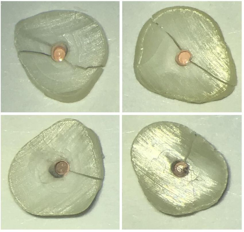
Photographs of the fractured discs were taken (Appendix II, Figure 7 - 11).
21Figure 3: Custom-made stainless-steel pistons were used to apply the load with the MTS 858 Mini Bionix® II
at a constant crosshead speed of 1mm/min directly on the gutta-percha until fracture of the section
occurred.
Figure 4: TestWorks® 4 Software running during load.
22The stress generated by the gutta-percha on the internal canal wall (Figure 5) was
calculated by using the hoop stress formula for thick-walled cylinders (Lame’s
Theorem):
Figure 5: Thick walled cylinder.
Where
σh = hoop stress, i.e. stress in circumferential direction (MPa)
Pi = internal pressure
Po = external pressure
ri = internal radius
ro = external radius
r = radius at point of interest (usually ri)
Statistical Analysis
One – way ANOVA with a 95% multiple range test was used to compare hoop stresses
at failure for all groups (SPSS, TBM). Linear regression was used to examine failure
load versus dentin wall thickness (SigmaPlot 13.0, Systa Software).
23IV. Results
The raw data with respect to the amount of stress exerted by the gutta-percha on
the internal tooth wall with or without sealer types is attached in the Appendix I, Table 4.
Table 1 shows the One – way ANOVA analysis, where there were significant
differences among the groups (p < 0.05). Table 2 shows when multiple comparisons
were made, results showed no significant difference between groups 1 and 2 (p <
0.928), groups 1 and 3 (p < 0.927), groups 1 and 4 (p < 1.000), groups 2 and 3 (p <
0.479), groups 2 and 4 (p < 0.845) and groups 3 and 4 (p < 0.973). Group 5 showed a
significant difference between all the groups (p < 0.05).
Mean failure stresses and standard deviations are presented in table 3 and figure
6. The Tetranite® group had the highest mean failure stress value, which was
statistically significant. The BC Sealer and gutta-percha group had the lowest mean
failure stress value, but this was not significant.
Table 1. One - way ANOVA analysis.
______________________________________________________
stress
Sum of Mean
Squares df Square F Sig.
Between Groups 1555626.585 4 388906.646 25.886 .000
Within Groups 540855.687 36 15023.769
Total 2096482.272 40
24Table 2. Multiple Comparisons
___________________________________________________________
Dependent Variable: stress
Tukey HSD
95% Confidence Interval
Mean Lower Upper
(I) group (J) group Difference (I-J) Std. Error Sig. Bound Bound
1 2 -50.85357 63.43677 .928 -232.9698 131.2627
3 45.08333 55.94597 .927 -115.5281 205.6948
4 12.08611 59.55904 1.000 -158.8978 183.0700
5 -579.12500* 69.87650 .000 -779.7286 -378.5214
2 1 50.85357 63.43677 .928 -131.2627 232.9698
3 95.93690 58.29437 .479 -71.4164 263.2902
4 62.93968 61.77022 .845 -114.3922 240.2715
5 *
-528.27143 71.77051 .000 -734.3124 -322.2304
3 1 -45.08333 55.94597 .927 -205.6948 115.5281
2 -95.93690 58.29437 .479 -263.2902 71.4164
4 -32.99722 54.04894 .973 -188.1626 122.1682
5 *
-624.20833 65.24366 .000 -811.5118 -436.9048
4 1 -12.08611 59.55904 1.000 -183.0700 158.8978
2 -62.93968 61.77022 .845 -240.2715 114.3922
3 32.99722 54.04894 .973 -122.1682 188.1626
5 *
-591.21111 68.36711 .000 -787.4815 -394.9407
5 1 579.12500* 69.87650 .000 378.5214 779.7286
2 *
528.27143 71.77051 .000 322.2304 734.3124
3 *
624.20833 65.24366 .000 436.9048 811.5118
4 *
591.21111 68.36711 .000 394.9407 787.4815
*. The mean difference is significant at the 0.05 level.
Group 1: No Sealer and gutta-percha
Group 2: AH Plus Sealer and gutta-percha
Group 3: BC Sealer and gutta-percha
Group 4: Tetranite® and gutta-percha
Group 5: Tetranite®
25Table 3. Mean Failure Stress (MPa) and Standard Deviations
____________________________________________________________
Tukey HSDa,b
Subset for alpha = 0.05
group N 1 2
3 12 255.0917
4 9 288.0889
1 8 300.1750
2 7 351.0286
5 5 879.3000
Sig. .556 1.000
Means for groups in homogeneous subsets
are displayed.
Figure 6: Failure Stress of Each Group
26V. Discussion
Endodontic treatment is completed by instrumenting, disinfecting, and obturating
the root canal space. Today with the use of the most advanced rotary file instruments
the goal of instrumentation can be accomplished with fewer procedural errors and more
predictability than with the stainless-steel hand files (Hargreaves et al., 2011). With the
introduction of NiTi rotary files in the 1980s and the now thermomechanically-treated
NiTi instruments, endodontics is advancing with each given day.
An increasing number of rotary nickel-titanium (NiTi) file systems have been
marketed by various manufacturers. These systems differ from one another in the
design of the cutting blades, body taper, and tip configuration. Despite the obvious
clinical advantages of these techniques over hand instrumentation, the influence of the
design of the cutting blades is still controversial (Peters et al., 2004; Bergmans et al.,
2002) and could generate increased friction and stresses within the root canal (Blum et
al., 2003). Rotary instrumentation requires less time to prepare canals as compared
with hand instrumentation but result in significantly more rotations of the instruments
inside the canal (Pasqualini et al., 2008). This may cause more friction between the files
and the canal walls.
With using NiTi instruments, a variable degree of rotational force is applied to
root canal walls which can lead to the creation of microcracks or craze lines in root
dentin. The extent of a defect may be related to various contributing factors such as the
tip design, cross-sectional geometry, taper, pitch, and flute form (Yoldas et al., 2012).
The complexity of root canal anatomy, remaining dentinal wall thickness and canal
27diameter after preparation (Rundquist et al., 2006), may also influence the stress
concentration. In addition, the age-related change in microstructure of dentin leading to
progressive dentinal sclerosis may correspond to lower resistance to damage initiation
and propagation (Mireku et al., 2010). The more dentin removed the more chance for a
fracture. The craze lines these could later propagate into vertical root fracture if the
tooth is subjected to repeated stresses from endodontic or restorative procedures (Bier
et al., 2009). It has been shown that the use of nickel–titanium rotary instrument
systems were associated with inducing microcracks in root dentin (Saha et al.,
2017). These microcracks are secondary to endodontic treatment which may eventually
propagate through the remaining dentin and lead to a vertical root fracture.
It is well known that endodontic sealers are used to achieve a satisfactory seal
between the gutta-percha and dentin (Pascon et al., 1990). Having a sealer that
enhances dentin strength would be advantageous. Currently there is a knowledge gap
in endodontics where there is no method to test whether endodontic sealers enhance
dentin strength, which in turn could possibly lessen the propagation of these
microcracks.
In this study we prepared tooth discs obturated with either gutta-percha and no
sealer, AH Plus Sealer and gutta-percha, EndoSequence Bioceramic Sealer and gutta-
percha, Tetranite® and gutta-percha, or Tetranite® and no gutta-percha. These discs
were essentially thick-walled cylinders. Hoop stress was applied to these cylinders until
fracture occurred. A comparison was then made between the fracture loads of each
group. The results of our study do not support the original hypothesis that there was no
difference in fracture loads among the groups (Table 1). Tetranite®/gutta-percha group
28showed to have the highest mean failure stress and the difference was statistically
significant (Table 3). BC Sealer/gutta-percha had the lowest mean failure stress, but
this was not significant compared to groups 1, 2 or 4 (Table 3).
In this study we used 0.5% Sodium Azide solution to store our extracted teeth.
Human teeth used for research and teaching purposes are a potential source of
bloodborne pathogens, according to the Bloodborne Pathogens Standard of the
Occupational Safety and Health Administration (OSHA) (Recommended infection-
control practices for dentistry, 1993). Therefore, sterilization or disinfection of extracted
teeth before their in vitro use is recommended. The optimal storage conditions for
dentin are controversial (Mitchem et al., 1986). Researchers have addressed different
methods for tooth storage such as freezing (Tonami et al., 1996), refrigerating, or
storing at an ambient temperature. Others have described storage of teeth in distilled
water, a physiologic solution, chloramine, formalin, or thymol (Lee et al., 2007; Tosun et
al., 2007; Goodis et al., 1993; Ziskind et al., 2003).
Sodium hypochorite (NaOCl) was not used a storage medium because storage in
NaOCl results in significantly lower bond strength than that of the other treatment
specimens (Mobarak et al., 2010). Chloramine is a close analogue to sodium
hypochlorite, but unlike bleach, it does not affect collagen (O’Brien et al., 1998). It has
been used by several investigators for disinfecting teeth (O’Brien et al., 1998; Haller et
al., 1993; Jörgensen et al., 1985; Munksgaard et al., 1988; Oilo et al., 1990). Various
types of media and methods have been used to keep extracted teeth moist and kill the
bacteria in them. Studies have also been performed to investigate the effect of
autoclaving, boiling, (Tosun et al., 2007) and gamma irradiation (Brauer et al., 2008;
29Pashley et al., 1993) on the integrity of dentin. No significant difference was found in
bond strengths to the enamel of disinfected or sterilized teeth (Shaffer et al., 1985) or to
those stored wet for up to 5 years (Williams et al., 1985).
Dentin moisture in extracted human teeth lacks dentinal fluid. Dentin surfaces
that are moist improve bond strengths when using adhesive systems (Tay et al., 1998;
Van Meerbeek et al., 1998). As a storage medium, Sodium Azide inhibits bacterial
growth in teeth due to a mechanism involving metal ion complexation and displacement
from enzymes (Komabayashi et al., 2009). Sodium Azide increases dentin moisture
within 24 hours. Soaking root dentin in Sodium Azide solution beyond 1 day does not
further increase dentin moisture (Komabayashi et al., 2009). Cross-linking of collagen
when using Sodium Azide is less expected because it is not a fixative.
During preparation of the root canal it is known that a smear layer is created
during cleaning and shaping that covers the instrumented root canal walls (Torabinejad
et al., 2002). This smear layer contains inorganic and organic substances as well as
fragments of odontoblastic processes, microorganisms and necrotic debris. Intracanal
irrigants and medications are used during root canal treatment to reach the natural
complexities and remove the smear layer. Intracanal irrigants exert their effects
mechanically and chemically. Mechanical effects of irrigants are generated by the back
and forth flow of the irrigation solution during cleaning and shaping of the infected root
canals, significantly reducing the bacterial load. Studies show that irrigants that
possess antibacterial properties have clearly superior effectiveness in bacterial
reduction and elimination when compared with saline solution (Byström et al., 1981;
Siqueira at al., 1997). When this layer is removed, the surface area is improved due to
30the increased number of exposed dentinal tubules resulting in better adaptation of the
sealer to dentin by forming sealer tags (Sayin et al., 2007).
AH Plus sealer generated a stronger bond to dentin after removal of the smear
layer (Eldeniz et al., 2005). The smear layer contains moisture. When the smear layer
is not removed, several other sealers demonstrate better bonding to the dentin because
of the remaining moisture, and it might possibly act as a coupling agent by helping the
adaptation quality of hydrophilic materials to the root canal walls (Lalh et al., 1999;
Yildirim et al., 2008). Endosequence BC Sealer is one such hydrophilic material. BC
sealer uses the moisture in the smear layer and creates a hydroxyapatite-like
precipitation while setting, which adheres to dentin chemically (Dawood et al., 2017).
Removal of the smear layer could therefore create a reduction of the BC sealer
adaptation to the canal walls.
In this study, we removed the smear layer for each group, which may have
affected the BC sealer group due to the missing moisture in the smear layer. McComb
was the first to demonstrate the removal of the smear layer with EDTA and showed
canals rinsed with EDTA were free of a smeared layer and superficial debris. (McComb
et al., 1975). Acid exposes surface collagen and removes peritubular dentin from the
top of the tubules (Pashley et al., 1984). Canals rinsed with EDTA creates a zone of
demineralized collagen matrices in eroded dentin and around the dentinal tubules.
Demineralized dentin zones create the opportunity for dentin hybridization by infiltration
of hydrophilic adhesives/sealers. Collapse leads to adhesion/bonding issues (Tay et al.,
2006). Coronal and middle sections are more eroded with EDTA than the apical
erosion of the root canal (Torabinejad et al., 2003).
31Our smear layer was removed with sequential use of 0.5% NaOCl and 17%
aqueous EDTA, with EDTA being the final irrigant which was left in the canal for 1
minute and a final rinse with sterile distilled water to remove residues of the solutions.
To effectively clean and disinfect the root canal system, an irrigant should be able to
disinfect and penetrate dentin and its tubules, offer long-term antibacterial effect,
remove the smear layer, and be nonantigenic, nontoxic and noncarcinogenic. In
addition, it should have no adverse effects on dentin or the sealing ability of filling
materials (Torabinejad et al., 2002). Sodium hypochlorite is the most commonly used
root canal irrigant. Advantages of NaOCl include its ability to dissolve organic
substances present in the root canal system and its affordability. The major
disadvantages of this irrigant are its cytotoxicity when injected into periradicular tissues,
foul smell and taste, ability to bleach clothes and ability to cause corrosion of metal
objects (Gomes et al., 2001). In addition, it does not kill all bacteria, (Sigueira et al.,
1997; Sjogren et al., 1997; Shuping et al., 2000; Shabahang et al., 2003), nor does it
remove all of the smear layer (McCome et al., 1975). It also alters the properties of
dentin (Sim et al., 2001; Grigoratos et al., 2001).
Chelating agents such as ethylenediaminetetraacetic acid (EDTA), citric acid and
tetracycline are used for removal of the inorganic portion of the smear layer
(Torabinejad et al., 2002). NaOCl is an adjunct solution for removal of the remaining
organic components. Irrigation with 17% EDTA for one minute followed by a final rinse
with NaOCl is the most commonly recommended method to remove the smear layer
(Johnson et al., 2009). Longer exposures can cause excessive removal of both
32peritubular and intratubular dentin (Calt et al., 2000). EDTA has little or no antibacterial
effect (Torabinejad et al., 2003).
One limitation of this study was not keeping track of whether the tooth sections
came from the apical, middle or coronal thirds. The dentin in the apical third has fewer
number of dentinal tubules and they have a reduced diameter, which in affect has a
reduced sealer area, then the coronal part. The coronal part of the root canal system
has a more intricate tubular structure. The coronal third has a higher number of dentinal
tubules and the diameter of the tubules is larger and produces better infiltration of sealer
compared to the apical counterpart (Carneiro et al., 2012; Nagas et al., 2011). Our
seemingly large standard deviations would be consistent with this.
Future Studies
Incorporation of all the sealers on the market could be used or upcoming sealers
which have not been released for future studies. Keeping track of the location of the
tooth sections, apical third, middle third, or coronal third may change the outcome.
Tooth selection with more circular shape should be used. Increasing the numbers of
samples and obturating canals with sealer types only could influence results.
VI. Conclusion
Application of a hoop stress provides the field of endodontics a method to test
whether sealers enhance dentin strength. Currently there is a knowledge gap in
endodontics where there is no method to test whether endodontic sealers enhance
dentin strength, and this shows it is possible to do so. Tetranite® enhanced dentin
strength in this study. The hypothesis was not confirmed, and the results showed there
was a significant difference in fracture stress among the groups and Tetranite®.
33VII. Appendix
Appendix I: Raw data
R0 R1 Sealer Force (N) Piston Pressure (MPa) Strength of Dentin (MPa)
Group 1 1.96mm 0.36mm GP 110.9 0.68mm 326.2 326.2
2.68mm 0.36mm GP 120.1 0.68mm 353.2 353.2
2.32mm 0.39mm GP 104.6 0.68mm 307.7 307.6
2.76mm 0.64mm GP 139.3 0.74mm 376.5 376.5
3.02mm 0.27mm GP 114.7 0.74mm 310 310
3.5mm 0.44mm GP 83.7 0.74mm 226.2 226.2
2.87mm 0.57mm GP 81 0.74mm 218.9 218.9
2.2mm 0.48mm GP 123 0.87mm 282.8 282.8
Group 2 2.73mm 0.38mm AH/GP 100.7 0.68mm 296.2 296.2
1.76mm 0.41mm AH/GP 53.5 0.68mm 157.4 157.4
2.2mm 0.4mm AH/GP 136.4 0.68mm 401.2 401.2
2.35mm 0.45mm AH/GP 161.4 0.74mm 436.2 436.2
2.95mm 0.45mm AH/GP 87.5 0.74mm 236.5 236.5
1.67mm 0.22mm AH/GP 180.5 0.74mm 487.8 487.8
3.05mm 0.75mm AH/GP 192.2 0.87mm 441.8 441.8
Group 3 3.3mm 0.35mm BC/GP 76.4 0.68mm 224.7 224.7
2.93mm 0.53mm BC/GP 92.7 0.68mm 272.6 272.6
2.75mm 0.26mm BC/GP 98.6 0.68mm 290 290
2.38mm 0.73mm BC/GP 122.3 0.68mm 359.7 359.7
2.0mm 0.44mm BC/GP 76.9 0.68mm 226.2 226.2
2.28mm 0.43mm BC/GP 91 0.68mm 267.7 267.6
1.75mm 0.25mm BC/GP 89.2 0.68mm 262.4 262.4
2.2mm 0.2mm BC/GP 88.3 0.74mm 238.7 238.7
2.45mm 0.62mm BC/GP 68.8 0.74mm 186 185.9
2.24mm 0.45mm BC/GP 60.7 0.74mm 164.1 164
2.45mm 0.55mm BC/GP 125.1 0.74mm 338.1 338.1
4.1mm 0.85mm BC/GP 100.6 0.87mm 231.3 231.2
Group 4 2.4mm 0.51mm TN/GP 134.1 0.68mm 394.4 394.4
2.88mm 0.35mm TN/GP 108 0.68mm 317.7 317.6
2.64mm 0.52mm TN/GP 111 0.68mm 326.5 326.5
2.82mm 0.34mm TN/GP 113 0.68mm 332.4 332.3
2.2mm 0.49mm TN/GP 80.4 0.68mm 236.5 236.5
2.31mm 0.53mm TN/GP 109.5 0.74mm 296 295.9
4.21mm 0.84mm TN/GP 155 0.81mm 191.4 191.3
2.7mm 0.44mm TN/GP 161.6 0.81mm 199.5 199.5
1.5mm 0.45mm TN/GP 121 0.81mm 298.8 298.8
Group 5 2.19mm 0.44mm TN 235.3 0.68mm 692.1 692.1
1.8mm 0.34mm TN 336.1 0.68mm 694.4 694.4
3.24mm 0.72mm TN 292 0.74mm 789.2 789.2
2.7mm 0.66mm TN 520 0.74mm 1405 1405
2.21mm 0.5mm TN 330.4 0.81mm 815.8 815.8
Table 4: Data collected.
34You can also read