Dental Wear Study in a 14th Century Skull of the Sao Tribe, Cameroon
←
→
Page content transcription
If your browser does not render page correctly, please read the page content below
Coll. Antropol. 30 (2006) 1: 13–24
                                                                                                         Original scientific paper

Dental Wear Study in a 14th Century
Skull of the Sao Tribe, Cameroon
Sylvain Santoni, Laurent Jean Sakka and Jean-Marc Garcier
Department of Anatomy, Faculty of Medicine, University of Auvergne, Clermont-Ferrand, France

ABSTRACT

   The aim of this work was to study the wear affecting the almost complete dentition of a Sao individual fossil from
Cameroon prehistory (XIVth century). Occlusal surfaces of the fossil fragile pieces were plaster replicated with an origi-
nal technique adapted from usual dental impression methods (silicon elastomer polymerising by addition). Axial macro-
photographs of both sectional dental casts and original pieces made it possible to produce drawings of the occlusal areas
on transparencies in order to superimpose the lateral hemiarch counterparts in their optimal intercuspal position. The
study of interarch contacts was completed by confronting and observing the occluding position of hemiarch replicas. The
occlusal analysis revealed that the wear extent was equivalent on left and right molars. Hall’s occlusal wear index and
Van Reenen and Reinach’s classification of proximal wear allow assessment of the degree of wear extent on premolar and
molar sections in relation to the side or the arch observed2,4. The even bilateral proximal and occlusal wears observed on
the different kinds of homologous teeth appeared as the main contributor to this well-balanced interarch occlusion. The
mandibular incisor losses and the particular type of wear affecting lower canines led to the conclusion of the presence of a
labret, a great number of which was found in the area. According to Miles’ method of age assessment based on tooth wear,
the pieces studied belonged to an individual between 30 and 40 years old5.

   Keys words: dental anthropology, dental wear, occlusal wear index, proximal wear index, Miles’ method

Introduction

    The aim of this work is the study of the wear affecting         In the Sao area, numerous »Labrets« were discovered.
an almost complete fossil dentition of a Sao individual         Do the absence of the lower incisors and the special wear
living in prehistoric Africa. In 1973, a mission led by An-     of the canines on the studied jaws mean that our fossil
nie and Jean-Paul Leboeuf unearthed the almost com-             wore a labret? Quantifying dental wear is also a means to
plete skeleton of a Sao individual living in the XIVth cen-     approximate the fossil age at the time of death. The ex-
tury in the Chari-Logone region (Cameroon), a vast hilly        tent of occlusal wear can be indexed on the basis of molar
area where settled ancient inhabitants, collectively nam-       eruption. Miles’ method was chosen to estimate the
ed Sao1. Sao people seem to have disappeared in the             crown height loss of the lower molars5.
XIVth century.
    Twenty-six permanent teeth constitute the exten-            Tooth wear, a general and ancient phenomenon
sively abraded dentition of this rare fossil. On the upper          Whether the result of abrasion or attrition6, wear is a
arch two right incisors are missing and on the mandibu-         generally observed process frequently described in den-
lar arch the four incisors are missing. The present study       tal anthropology7. Tooth attrition is constantly observed
describes, according to Hall’s classification adapted from      in the course of evolution8,9, and gives an information on
Molnar’s scale, the importance and the location of the          the first mammals’ diet10. It has also been helpful in con-
wear process, according to the presence of dentine patch-       firming the existence of a main stage in the evolution of
es2,3. Proximal wear is assessed by the Van Reenen and          the equids11. This wear process was observed in current
Reinach method according to the shape and outline of            mammals12, primates13, chimpanzees14 and hominoids15.
the proximal aspects of the tooth occlusal tables4.             It was studied to enlighten dental sexual dimorphism16

Received for publication May 31, 2005

                                                                                                                              13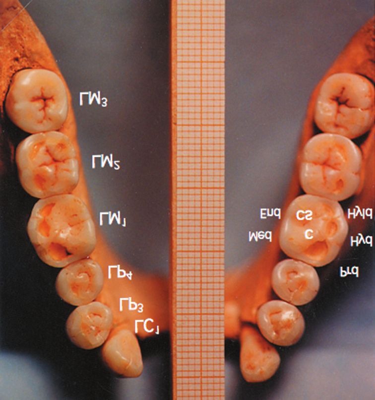
S. Santoni et al.: Dental Wear Study of the Sao Tribe Skull, Coll. Antropol. 30 (2006) 1: 13–24

and to underline d’Amico’s concept of the canine prepon-            In Sinanthropus, Weidenreich (1937) distinguished eight
derant role in the Californian Maidu Indians17.                     degrees62. With the Davies and Pedersen four-stage sca-
    It is necessary to distinguish attrition from enamel            le64, Lysell (1958) gave a numeric value for the occlusal
cracking which should be related to forceful use of the             surface wear and calculated a mean63. Murphy (1959) de-
teeth as additional tools. The presence of abrasive parti-          scribed in a series of successive drawings the different
cles in food increases the wear process17,18. The tooth po-         patterns of wear encountered in Australian Aborigines’
sition along the arch also seemed to be of importance19,20.         jaws. Seven degrees were displayed for the anterior teeth
Dental attrition seems to be related to the diet21. Wear            and nine for the premolar-molar sections37. Baron, Lai
has also been observed in Australopithecus22,23 and more            Son Chan Thu, and Mailland (1972) set up a more precise
particularly studied in A. africanus by microanalysis of            scale for each cusp with eight indices for enamel and
dental tissues24. Microscopic views of dental surfaces be-          eight for dentine65. Their scale was used in other studies
longing to gracile forms of individuals with tender diets           on bruxism66 and on Aborigines67. In a prehistoric popu-
composed of fruits and leaves differ from those with hard           lation of South American Indians, Scott (1979) divided
grain diets as encountered with more robust forms of in-            the occlusal surface into four quadrants. Each quadrant
dividuals.                                                          wear was appreciated in a ten-degree scale. The measure
    The type of attrition in Homo and Australopithecus is           of the occlusal surface wear was finally estimated in a
thought to depend on the dental arch width and the lat-             forty degree resulting scale68,69. He compared his obser-
eral excursions of the mandible25. In neolithic popula-             vations with the eight degrees Molnar’s classification3
tions, the wear observed is very destructive for the dental         and with the nine degrees Hall’s2 classification adapted
arches26–29. On the contrary, in a group of Sudanese indi-          from Molnar. Hall used the descriptions proposed by
viduals of the same period, the wear is considered lighter          Molnar and added an intermediate 2.5 level where the
and associated with periodontal pathologies30. Wear is              cusp had not disappeared. Molnar’s scale is commonly
also recorded in populations living in the Sudan between            used in anthropology70. Benfer and Edwards (1991) con-
700 B.C. and 400 A.D.31, in Eastern France between IVth             sider the crown height as the average of the distance be-
and VIIth centuries32 or in European populations of the             tween the tips and the cemento-enamel junction of the
Middle Ages, in Xth and XIth centuries33,34.                        four molar cusps71. Data obtained from different observ-
                                                                    ers and the use of a double scale taking into account both
    The state of attrition was compared in Australian Ab-
                                                                    the speed and the degree of wear could be beneficial72. A
origines, Anglo-Saxons, Mongols and West Africans, who
                                                                    volumetric »attritional index« was proposed by Abreu
all lived in XIXth century35. Some present-time popula-
                                                                    Tabarini73. In present-day populations, occlusal wear is
tions who have kept ancestral customs, like the South Af-
                                                                    not sufficient to apply the preceding scales. Therefore,
rican Bushmen36 or the Australian Aborigines37–41 are al-
                                                                    the evaluation of wear facets by Gourdon and Woda43 is
ways quoted as references. The chronology of the crown
                                                                    preferred74. A new index is used by Hooper, Meredith and
wear process has been reported in the form of draw-
                                                                    Jagger75.
ings21.

Contemporary man                                                    Proximal wear
   In modern populations, attrition is less frequent and               In mammals, proximal and occlusal wears develop
less severe42–45. The effects of abrasion appear in the             together39,63,76,. Wolpoff (1971) described this kind of
form of facets on the occlusal table where 93% of inter-            tooth wear in groups as different as australopithecines,
arch contacts are established in what is called the Maxi-           chimpanzees, Australian Aborigines and American In-
mal Intercuspal Position (MIP) or centric occlusion46.              dians77. Poitrat-Targowla (1977) studied this type of
Through electron microscopy, the striae that run along              wear in Ibero-Maurusian populations who lived 10.000 to
the wear facets are visible on the surface of replicates            12.000 years ago18. After studying twenty medieval
made of hardened polyvinyl acid47 or plaster47–55. Buccal           dentitions from Northern Sweden, Lysell (1958) argued
and lingual arch surfaces have been observed through a              that attrition in all age groups was more marked for
microscope56 or on digital macrophotos57. Examination of            mandibular incisors and molars, and maxillary premo-
the number, orientation and type of striae made it possi-           lars63. Van Reenen and Reinach (1988) studied proximal
ble to infer the nature of food chewed. A confocal reflec-          wear in Bushmen4.
tion microscope giving a 3D image58 or a profilometer59                Mesial migration is probably not related to occlusal
should have offered a more precise and thus more objec-             wear78. On facets deprived of enamel of neanderthalian
tive assessment of the surface.                                     teeth, striae were identified through electron microsco-
                                                                    py79.
Occlusal wear measurements                                             Proximal wear reduced the mesiodistal length of each
   Different scales have been conceived in the past cen-            tooth. In Aborigines, Begg (1954) estimated at about 14.7
tury60. Broca in 1879 described five numéros descriptifs            mm the mean reduction of mandibular arch length76, but
(degrees) of dentition abrasion extending from the oc-              this was refuted80. Murphy39 estimated on the same
clusal surface to the cemento-enamel junction61. In 1935,           skulls that this reduction amounted to 3.6 mm and
Perier described four degrees, which he did not consider            showed remarkable symmetry on each side from P3 to
perfectly adapted to his study of 150 Bushmen’s skulls36.           M3.

14
S. Santoni et al.: Dental Wear Study of the Sao Tribe Skull, Coll. Antropol. 30 (2006) 1: 13–24

Wear as evidence of function                                     dentition). Cusp denomination is adapted from the Cope
    Tooth wear is a dental trait in close relation with          and Osborn (1895) theory on mammalian molar cusps118.
mastication81,82 and particularly with its neurophysiolo-        This was extrapolated to human molars and premol-
gical component83,84. Interarch contacts correspond first-       ars119,121. Cusps are named cones for the maxilla and
ly to the chewing cycle, which is shaped in its upper part       conids for the mandible.
when going towards centric occlusion (cycle in) and when             At the maxilla, the lingual (palatal) cusp of a premolar
leaving it (cycle out)41. Secondly, the cycle is also guided     and the mesiolingual cusp of a molar are protocones. The
by the temporomandibular joint TMJ85–87 and its possible         buccal cusp of a premolar and the mesiolingual cusp of a
associated pathologies88,89. This was the starting point of      molar are paracones. The distobuccal cusp of a molar is a
studies linking dental anatomy, wear, mastication move-          metacone. On the molars, the protocone, paracone and
ments and diet90–95. But in platyrrhinians, for instance, a      metacone form the trigon. The fourth distolingual cusp is
wide range of dental forms is independent from the diet          called the hypocone or heel.
and chewing behaviours96. The toughest foods are pro-                At the mandible, the buccal cusp of a premolar and
cessed with more lateralized movements of the mandible           the mesiobuccal cusp of a molar are protoconids. The
as was observed in Polynesian Morioris, Maoris97 and             distobuccal cusp and the small distal cusp of a molar are
American Indians81,98. Differences were noted between            respectively a hypoconid and a hypoconulid. The mesio-
hunger-gatherer populations and agriculturalists. Occlus-        lingual cusp and the distolingual cusp of the molar or the
al wear planes have different angulations99. Dental wear         second premolar are respectively a metaconid and an
could represent a mosaic-type evolution100. Moreover,            entoconid. The first premolar only has a metaconid. The
Sakka101–105 underlines a relative dissociation between          protoconid and the metaconid form the trigonid. The
form and function »in the environment of the stoma-              hypoconid, the hypoconulid and the entoconid form the
tognathic system (morpho-functional set) particularly            talonid. The occlusal surface of a mandibular tooth may
represented by the masticatory muscles, their peripheral         look upwards and slightly outwards (ad vestibulum) or
and central innervations, their vasculature, the max-            upwards and slightly inwards (ad linguam). The occlusal
illary and mandibular bony structures, the teeth and             surface of a maxillary tooth may look downwards and
their functional capacities«105.                                 slightly outwards (ad vestibulum) or downwards and
                                                                 slightly inwards (ad palatum). If the occlusal surface
Age at the time of death                                         looks directly downwards or upwards its direction is
    In biological anthropology as in forensic dentistry,         called ad planum.
tooth wear study is one of the criteria taken into account
to determine the age at the time of death5,106–115. Lucy,
                                                                 The moulds and the casts
Pollard and Roberts (1995) recognised the value of Gus-             Moulding with three different viscosity silicon elasto-
tafson or Johanson’s methods116. But these methods only          mer and plaster casting techniques to make replicas has
addressed Scandinavian populations. Song and Jia (1989)          been described in an other paper. The reproducible accu-
studied a Chinese population and established a simple            racy of the method, i.e. the accuracy of the impression
mathematical formula linking wear and age117. The co-            material (irreversible hydrocolloid and elastomer), is lim-
-factors mentioned above did not allow the extension of          ited to 20 µm.
this method to other populations.                                   The examination is made on a cast (or replica) of the
                                                                 total mandibular arch and on 3 sectional replicas, i.e.
                                                                 occlusal, buccal and lingual surfaces of each maxillary
Materials and Methods                                            and mandibular hemiarch. The occlusal replica is sawed
                                                                 to separate the canine from the cuspid teeth.
   The material is constituted of 26 teeth in situ belong-
ing to a single individual:
                                                                 Observation of the samples and replicas
   ¿ 14 maxillary teeth. Six on the right: the canine, two
                                                                     The occlusal wear is analysed by examining the origi-
      premolars and three molars. Eight on the left: two in-
                                                                 nal samples and their casts with a magnifying glass.
      cisors, the canine, two premolars and three molars.
                                                                 Manual staining of the wear facets on replicas of the
   ¿ 12 mandibular teeth. The 4 incisors are missing.            casts and macrophotographs allows a precise description
                                                                 of the extent and intensity of the abrasion process. Ho-
Anthropological nomenclature                                     mologous teeth are compared by juxtaposing the real
   In this study, each tooth is designated by its first let-     tooth to its image replica viewed in a mirror. Left and
ter in capitals, followed by a number corresponding to its       right teeth are also compared by tracing the macropho-
order in the tooth series (incisors, canine, premolars, mo-      tographs on transparencies, allowing superimposition of
lars) increasing from mesial to distal position. The num-        each tooth with its homologous tooth. Comparisons be-
ber is an exponent to indicate an upper tooth and a suffix       tween homologous and contralateral teeth (LM1 and RM1
for a lower one. Letters R (right) and L (left) identify the     for instance) are carried out by superimposition of the
side on which the tooth is located. Premolars are named          first tooth contour on the reversed contour of the other
P3 for the first one, P4 for the second one (assuming that       tooth. The tracings are also compared to those published
P1 and P2 disappeared with the evolution of mammalian            by Miles5.

                                                                                                                              15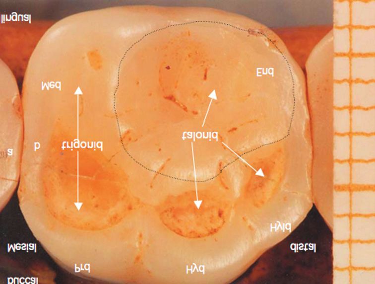
S. Santoni et al.: Dental Wear Study of the Sao Tribe Skull, Coll. Antropol. 30 (2006) 1: 13–24

Wear assessment                                                                             TABLE 1
                                                                      SAO TEETH OCCLUSAL WEAR EVALUATED ACCORDING TO
   Measurements are made with a calliper rule with                    HALL (1976) SHOWING COMPARATIVE DATA BETWEEN LEFT
curved ends with a 0.1 mm precision, which is very close                                AND RIGHT TEETH
to the 0.2 mm precision of a dental practitioner.
                                                                                            Comparison
                                                                                  Hall’s
a) The level of occlusal wear is assessed according to                                      between left          Tipping of
                                                                                  Scale
   Hall’s classification adapted from Molnar’s scale (Ta-                                     side and          occlusal plane
                                                                                  index
   ble 1). This scale allows separate assessments of inci-                                   right side
   sor, canine, premolar and molar wear. Hall established                LI1         4
   eight degrees of abrasion taking into account the loca-               LI2         3
   tion, number and extent of dentine patches. The wear                  C1          4            L
   observed on the Sao fossil is ranked between degrees
                                                                         C1          4            L
   two and four.
                                                                         P3          2,5          R             Ad palatum
b) Proximal wear is assessed according to the six stages
                                                                         P4          3            R             Ad palatum
   of the Van Reenen and Reinach scale. The area of pro-
   ximal wear extends up to the marginal crest of the                    P3          3            R             Ad linguam
   tooth occlusal surface. The outline of the occlusal sur-              P4          3            R             Ad vestibulum
   face could be convex when not abraded (degree one) or                 M1          3            L             Ad palatum
   concave when abraded (degrees three to five). Four de-                M2          3            L             Ad planum
   grees are enough to evaluate the Sao tooth wear.
                                                                         M3          2            L             Ad vestibulum
c) The mandibular intercanine shortest distance is 16                    M1          4            R             Ad vestibulum
   mm. This is the chord of an arc where the four missing
                                                                         M2          4            L             Ad planum
   incisors should be positioned. A fine wax thread is ap-
                                                                         M3          2            L             Ad linguam
   plied along the tooth buccal surfaces from LP4 to RP4
   to give a harmonious contour at the level of the miss-           Letter L (left) or R (right) point out the side of the arch with
   ing incisors. It is then cut at the level of the mesial sur-     greater wear. For each cusped tooth, the wear index is correlated
   face of each canine. The resulting measure on the arch           with the tipping of the occlusal plane. Hall’s scale is adapted
   is 19 mm. This dimension is close to the space com-              from Molnar’s, with the addition of the 2.5 index. From Hall
   monly occupied by four incisor crowns measured at tip            (1976, appendix 1 page 75). Index reading is as follows: 2 – for
   level (22 mm).                                                   molars, wear facets present, no observable dentine; 2, 5 – small
                                                                    dentine patches, cusp pattern not obliterated; 3 – for incisor and
                                                                    canine, cusp pattern obliterated. dentine patches present. For
Age at the time of death                                            premolar and molar: cusp pattern partially or completely oblit-
   Age at the time of death was assessed by Miles’                  erated; small dentine patches. 4 – For incisor and ca- nine, dent-
method5. The measurements of the fossil teeth were                  ine patch (minimal). For premolar: two or more dentine patches;
taken. A set of human teeth of equivalent sizes and forms           secondary dentine may be slight. For molars: three or more lar-
showing no sign of wear120was selected. On the fossil and           ge dentine patches; secondary dentine, none to slight. Degree
                                                                    2.5 corresponds to the second upper bicuspid wear (RP3). This
on the set of teeth without wear the crown heights on the
                                                                    allows the difference from the wear of the con- tralateral tooth
buccal side of the mandibular molar were measured. The              to be shown. Incisors and canines have an index, which is as high,
loss of crown height was calculated by the difference betwe-        or even higher than that of bicuspids and molars, whereas abra-
en the two preceding values. A precision calliper with              sion had less impact on their height. Wear is more important on
curved ends was used. This crown height differences take            M1 and M2 than on their maxillary counterparts. This is due to a
into account the chronology of crown eruption109,110,121,122.       different cusp pattern (thick transverse enamel ridge at the
Therefore, according to Miles, it would take a 6 years              maxilla) and to the occlusal relation between buccal and lingual
time lapse for the first molar to attain the wear observed          cusps (overjet). The right bicuspids are more abraded than the
                                                                    left ones. The left molars are more worn than the right ones.
on the second molar in 6.5 years, and a in 7 years on the
                                                                    The wear index corresponding to each tooth can be specified by
third molar, if we assume that its eruption occurred at 18          the disposition of the occlusal plane. The Latin expressions de-
years of age.                                                       scribing the tipping of the occlusal plane i.e. ad palatum, ad
                                                                    linguam, ad vestibulum124 and ad planum88, indicates the trans-
                                                                    versal slant of the occlusal surface. For example the worn sur-
Results                                                             face of M1 looks downwards and slightly inwards (ad palatum).
   The dentition observed was that of a Sao individual
unearthed in January 1978 by the Annie and Jean-Paul
Leboeuf mission in the United Republic of Cameroon.                 mm wide and 18 to 32 mm thick and were either from
The Region was described as a vast area with hillocks of            massive or hollowed wood. The external part was convex
different sizes corresponding to the settlements of the for-        while both lateral parts were concave to fit the canine
mer inhabitants collectively named Sao1. The dentition is           collar shapes. Thus, we assume that the loss of the four
almost complete consisting of 26 fossil teeth concerned             mandibular incisors result from ritual tribal mutilation.
by proximal and occlusal wear. Around the site of the Sao           Because of their socket persistence, both right maxillary
fossil a lot of labrets were discovered. They were 22 to 37         incisors were lost post-mortem. Wear is noticeable on

16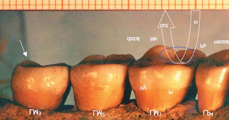
S. Santoni et al.: Dental Wear Study of the Sao Tribe Skull, Coll. Antropol. 30 (2006) 1: 13–24
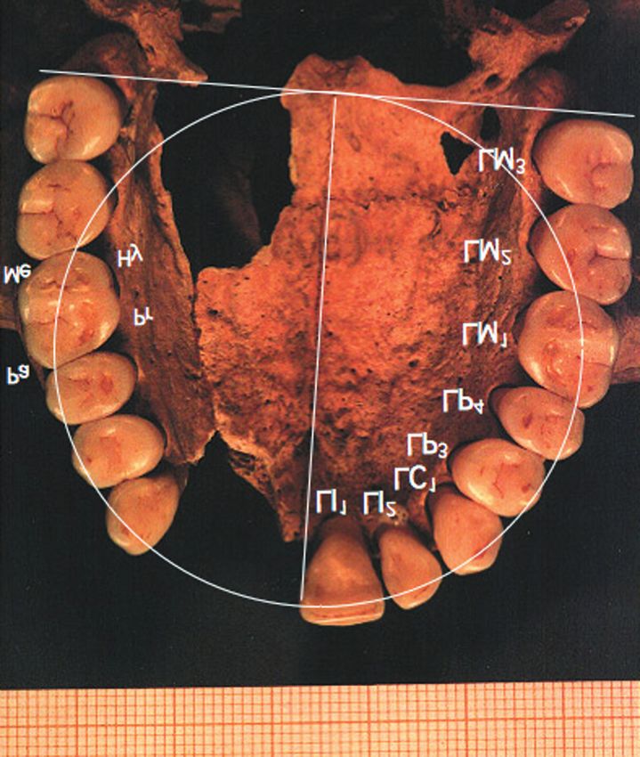
Fig. 2. Occlusal view of the Sao fossile lower arch. The incisors
were probably eliminated prior to insertion of a labret. The arch
is almost V-shaped or ellipsoid type. Anthropological nomencla-
Fig. 1. Occlusal view of the Sao fossile upper dental arch. The ture symbols identifying the first right mandibular molar (RM1)
arch disposition is of parabolic type. Dental wear resulting from cusps: Prd – protoconid, Med – metaconid, Hyd – hypoconid, Hyld
attrition seems equally shared between the anterior and lateral – hypoconulid, End – entoconid. Odontological nomenclature: cf
parts of the arch. A sagittal line corresponding to the diameter of – central fossa, cs – central slope (internal), sc – supporting cusp
a drawn circle covers the intermaxillary suture. A second line (buccal), gc – guiding cusp (lingual).
perpendicular to the first one is drawn tangentially to the two
distal surfaces of the third molars. The circle diameter is the dis-
tance between the occlusomesial angle of LI1 (maxillary left me-
dial incisor) and the posterior line. The circle underlines the
¿ The same degree of wear affects the four remaining
presence of an anterior overjet at arch labial level. The presence maxillary anterior teeth i.e. both canines and the
of interdental spaces can also be noted. Both homologous land- left incisors
marks LM1 and RM1 are on the circle line. Anthropological
¿ The left upper medial incisor (LI1) shows wear loss
nomenclature symbols identifying the right first upper molar
(RM1) cusps: Pr – protocone, Pa – paracone, Me – metacone, (1 mm) on its occlusal aspect
Hy – hypocone. ¿ The left upper lateral incisor (LI2) has a wear facet
erasing half of its distal marginal ridge
¿ The left canines are more occlusally abraded than
occlusal and proximal (mesial and distal) surfaces of all
the right ones. The cusp tips of the lower canines
the teeth, on labial and mesiolabial crown and root parts
are erased forming a flattened area. The lingual as-
of the mandibular canines, and on the buccal aspects of
pects of the maxillary canines are levelled even
the lower molar cusps at the time of death.
where no occlusal contact exists in centric occlusion
¿ The premolars has the same degree of wear at cusp
tip level except for the right first upper bicuspid
Occlusal wear (RP3) which is more abraded. The right premolars
look more abraded than the left ones
The crown height of all fossil teeth was reduced by at-
trition and this process concerns all the cusps. Wear val- ¿ The occlusal surfaces of the lower molars are dis-
ues assessed according to Hall’s scale (1976) are pre- posed in a 3-segment helicoid shape due to their dif-
sented in Table 1. Wear differences between right and ferent individual slants. This helicoid shape begin-
left homologous teeth are recorded and the most worn ning at the levelled occlusal surface of premolars
side is noted. The tipping of the occlusal plane for each wear decreases from the first molar to the third one
crown is indicated according to its orientation (ad ves- (M1 to M3), maintaining a posterior-anterior Von
tibulum, ad linguam or ad palatum). It is noted that: Spee curve (Figure 3)
¿ Most of the occlusal contour is maintained (Figure ¿ The mandibular molars are more abraded than the
1 and Figure 2) maxillary ones
17
S. Santoni et al.: Dental Wear Study of the Sao Tribe Skull, Coll. Antropol. 30 (2006) 1: 13–24

Fig. 3. Buccal view of the lower right mandibular hemiarch of
the Sao fossile. The wear process has levelled and smoothed the
occlusal surface. At molar level, the intensity of wear decreases
from the first molar (RM1) to the third one (RM3). This corre-
sponds to the tooth chronological eruption and orientation. The
orientation of molar occlusal surfaces is helicoidal. M1 occlusal
surface is looking ad vestibulum, M2 ad planum, and M3 ad            Fig. 4. First right mandibular molar (RM1) of the Sao fossile.
linguam. The Von Spee curve (CS) is the result of both tooth erup-   (occlusal view). All the cusps have been worn off. This process is
tion and tooth abrasion and combines with a variable Wilson curve    more pronounced on buccal cusps (supporting cusps) than on lin-
(transversal curve) to form an arch of helicoid disposition (see     gual ones (guiding cusps). Dentine patches are showing through the
                            Figure 5).                               enamel. The occlusal wear here visible is evaluated as stage 4 on
                                                                     Hall’s scale. The enamel surface has been reshaped by wear giv-
                                                                     ing the talonid a mortar like shape (contoured area) in which the
     ¿ In comparison to M2 and M3, the first mandibular              M1 protocone acts as a pestle at the uppermost part of the chewing
       molar (M1) wear reaches dentine level creating                cycle. Intra-arch contacting zones adopt a congruent shape i.e.
       more numerous and wider dentine patches, espe-                the RM1 mesial zone of contact (a) being slightly concave (stage 3
                                                                     of Van Reenen and Reichnach) whereas the RP4 distal one (b) is
       cially on the buccal side
                                                                     convex (stage 1). This interlocking maintains arch cohesion and
     ¿ The first right lower molar (RM1) is more abraded                       compensates for less interarch intercuspation.
       than the left one (LM1),
     ¿ On M1 the loss of enamel of the cusp decreases in                ¿ Maxillary molars keep their occlusal oblique ridge
       the following order: hypoconulid, hypoconid, pro-                  but this structure become less acute or somewhat
       toconid, entoconid and metaconid (Figure 4),                       flat (Figure 5)
     ¿ The uneven wear of the M1 occlusal surface is less               ¿ The occlusal surface of M1 is made of two concave
       noticeable at metaconid level, suggesting a mor-                   areas (trigon and hypocone) separated by a rounded
       tar-like inside wall. This is less visible on M2.                  enamel ridge oriented transversally and obliquely
                                                                          linking metacone with protocone. The mesial part,
     ¿ The concave disposition of the entoconid and part                  larger and more abraded, represents the trigon in
       of the M1 metaconid suggest a kind of mortar in                    which M1 hypoconid comes into contact. The distal
       which the protocone of the opposite tooth actes as a               part is the hypocone (heel) worn by the contacts
       pestle. The same disposition is noted for M2 and M3,               with M2 metaconid and protoconid. The ridge be-
     ¿ On M1 the central slope of the metaconid, occlusally               comes flat on M2 and really bowl-shaped on M3.
       concave, is continued buccally in the form of an                 On the whole, the tooth occlusal abrasion was more
       enamel strip. It is a remnant of the protoconid and           marked on the left molars and on the right premolars
       hypoconid central slope separating trigonid and               compared to the opposite side.
       talonid. It then extended distally with the shape of
       a lingually concave arc separating the buccal and             Proximal wear
       lingual aspects of the talonid. On M2 and M3, with a
                                                                        Proximal wear is observed on all proximal surfaces
       similar occlusal aspect, that shape is less marked
                                                                     except on the distal surface of the third molars and the
     ¿ Occlusal grooves are partially erased on M1 and               lower canines. Wear is assessed with the method of Van
       their Y shape disposition (Dryopithecus pattern) is           Reenen and Reinach4. These values are shown in table 2.
       not easy to make out. The cross disposition of                Mesial and distal surfaces are compared:
       occlusal grooves on M2 and particularly on M3 is                 ¿ In premolars, few differences are observed between
       still visible                                                      mesial and distal surfaces. On the lower premolars
     ¿ The left second and third lower molars (LM2 and                    wear has the aspect of a yellowish oval area and a
       LM3) were more abraded than their right counter-                   dentine patch is clearly visible on RP4 (right second
       parts (RM2 and RM3). Consequently the maxillary                    lower bicuspid)
       molars are more abraded on the left side than on                 ¿ A section of the cast arch between lower canine and
       the right side                                                     first bicuspid enables the appreciation of the contig-

18
S. Santoni et al.: Dental Wear Study of the Sao Tribe Skull, Coll. Antropol. 30 (2006) 1: 13–24
TABLE 2
SAO TEETH PROXIMAL WEAR EVALUATED ACCORDING TO VAN
REENEN AND REINACH (1988). COMPARISON BETWEEN
MESIAL AND DISTAL SURFACES
Mesial surface Distal surface
LI1
LI2
C1
C1
P3 1 1
Fig. 5 Lingual and partly occlusal view of the upper left max- P4 2 2
illary hemiarch of the Sao fossile. Lingual cusps (supporting P3 1 2
cusps) are more abraded than buccal ones (guiding cusps), as can P4 2 2
be seen particularly on first (LM1) and second (LM2) molars. The
occlusal surface is orientated ad palatum for LM1, ad planum for M1 4 1
LM2 and ad vestibulum for LM3. This helicoid disposition of the M2 3 1
upper molar segment is complementary to the opposed mandibu- M3 1
lar one. The big arrow shows the displacement of the opposite
mandibular structure during the uppermost part of the classic M1 3(R) 4(L) 1(R) 2(L)
chewing pattern before (»cycle in«), during and after (»cycle out«) M2 4 1
the mandible reaches centric occlusion. For instance, the LM1 M3 1
hypoconid meets the LM1paracone (Pa) during the cycle in and
glides along the mesial aspect of the oblique enamel ridge during The two first mandibular molars are differentiated by the let-
the cycle out. The trigon fossa widens and levels out into a mor- ters L (left) or R (right). The Van Reenen and Reinach classifica-
tar-like shape in which the M1 hypoconid acts as a pestle (circle). tion was only applied to Bushmen’s molars, but for the Sao teeth
The small arrow shows the posterior abutment of the LM3 (salient it was also used for bicuspids. The oscillating movements occur-
ridge of the LM3 metacone) as distal stopper. ring between the teeth produce this proximal wear. The mesial
surface of a tooth comes into contact with the distal surface of
the more forward tooth. Mesial surfaces are flatter whereas dis-
uous proximal surfaces exposed. There is no visible tal ones are more convex. These different values represent the
wear in these places outline of the worn marginal ridges. The index diffe- rence be-
tween mesial and distal surfaces is greater in the molar section
¿ On the premolar-molar section, wear is more im- than in the premolar section. On both mesial and distal sides the
portant mesially than distally (Figure 4), first left mandibular molar (LM1) is more abraded than its
¿ Proximal wear of the molars was more marked on contralateral counterpart. (1 – convex contact area. 2 – straight
the left side. contact area. 3 – slight concave contact area. 4 – definite concave
contact area).
Canine wear
Wear attack was localised on the labiomesial part of
the crown and root cervical portions (Figure 6). This and to the possible abrasion and wear against the lower
shaping action results from the presence of the labret canines. They could also help to preserve the highly regu-
which modified the previous convex, anterior surfaces of lar shape of the arch.
the lower canines into a flat and perfectly polished sur-
face. Wear is symmetrical on each lower canine. It does- Age of the fossil at the time of death
n’t reach the lingual aspect of the canines. A recession of The time of death is deduced from the abrasion of the
the medial adjacent alveolar ridge shows part of the lower molars. The first lower molar crown wear is as-
rather short canine root. sessed buccally at 3.1 mm on the left side and 3.0 mm on
the right side. The wear of the second lower molar was
The missing incisors 2.5 mm on the left and 2.3 mm on the right. Thus the dif-
The lower arch portion without incisors corresponds ference between first and second lower molars is 0.6 mm
to a 19 mm space on the alveolar ridge. A 1 mm gap on on the left and 0.7 mm on the right. This difference cor-
each side results from the presence of a diastema be- responds to 6.5 years of wear, in relation with the time
tween bicuspid and cuspid. That explains the 21 mm in interval between first and second molar eruptions. Sup-
the evaluation of the lower arch dimension. This is close posing a regular wear time-rate identical on both sides,
to the 22 mm evaluation of the mesial-distal width of the the 3.1 mm wear on the left side should correspond to
four missing incisors, taking into account mean mea- 33.5 years of life. This duration, added to the mean time
surements at tip level. The latter value corresponds to of first molar (M1) eruption, gave a total of 39.5 years at
the width of some of the labrets found near the fossil. Af- the time of death. The same calculation for the right side
ter a first mesial migration of the teeth, the labret con- gave 33.8 years. It was therefore reasonable to assume
tributed to the preservation of their mesiolabial wear that the age of the Sao fossil was between 30 and 40.
19S. Santoni et al.: Dental Wear Study of the Sao Tribe Skull, Coll. Antropol. 30 (2006) 1: 13–24
more accurate than the scales quantifying the massive
dental attrition occurring in ancient populations and
somewhat less accurate than the scales designed for con-
temporary populations. Scott’s scale68,69 could have been
used being more accurate than Molnar’s. Actually, the
main use of Scott’s scale is statistical analyses, which is
not the objective of our study. Hall’s scale is simple. De-
gree 2.5 does not exist in Molnar’s scale and in our study
it corresponded to the first upper bicuspid wear (RP3). It
was then possible to make the difference in the wear of
contralateral teeth. With Molnar’s scale, the value would
have been the same that is 3. The use of a scale reduces
the occurrence of a possible bias. For each tooth series,
except for lower incisors, the continuous shape progres-
sion and the precise description made it possible to un-
derline the role of each tooth or group of teeth. This was
Fig. 6. Mandible of the Sao fossile. Labial and mesial view of the a clue to determine the preferred chewing side.
canine. The four lower incisors were lost ante-mortem. This could
probably be the result of a ritual practice before insertion of a The wear was analysed on original samples and repli-
labret, akin to those found in the Chari and Logone area. Involu- cas. On macrophotographs a precise colour scale helped
tion of the alveolar process followed, leaving a bone ridge with in the evaluation of the intensity of wear. It would have
concave outline. Extension of the alveolar regression is also notice- been useful to compare these records to digital ones. The
able at the canine labial and mesial aspects, thus showing the non-reflecting surfaces of the replicas (in comparison
worn part of the crown and the root in probable relation with the
with the surfaces of the original samples) helped in as-
use of a labret (arrows). Both canine attritioned tips are beyond the
level of the occlusal plane as a result of insufficient blocking con-
sessing the extent of wear areas. Painting the surfaces
tacts from opposite teeth. The labret has undoubtedly played the made it easier to reveal the contours and extent of
role of a retainer as the intercanine distance has been maintained. dentine patches. Computer science should open up re-
search paths and well-adapted software would be useful.
The samples studied are a perfect example of what
Evaluation of wear on the second and third molars
Begg called attritional occlusion76, also described by Camp-
could also help corroborate the first estimation of the fos-
bell123, Beyron41 and Tobias25 in Australian Aborigines.
sil’s age. For instance the 1 mm wear of the second molar
Ackermann124,125 used to mention unequal and dyschron-
crown would have corresponded to 15 years of wear.
ic wear of the teeth. Abrasive processes create planes vis-
Given that the second molar eruption occurs between 18
ible on the arch and the occlusal surface of each tooth.
and 20, we estimate the death between 30 and 40 years of
Two planes are visible on M1 (trigon and hypocone) and
age. The wear reduced the crown height by 1 mm at third
M2 (trigonid and talonid). The angulations between each
lower molar level and perhaps even more at entoconid
tooth occlusal plane and the mean overall arch occlusal
level. If the third molar has erupted around 18–20 years
plane are always difficult to determine2,99, as for the as-
of age its wear would correspond to a function span of 15
sessment of the Von Spee Curve126. However the quanti-
years. This corroborate the preceding deduction.
fication of the wear (depth, area) and its chronology
However the wear process was slowed down by the could be studied with better accuracy through mode-
presence of other teeth and there is no data available to lisation73. The structure of enamel and the modelisation
indicate the third molar time of eruption. Miles’ Table in- of the wear process should stimulate further research.
dicates that according to the wear aspects of the three
In the arch, the interdental attrition process was cor-
mandibular molars the age of the fossil was at least 30.
related with the mesial drift of the teeth77,88,127. Accord-
The slight wear of the upper incisors (1 mm) corre-
ing to Kubein and Kruger128, and Lasserre28 the oscillat-
sponded to the time they were in anterior rubbing con-
ing movements between the teeth produce this proximal
tact with their counterparts. The avulsion of the lower
wear associated with occlusal wear in the posterior sec-
incisors, possibly around 13 to 14 years of age, could have
tions. The contact zone migrates occlusally, as was noted
stopped the wear of the upper incisors and accelerated
by Hillson21. In a posterior-anterior relation, convex pro-
the wear of the posterior teeth. If the labret were more
ximal surfaces are confronted in a horizontal plane.
than 15 mm thick the upper incisor wear would have
Through abrasion they become flat and progressively ac-
continued.
quire an S-shape outline (stage 6 of Van Reenen and
Reinach). It is as if, as the occlusal interlocking flattens
and disappears, a new intra-arch tooth by tooth articula-
Discussion tion appeared increasing the dentition stability. Conspic-
uously on the Sao teeth, the distal proximal surfaces
Dental wear have remained convex as observed by Kubein and Kru-
Hall’s Scale adapted from Molnar seemed suitable to ger. The flatter or hollow mesial proximal surface would
describe and assess the mean wear in one individual. It is have come into contact with the more convex distal prox-
20S. Santoni et al.: Dental Wear Study of the Sao Tribe Skull, Coll. Antropol. 30 (2006) 1: 13–24
imal surface of the mesial tooth129. Some factors should senting of the cusp height loss in function of age is not
be quantified according to the chronology of tooth erup- linear. Wear process cannot be considered as a continu-
tion, the convex shape of proximal surfaces, the thick- ous and constant phenomenon throughout life. Dental
ness and quality of enamel and dietary habits. These fac- eruption is followed by a progressive occlusal confronta-
tors also play a role in the characteristics of occlusal tion concerning, sometimes distally, sometimes mesially,
wear. M1, P3 and P4, then M2 and finally M3, thus increasing
The reduction of the mesial-distal crown length is the interarch confronted surfaces. Therefore, wear pro-
linked to the mesial drift, which reduces the arch length. cess of the first erupted teeth is slowed down. Wear
Campbell, Murphy and Begg’s data on Australian Ab- would have followed 3 stages. The first stage was repre-
origines or Drennan’s data130 on Bushmen are different sented by M1 wear up to 11 or 12 years of age in the case
according to Wolpoff77. The measurements given by Mur- of mixed dentition. The second one was dominated by the
phy were of relative value to quantify the mesial drift39. premolars and M2 wear up to 18 years of age and could
Slightly more than a mean 0.4 mm could have been lost have been accelerated by early avulsion of the mandibu-
on each proximal surface. These figures are far from lar incisors. The third one was slowed down by the pres-
those of a modern population. In adults, Harris131 found ence of permanent teeth, but accelerated by the presence
a very slight reduction of arch length amounted to a of a larger exposed dentine surface compared to enamel
mean 0.25 mm between the distal surface of M2 and the on the early erupted teeth.
mesial surface of C1. Nevertheless these figures depend Thus it seems difficult, from the dental remains dat-
on of the kind and the number of samples, as was under- ing back to African prehistory, to give this human fossil
lined by Corruccini80 when quoting Dawes132. A mesial the age at the time of death. Occlusal and proximal wears
drift occurred on the Sao teeth. Proximal wear concerns were not the sole criteria to be taken into account. To as-
all the teeth except the anterior ones at the mandible by sess the age of the subject, the presence of secondary
absence of contact between canines and premolars, at the dentine, the cement layer, the relative thickness of dental
maxilla except the three left anterior teeth separated by tissues or their composition137, and the root translucence
diastemas. When swallowing, the tongue protrudes through must be considered138. This implies penetrating and part-
the space left by the lack of mandibular incisors and pro- ly destroying the dental structures? This explains why
duces a forward hyperpressure on the upper incisors. the above-mentioned criteria are not assessed on the Sao
Therefore, these diastemas directly derive from the pres- teeth. It was decided to preserve the pieces. The persis-
sure of the tongue. tence of a neat pulp chamber and an almost intact alveo-
lar crest observed on radiographs confirm that the pieces
Estimation of dental age in relation belonged to a young adult. The study of the skeletal
to dental wear characteristics102,139 could bring forth further informa-
According to Vallois107 or Dalhberg133 fossil men gen- tion. The chronology of dental abrasion and the search
erally had short lives. Determining the age at the time of for its aetiology are thus not easy to establish. To trace
death through the study of tooth wear26 was difficult due back to a past activity is impossible to reproduce in the
to the subjectivity of observation (assessment errors), in- present day20. It would have been interesting to compare
terrelation between physical phenomena (dental tissue this type of wear to the one found in today’s older indi-
anisotropy, saliva lubrification), complex kinematics and viduals, and in those suffering from bruxism. But such
dynamics134,135, role differences between supporting cusps severe wear is very rarely encountered nowadays.
and guiding ones, dental axes orientation, TMJ mor-
phology87, diet and chewing habits related to age, social
and cultural habits, etc… all factors leading to different Conclusion
results according to the authors. Modifications of the crown shape are partly related to
The models of mandibular molar wear of the Sao fos- wear. This ancient and general phenomenon in the co-
sil were compared to those presented in Miles’ table. urse of evolution is less frequently observed today. In
Even though Miles’ method is still considered reliable112 that respect, the study of an almost complete dentition of
some criticism are put forward136. The age of the fossil a prehistoric African individual is of particular interest.
deduced from these models could have been around 30. Moulding fragile pieces such as fossilized teeth to obtain
These models present an instant picture at a given time replicas was suitably achieved with a method adapted
of the occlusal surface, as in the models of Murphy’s or from the impression procedures used in dentistry. The
Molnar’s Tables. No author associates an age value to moulding of the occlusal surface alone was performed
the models. Moreover Miles’ Tables only represents man- without exerting any pressure on the original pieces with
dibular molar occlusal surfaces viewed occlusally. They three different viscosities of silicon elastomer. This al-
do not take into account cusp height loss. Some auth- lowed solid replicas of high precision. The observation of
ors5,108 estimate a 1 mm wear reduction for a 6–12 year the original pieces and their replicas through a magnify-
period, but this had never been demonstrated. ing glass was completed with the examination of the cor-
The metric study would have led to the conclusion responding macrophotographs and transparencies. Oc-
that the Sao teeth had sizes fitting those of mean values clusal wear was assessed using Hall’s scale while proximal
of contemporary human teeth. However the curve repre- contact wear was assessed according to the Van Reenen
21S. Santoni et al.: Dental Wear Study of the Sao Tribe Skull, Coll. Antropol. 30 (2006) 1: 13–24
and Reinach scale, thus detailing this perfect model of had evolved to be replaced by a complementary adapta-
typical helicoid wear. tion (helicoid shape) at intra- -arch level. Miles’ method
It appeared that occlusal wear is more marked on the makes it possible to assume that the Sao fossil died be-
left molar side and on the right premolar side compared tween 30 and 40. This does not invalidate the results ob-
to the opposite side. The wear facets noticed on the upper tained with Murphy’s diagrams. In the case of bone and
incisors are probably due to the contacts established with tooth pieces scattered on the ground and belonging to
the opposed lower incisors prior to their avulsion. The one or more individuals, the study of tooth attrition com-
unusual root and crown wear of the lower canine is re- pletes the occlusal analysis. Such elements combined
lated to the labret that plays the role of a retainer in spite with those coming from the metric and morphological
of the mesial drift of posterior teeth. studies are true markers and, as such, a great help in
The wear observed on the Sao teeth reflects his diet identifying the samples and reassembling them.
and masticatory habits. This is why the permanent teeth
were probably submitted to the abrasion process as soon
as the first molars erupted. The type of wear is specific to Acknowledgements
each tooth, even to each cusp, and reflects the way antag-
onistic teeth made contact and interacted. In spite of the This work is a part of the Sao skull study directed by
mandibular incisor loss, the arches stayed balanced and Pr. Michel Sakka, M.D., Dr. Sci., anatomist. We are grate-
stable in the arch distal parts, particularly at molar level. ful for his comments, criticism and his practical help
Proximal wear contributed to maintain interdental abut- along this research. Thanks are also due to Dr. Pierre
ment as if the dental interlocking inter-arch disposition Bourdiol, orthodontist.
REFERENCES
1. LEBEUF, J.-P.: Archéologie tchadienne, les Sao du Cameroun et du F. TWIESSELMANN, Bull. Group. Int. Rech. Sci. Stomatol., 7 (1964) 11.
Tchad. Actualités scientifiques et industrielles n° 1295. (Hermann, Paris, — 35. LAVELLE, C. L. B., J. Dent. Res., 49 (1970) 822. — 36. PERIER, A.
1962). — 2. HALL, R. L., Am. J. Phys. Anthropol., 45 (1976) 69. — 3. L., L'Odontologie, 28 (1935) 687. — 37. MURPHY, T. R., Am. J. Phys. An-
MOLNAR, S., Am. J. Phys. Anthropol., 34 (1971) 175. — 4. VAN REENEN, thropol., 17 (1959) 167. — 38. MURPHY, T. R., Am. J. Phys. Anthropol.,
J. F., S. G. REICHNACH: Interstitial and Occlusal Wear of First Molar 17 (1959) 179. — 39. MURPHY, T. R., Brit. Dent. J., 116 (1964) 483. — 40.
Teeth in San (Bushmen). In: Proceedings. (VIIth International Sympo- MURPHY, T. R,. Arch. Oral. Biol., 9 (1964) 269. — 41. BEYRON, H., Acta
sium on Dental Morphology, Paris, 1988). — 5. MILES, A. E. W., Den- Odontol. Scand., 6 (1964) 598. — 42. GOURDON-LEVADOUX, A.- M.,
tition in the Assessment of Individual Age in Skeletal Material. In: BROTH- Etude macro-anatomique de l'abrasion dentaire dans une population oc-
WELL, D. R. (Ed.): Dental Anthropology. (Pergamon Press, Oxford, Lon- cidentale moderne. PhD Thesis. In French. (Université de Clermont-Fer-
don, New York, Paris, 1963). — 6. KLATSKY, M., J. Am. Dent. Assoc., 26 rand I, Clermont-Ferrand, 1982). — 43. GOURDON, A.-M., A. WODA,
(1939) 73. — 7. KAIFU, Y., K. KASAI, G. C. TOWNSEND, L. C. RICH- Cah. Proth., 43 (1983) 91. — 44. SILLNESS, J., M. BERGE, G. JOHANN-
ARDS., Am. J. Phys. Anthropol., Suppl. 37 (2003) 47. — 8. ROSE, J. C., P. SSEEN, Acta Odontol. Scand., 53 (1995). 331. — 45. BOURDIOL, P., L.
S. UNGAR, Gross dental wear and dental micro-wear in historical per- MIOCHE, Arch. Oral Biol., 45 (2000) 691. — 46. WODA, A., L'Orthodon-
spective. In: ALT, K. W., F. W. ROSING, M. TESCHLER-NICOLA (Eds): tie Française, 57 (1986) 501. — 47. DAHLBERG, A. A., W. KINZEY, Bull.
Dental Anthropology. Fundamentals, limits, and prospects. (Springer, Group. Int. Rech. Sci. Stomatol., 5 (1962) 242. — 48. GRUNDY, J. R., Brit.
Wien, New York, 1998). — 9. YOUNG, W. G., J. Dent. Res., 77 (1998) Dent. J., 130 (1971) 113. — 49. GRIMBERT, L., Act. Odont. Stomatol.,
1860. — 10. CROMPTON, A. W., F. A. JENKINS, Biol. Rev., 43 (1968) 106 (1974) 361. — 50. GRIMBERT, L., J. Biol. Bucc., 2 (1974) 23. — 51.
427. — 11. MCFADDEN, B. J., N. SOLOUNIAS, T. E. CERLING, Science, RAJAONA, Z. J.: Etude par la méthode des répliques de la spécialisation
283 (1999) 824. — 12. BARON, R., M. LEJEUNE, M. KLAPISZ-WOLIK- fonctionnelle des facettes d'usure. PhD Thesis. In French. (Université de
OW, Sci. Rech. Odonto-stom., 2 (1972) 25. — 13. MILLS, J. R., The Dental Clermont-Ferrand I, Clermont-Ferrand, 1982). — 52. ROSE, J. J., Am. J.
Practitioner, 6 (1955) 47. — 14. GORDON, K. D., Am. J. Phys. Anthropol., Phys. Anthropol., 62 (1983) 255. — 53. BROMAGE, T. G., J. Microsc., 137
59 (1982) 195. — 15. MAIER VON, W., G. SCHNECK, Z. Morphol.. An- (1985) 209. — 54. WODA, A., A.-M. GOURDON, M. FARAJ, J. Prosth.
thropol., 72 (1981) 127. — 16. DEAN, M. C., A. D. BEYNON, Z. Morphol. Dent., 57 (1987) 85. — 55. TEAFORD, M. F., O. J. OYEN, Am. J. Phys.
Anthropol., 78 (1991) 425. — 17. D'AMICO, A., J. South. Calif. Dent. As- Anthropol., 80 (1989) 73. — 56. UNGAR, P. S., M. F. TEAFORD, Am. J.
soc., 26 (1958) 239. — 18. POITRAT-TARGOWLA, M. J., Bull. Mém. Soc. Phys. Anthropol., 100 (1996) 101. — 57. PEREZ-PEREZ, A., J. M. BER-
Anthrop. Paris, 4 (1977) 389. — 19. FORMICOLA, V., Bull. Mém. Soc. MUDES DE CASTRO, J. L. ARSUAGA, Am. J. Phys. Anthropol., 108 (1999)
Anthrop. Paris, 3 (1986) 37. — 20. LUKACS, J. R., Dental Morphology 433. — 58. BOYDE, A., M. FORTELIUS, Scanning, 13 (1991) 429. — 59.
and Odontometrics of early Agriculturalists from Neolithic Mehrgarh, WALKER, P. I., E. H. HAGEN, Am. J. Phys. Anthropol., 18 (1994) 203. —
Pakistan. In: Proceedings. (VIIth International Symposium on Dental 60. WALKER, P. L., G. DEAN, P. SHAPIRO, Estimating age from tooth
Morphology, Paris, 1988). — 21. HILLSON, S.: Dental Anthropology. wear in archaeological populations. In: KELLEY, M., C. S. LARSEN (Eds):
(Cambridge University Press. Cambridge, 1996). — 22. GRINE, F. E., Advances in Dental Anthroplogy. (Wiley-Liss, New York, (1991). — 61.
Sca. Microsc., 1 (1987) 647. — 23. GRANAT, J., E. GENET-VARCIN, J. L. BROCA, P., Bull. Soc. Anthropol. Paris, 2 (1879) 128. — 62. WEIDEN-
HEIM, Evolution de la denture permanente des Hominidés. In: Encyc- REICH, F.: The dentition of Sinanthropus Pekinensis: A comparative od-
lopédie Medico-Chirurgicale, Stomatologie, 22003 S 10. (EMC, Paris, 1992). ontography of the Hominids. (Palaeontologia Sinica, 1937). — 63. LYS-
— 24. SPONHEIMER, M., J. A. LEE-THORP, Science, 283 (1999) 368. — ELL, L., Acta. Odontol. Scand., 16 (1958) 267. — 64. DAVIES, T. G. H., P.
25. TOBIAS, P. V., Am. J. Phys. Anthropol., 57 (1980) 173. — 26. SIFFRE, O. PEDERSEN, Brit. Dent. J., 99 (1955) 35. — 65. GENON, P.: Contribu-
A., J. Dent. Belge, 14 (1922) 213. — 27. GISCLARD, L. F., J. LAVERGNE, tion à l’étude du bruxisme chez les enfants de quatre à huit ans. PhD
Act. Odont. Stomatol., 91 (1970) 391. — 28. LASSERRE, J. F. : Usure Thesis. In French. (Université R. Descartes, Paris, 1972). — 66. GENON,
dentaire d'hier et aujourd'hui. (XIèmes Journées du Collège National P., Quintescence Int., 5 (1974) 55. — 67. SERVIERE, F. F., Introduction au
d'Occlusodontologie, Paris, 1994). — 29. GAMBAROTTA, J. P., Bull. concept d'occlusion attritionnelle de Begg. PhD Thesis. In French. (Uni-
Mém. Soc. Anthrop. Paris, 7 (1995) 21. — 30. ZUHRT VON, R., Z. Mor- versité R. Descartes, Paris, 1976). — 68. SCOTT, E. C., Am. J. Phys. An-
phol. Anthropol., 59 (1967) 36. — 31. GUENET, S., F. JANOT, B. TAVER- thropol., 51 (1979) 203. — 69. SCOTT, E. C.. Am. J. Phys. Anthropol., 51
NIER, Inf. Dent., 40 (1998) 3195. — 32. JANOT, F., C. STRAZIELLE, Info. (1979) 213. — 70. ZEITOUN, V., D. AMBROISE, Paléo, 7 (1995) 13. — 71.
Dent., 4 (1993) 209. — 33. BRABANT, H., A. SAHLY, M. BOUYSSOU, BENFER, R. A., D. S. EDWARDS, The principal axis method for measur-
Bull. Group. Int. Rech. Sci. Stomatol., 4 (1961) 382. — 34. BRABANT, H., ing rate and amount of dental attrition: Estimating juvenile or adult
22S. Santoni et al.: Dental Wear Study of the Sao Tribe Skull, Coll. Antropol. 30 (2006) 1: 13–24
tooth wear from unaged adult teeth. In: KELLEY, M. A., C. S. LARSEN nèse du crâne et Origine de l'homme. Table Ronde internationale du 24
(Eds): Advances in Dental Anthropology. (Wiley-Liss, New York, 1991). — au 27 juin 1980, C.N.R.S, Paris, 1983). — 105. SAKKA, M.: Les Origines
72. JOHANSON, A, T. HARALDSON, R. OMAR, S. KILIARIDIS, G. E. de l'Homme. Un autre regard. (Messidor, Paris, 1991). — 106. VALLOIS,
CARLSSON, J. Oral Rehab., 20 (1993), 125. — 73. ABREU TABARINI, H. V., L'Anthropologie, 47 (1937) 499. — 107. GUSTAFSON, G., J. Am.
H. S., Bull. Tokyo Med. Dent. Univ., 42 (1995) 31. — 74. KIM, S. K., K. N. Dent. Assoc., 41 (1950) 45. — 108. ZUHRT VON, R., Deutsche Zahn-Mund-
KIM, I. T. CHANG, S. J. HEO., J. Oral Rehabil., 28 (2001) 1048. — 75. kieferheilkunde, 25 (1955) 1. — 109. BROTHWELL, D. R., The Macro-
HOOPER, S. M., N. MEREDITH, D. C. JAGGER, J. Oral. Rehabil., 31 scopic Dental Pathology of Some Earlier Human Population. In: BROTH-
(2004) 206. — 76. BEGG, P. R., Am. J. Orthod., 40 (1954) 298. — 77. WOL- WELL, D. R. (Ed.): Dental Anthropology. (Pergamon Press, Oxford, London,
POFF, M. H., Studies in Anthropology, 2 (1971) 1. — 78. ROUX, D., C. NewYork, Paris, 1963). — 110. LEGOUX, P.: Détermination de l'âge den-
CHAMBAS, B. NORMAND, A. WODA, Arch. Oral. Biol., 35, 1 (1990) 17. taire de fossiles de la lignée humaine. (Maloine, Paris, 1966). — 111.
— 79. VILLA, G., G. GIACOBINI, Am. J. Phys. Anthropol., 96 (1995) 51. JOHANSON, G., Odontol. Rev., 22 (1971) 1. — 112. LOVEJOY, C. O., Am.
— 80. CORRUCCINI, R. S., Am. J. Orthodont. Dentofac. Orthoped., J. Phys. Anthropol., 68 (1985) 47. — 113. ROSING, F. W., S. I. KVAAL,
97 (1990) 349. — 81. HINTON, R. J., Am. J. Phys. Anthropol., 57 (1982) Dental age in adultes – A review of estimation methods. In: ALT, K. W., F.
103. – 82. LAURET, J. F., M. G. LE GALL, Pract. Periodont. Aesth. Dent., W. ROSING, M. TESCHLER-NICOLA (Eds): Dental Anthropology. Fun-
8 (1996) 807. — 83. PUECH, P. F.: Etude de la surface des Couronnes damentals, limits, and prospects. (Springer, Wien, New York, 1998). —
dentaires en Anthropologie. Apport de la Technique des »Répliques». 114. LAMENDIN, H., E. BACCINO, J. F. HUMBERT, J. C. TAVERNIER,
PhD Thesis. In French. (Université d’Aix-Marseille II, Marseille. 1975). P. M. NOSSINTCHOUK, A. ZERILLI., J. Forensic. Sci., 37 (1992) 1373.
— 84. PINET, R.: Etude parodontologique d'un matériel fossile humain — 115. CRETOT, M., C. LABORIER, Y. PAUTRAT, J. PUJOL, Chir. Dent.
(époque Chalcolithique-Bronze) provenant du Languedoc oriental. PhD Fr., 717 (1994) 43. — 116. LUCY, D., A. M. POLLARD, C. A. ROBERTS, J.
Thesis. In French. (Université de Lyon, Lyon, 1981). — 85. MONGINI, F., Archaeo. Sci., 22 (1995) 417. — 117. SONG, H., J. JIA, Med. Sci. Law, 29
Acta Anat., 92 (1975) 292. — 86. MONGINI, F., J. Prosthet. Dent., 38 (1) (1989) 69. — 118. OSBORN, H. F., Int. Dent. J., 26 (1895) 389. — 119.
(1977) 539. — 87. RICHARDS, L. C., Am. J. Phys. Anthropol., 82 (1990) OLIVIER, G.: Pratique anthropologique. (Vigot Frères, Paris, 1960). —
377. — 88. GASPARD, M.: L'appareil manducateur et la mandibule. 120. WOELFEL, J. B., SCHEID, R. C.: Dental anatomy. Its relevance to
Première partie: Anatomie descriptive, Ontogenèse et phylogenèse de la dentistry. (Williams and Wilkins, Baltimore, Maryland, 1997). — 121.
mandibule humaine. Premier volume. (Prélat, Paris, 1978). — 89. SE- SMITH, P., Am. J. Phys. Anthropol., 37 (1972) 233. — 122. SMITH, B. H.,
LIGMAN, D. A., A. G. PULLINGER, W. K. SOLBERG, J. Dent. Res., 6 Am. J. Phys. Anthropol., 94 (1994) 307. — 123. CAMPBELL, T. D.,
(1988) 1323. — 90. KAY, R. F., Am. J. Phys. Anthropol., 43 (1975) 195. — Dentition and palate of the Australian aboriginal. (The Hassell Press,
91. KAY, R. F., COVERT, H. H., Anatomy and Behaviour of Extinct Pri- University of Adelaide, 1925). — 124. ACKERMANN, F.: Le mécanisme
mates. In: CHIVERS, D. J., B. A. WOOD, A. BILSBOROUGH (Eds.): Food des mâchoires naturelles et artificielles. (Masson, Paris, 1953). — 125.
Acquisition and Processing in Primates. (Plenum Press, New York, 1984). ACKERMANN, F., Rev. Fr. Odonto-Stomatol., 11 (1964) 1063. — 126.
— 92. JANIS, C. M., Predictions of Primate Diets from Molar Wear Pat- SENGUPTA, A., D. K. WITTAKER, G. BARBER, J. ROGERS, J. H. MUS-
terns. In: CHIVERS, D. J., B. A. WOOD, A. BILSBOROUGH (Eds.): Food GRAVE, Arch. Oral Biol., 44 (1999) 925. — 127. BRASH, J. C., Dent. Rec.,
Acquisition and Processing in Primates. (Plenum Press, New-York, 3 (1953) 460. — 128. KUBEIN, D., W. KRUGER, Z. W. R., 13 (1988) 626.
1984). — 93. CHIVERS, D. J., P. ANDREWS, H. PREUSCHOFT, A. BILS- — 129. GASPARD, M., Troubles de l’occlusion dentaire et S.A.D.A.M.
BOROUGH, B. A. WOOD, Concluding Discussion. In: CHIVERS, D. J., B. (PROCODIF, Paris, 1985). — 130. DRENNAN, M. R., Ann. S. Af. Mus., 24
A. WOOD, A. BILSBOROUGH (Eds.): Food Acquisition and Processing (1929) 61. — 131. HARRIS, E. F., Am. J. Ortho. Dent. Orthoped., (1997)
in Primates. (Plenum Press, New York, 1984). — 94. KINSEY, W. G., Am. 154. — 132. DAWES, B. E.: Dental arch crowding in prehistoric man, and
J. Phys. Anthropol., 88 (1992) 499. — 95. SELIGSOHN, D., Analysis of in indigenous racial groups of North America and Australia. M.S. Thesis.
Species-Specific Molar Adaptations in Strepsirhine Primates. In: SZAL- (University of Sydney, Sydney, 1986). — 133. DAHLBERG, A. A., Am.
AY, F. S. (Ed.): Contributions to Primatology. (S.Karger, Bâle, Munich, Assoc. Adv. Sci., 65 (1960) 357. — 134. WEISZFELD, G. A., L'usure occlu-
New York, 1977). — 96. ROSENBERGER, A. L., Am. J. Phys. Anthropol., sale: approche prévisionnelle tribologique. PhD Thesis. In French. (Uni-
88 (1992) 525. — 97. TAYLOR, R. M. S., Acta Anat., 53 (1963) 97. — 98. versité R. Descartes Paris,1990). — 135. WEISZFELD, G., Info. Dent., 5
HINTON, R. J., Am. J. Phys. Anthropol., 54 (1981) 555. — 99. SMITH, B. (1991) 271. — 136. KIESER, J. A., B. PRESTON, W. G. EVANS, J. Ar-
H., Am. J. Phys. Anthropol., 63 (1984) 39. — 100. SZALAY, F. S., Man, 10 chaeol. Sci., 10 (1983) 9. — 137. LAVERGNE, J., D. COLLANGETTES, F.
(1975) 420. — 101. SAKKA, M., Mammalia, 37 (1973) 478. — 102. SAK- HUERTA, Symbioses, 17 (1985) 2. — 138. LAMENDIN, H., Rev. Odonto.
KA, M., C. Acad. Sci. Paris, 277 (1973) 865. — 103. SAKKA, M., Bull. Stoma., 2 (1978) 111. — 139. BACCINO, E., D. H. UBELAKER, L. A. C.
Mém. Soc. Anthrop. Paris, 10 (1983) 285. — 104. SAKKA, M., De la mor- HAYEK, A. ZERILLI, J. Forensic Sci., 44 (1999) 931.
phologie évolutive. In: Proceedings. (Morphologie évolutive. Morphoge-
S. Santoni
Department of Anatomy, Faculty of Medicine, University of Auvergne, 28 place Henri Dunant,
63000 Clermont-Ferrand, France
e-mail: sylvain.santoni@wanadoo.fr
ISTRA@IVANJE ABRAZIJE ZUBA NA LUBANJAMA PLEMENA SAO (KAMERUN) IZ 14. STOLJE]A
SA@ETAK
Svrha ovog rada bila je istra`ivanje abrazije koja je izra`ena u podru~ju cijelog zubala kod fosilnih nalaza iz Kame-
runa koji potje~u iz 14. stolje}a. Okluzalne plohe zuba prou~avane su na sadrenim modelima fosilnih nalaza koji su
otisnuti kori{tenjem adicijskih silikona uobi~ajenom stomatolo{kom metodom. Kori{tenjem makrofotografija modela i
originalnih dijelova zuba bilo je mogu}e rekonstruirati izgled antagonisti~kog zubnog luka na foliji u svrhu pozicio-
niranja u habitualnoj okluziji. Zubni lukovi su fiksirani u zagrizu. Analizom okluzijskih ploha utvr|ena je podjednaka
abrazija na lijevim i desnim kutnjacima. Hallov indeks okluzalne abrazije te Van Reenenoav i Reinachova klasifikacija
interproksimalne abrazije kori{tene su u utvr|ivanju abrazije na pretkutnjacima i kutnjacima obaju zubnih lukova.
23You can also read