Infusion pump flow rates in central venous catheters: thrombus reflux and aspiration clot
←
→
Page content transcription
If your browser does not render page correctly, please read the page content below
cc-by-nc-sa 4.0                                                                                                                         artigo investigação
                                                                                              recebido: 10/12/2020; aceite: 02/03/2021; publicado on-line: 15/06/2021
                                                                                                                               https//doi.org/10.31877/on.2021.42.02

infusion pump flow rates in central
venous catheters: thrombus reflux and
aspiration clot
Perfusão através de infusor em cateteres venosos centrais: refluxo e aspiração de trombos

               Jose Manuel Martinez                                       Rita Capela
               MSc, CNS, Clinical Nurse Specialist, Infection Control     MSc, CNS, Clinical Nurse Specialist, Hematology-
               Department, Portuguese Institute of Oncology Porto         Oncology Department, Portuguese Institute of
               Master degree of Oncology at the Institute of Biomedical   Oncology, Porto
               Sciences Abel Salazar - University of Porto (ICBAS) and    Master Degree of Palliative Care at the Portuguese
               Thomas Jefferson University (USA)                          Catolic University
               jmmartinez@ipoporto.min-saude.pt                           i11124@ipoporto.min-saude.pt

               resumo: As taxas de perfusão contínuas elevadas podem levar a oclusão do cateter venoso central
               (CVC) através do refluxo e aspiração de trombos.
               Metodologia: Foi efetuado um estudo comparativo prospetivo, unicêntrico, incluindo todos os casos
               consecutivos de eventos obstrutivos em doentes hemato-oncológicos que usavam CVC (Hickman® nº7)
               desde agosto de 2018 a setembro de 2019 (Fase 1) e de outubro de 2019 a setembro de 2020 (Fase 2)
               no departamento de Onco-Hematologia do Instituto Português de Oncologia do Porto. Descrição das
               duas fases: Fase 1: Período observacional usando um esquema de QT à base de cisplatino (DHAP), com
               taxas de perfusão contínua ≥ 200 mL/hr. Fase 2: Período de intervenção usando o esquema de QT à
               base de cisplatino (DHAP), com taxas de perfusão contínua ≤ 200 mL/hr.
               Resultados: Ao longo do estudo foram realizados 39 esquemas de QT (DHAP), tendo-se identificado 43
               oclusões de CVC (fase 1, n=28 vs fase 2, n=15). O risco de oclusão associado a altos débitos foi maior
               na primeira fase (fase 1, n=11 vs fase 2, n=3, RR 3.313 [1.010 to 13.863], ≤0.05). Quando foi identi-
               ficada a aspiração de um trombo (n=5), o CVC foi sempre removido. Nenhum trombo por aspiração foi
               observado na fase 2.
               Conclusão: As taxas de perfusão contínuas elevadas podem aumentar o risco de oclusão do cateter
               venoso central (CVC) através do refluxo e aspiração de trombos.
               palavras-chave: Taxas de perfusão; Flush agressivo; Trombos de aspiração; Oclusão.

               abstract: Aggressive infusion pump flow rates can lead to central venous catheter (CVC) occlusion
               resulting from thrombus reflux into the CVC lumen.
               Methods: A single-center prospective comparative study was performed, including all consecutive cases
               of occlusion events in hematology oncology patients using a CVC (Hickman® nº7) since August 2018 to
               September 2019 (Phase 1), and October 2019 to September 2020 (Phase 2) at the Onco-Hematology
               Department of the Portuguese Institute of Oncology (Porto). Two phases were described: Phase 1:
               Observational period using a platinum-based regime (DHAP) undergoing continuous infusion pump
               rate ≥ 200 mL/hr. Phase 2: Intervention period using a platinum-based regime (DHAP) undergoing
               continuous infusion pump rate ≤ 200 mL/hr.
               Results: Overall, 39 DHAP regimens were reported with a total number of 43 occlusions identified in the
               study period (phase 1, n=28 vs phase 2, n=15). Occlusion risk associated with the infusion pump rates
               between phases was higher in phase 1 (phase 1, n=11 vs phase 2, n=3, RR 3.313 [1.010 to 13.863],

16          ON 42 > [JAN-JUN 2021]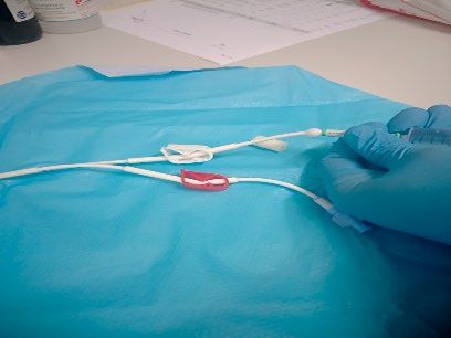
≤0.05). When aspiration clot (n=5) was identified, CVC always was removed. No aspiration clot was
         observed in phase 2.
         Conclusion: Aggressive infusion pump flow rates can increase occlusion risk resulting from thrombus
         reflux into the CVC lumen and aspiration clot.

         keywords: Infusion pump; Aggressive flushing; Aspiration clot; Occlusion.

   Introduction                                                showed that 169 (18.1%) patients reported thrombosis
     Central venous catheters (CVC) devices have been          events during the treatment or within 4 weeks after the
used in hematology oncology patients who require high          last dose, suggesting the necessity of studies of cancer
dose chemotherapy treatment (CT), transfusion support,         patients undergoing cisplatin-based regimes (Moore et
and blood sampling for an extended period of time              al, 2011). However, research into this issue remains scarce.
(Zakhour, Chaftari, Raad, 2016). Large osmolarity spec-             The continuous infusion pump flow rate and aggres-
trum drugs, multiple infusions, and perfusion volumes          sive flushing increase the risk of endothelial damage asso-
related to treatment and secondary complications (e.g.,        ciated with the continued vessel wall contact by the CVC
septicemia) lead to a higher continuous CVC use ratio          tip (Gunawansa, Sudusinghe & Wijayaratne, 2018).
( Joint Commission Resources, 2012).                           Moreover, it could increase the risk of a filling defect
     Catheter-related occlusion due to mechanical obs-         near the catheter tip or retrograde flow along the external
truction and catheter-related infection are the most           surface of the catheter leading to extrinsic fibrin sheath
important complications in the management of CVCs              formation (which causes up to 43% of catheter dysfunc-
(Baskin et al, 2009; Callister et al, 2015. Catheter-related   tion), (Beard, Gaines & Loftus, 2013). Blood withdrawal
occlusion can occur from two different sources: throm-         and flushing procedures (e.g., ineffective positive pressure
botic and nonthrombotic (Baskin et al, 2009; Cesaro et         and push-pause techniques) could increase the accumu-
al, 2004). A clot is considered the most common cause of       lation risk of blood deposits in the catheter lumen. In
thrombotic occlusion. Occlusion (partial and complete)         these cases, the fibrin deposit can progress to an intrinsic
should be considered when the capacity to withdraw             thrombosis, such as intraluminal thrombus or fibrin tail
blood is compromised and the ability to flush fluids is lost   formation on the catheter tip (Hadaway, 2005).
(Smith et al, 2017). Nonthrombotic causes such as cathe-            The aim of this study is to evaluate the occlusion
ter pinch-off, precipitation of drugs, and catheter migra-     events associated with a new platinum-based regimen
tion can result in the inability to aspirate blood as well     undergoing salvage with continuous infusion pump flow
(Smith et al, 2017). Catheter-related occlusions usually       rates.
are reported in the literature in the second and third week
after CVC placement (Napalkov et al, 2013).                       Methods
     Thrombotic occlusions could be associated with treat-         Selection and Description of Participants
ment-related factors such as bolus chemotherapy infu-              A single-center prospective comparative study was
sions, antigenic or platinum therapy (irritant and exfoliant   performed, including all consecutive cases of occlusion
drugs), chest radiation therapy, erythrocyte stimulating       events associated with a new platinum-based regimen
agents and parenteral nutrition (Kitchens, Konkle &            (DHAP) in hematology oncology patients using a CVC
Kessler, 2013). Moore and colleagues performed in 2008         since August 2018 to September 2019 (Phase 1), and
a large retrospective analysis of all patients treated with    October 2019 to September 2020 (Phase 2) at the Onco-
cisplatin-based chemotherapy for any type of malignancy        -Hematology Department of the Portuguese Institute of
at Memorial Sloan-Kettering Cancer Center. The study           Oncology (Porto).

                                                                                              ON 42 > ANO XIV ∙ JAN-JUN 2021   17
Patients older than 18 years old, with the new plati- Flushing frequency was reporting in periods ≤ 72 hrs
num-based regimen (DHAP) and CVC were included. in hospital admissions and periods ≤ 30 days after hospi-
tal discharge.
Phase and Platinum-based regime (DHAP) tech- Alteplase protocol was performed by syringe or stop-
nical information cock method declotting when complete occlusions were
Phase 1: Observational period using a platinum-ba- reported.
sed regime (DHAP) undergoing continuous infusion
pump rate ≥ 200 mL/hr. Data Analysis
Phase 2: Intervention period using a platinum-based Data analysis is conducted using IBM SPSS Statistics
regime (DHAP) undergoing continuous infusion pump for Windows (SPSS Inc., Version 24.0). A continuous
rate ≤ 200 mL/hr. To reduce the continuous infusion variable was reported by median and range. Categorical
pump rate, volume redistribution between perfusions was variables were reported as frequency and percentages.
performed. Normality tests reported a sample without normal distri-
DHAP: Dexamethasone (po or IV, day 1–4), High- bution, considering that, hypothesis tests were analyzed
-dose Ara-C-cytarabine (IV infusion over 2 hrs, day 2, by non-parametric tests. Relative Risk was performed by
every 12 hrs) and Platinol (IV infusion over 24 hrs, day 1). confidence interval of 95%. A p value of ≤ 0.05 was deter-
Infusion therapy volume including IV hydration therapy mined to be statistically significant.
and CT in the first 36 hrs ≥ 9000 mL.
Results
Occlusion definitions A total number of 43 occlusion events were identified
Occlusion was considered when the capacity to with- in the study period (phase 1, n=28 vs phase 2, n=15). The
draw blood was compromised and the ability to flush DHAP regime occlusion events were reported in 32.5%
fluids was lost (Smith et al, 2017). Partial occlusion (ina- (n=14) cases. Overall, 39 DHAP regimens were reported,
bility to aspirate blood, but ability to infuse fluids through with no identified significant DHAP regimen distribu-
the catheter lumen) and complete occlusion (inability tion differences between phases (phase 1, n= 21 vs phase
to aspirate blood and infuse fluids through the catheter 2, n=18, p>0.05). The occlusion risk associated with the
lumen) were considered. Catheter lock was considered infusion pump rates between phases undergoing DHAP
when the solution was injected into the catheter lumen regime was higher in phase 1 (phase 1, n=11 vs phase 2,
dead space until it was to be accessed again (Smith et al, n=3, RR 3.313 [1.010 to 13.863], ≤ 0.05).
2017). Flushing protocol was performed a median of 17 (6 to
22) days after discharge. In 78.6% (n=11) cases the occlu-
Technical Management CVC Hospital Policy sion event was reported after 30 days of CVC-life (median
The management of CVCs follows the CDC (2011) 43 days, 12 to 126). The CVC was removed in 64.3% (n=9)
guidelines. Double lumen Hickman® type catheters of occlusion events. Restoration of CVC patency under-
(Vygon®) are used (7 French, lumen No.1=0.6 and lumen going alteplase protocol was observed in 35.7% (n=5)
No.2=1.0). Control chest x-ray was always performed cases. A complete occlusion in the CVC 0.6 mm lumen
after CVC insertion. was observed in each case, and complete occlusion of the
Specific CVC flush and catheter care includes the use of CVC 1.0 mm lumen at the same time as occlusion of the
SASH technique ( Smith et al, 2017), catheter lock with 10 CVC 0.6mm lumen on 2 occasions. When aspiration clot
mL normal saline solution flush and anticoagulant (Fibri- (n=5) was identified, the CVC was always removed. No
lin®), positive pressure technique (Hadaway, 2005), volume aspiration clot was observed in phase 2.
of syringe used ≥ 10 mL, neutral split-septum needleless
connector (Bionecteur®), and push-pause flushing (± 2.5 Discussion
mL pulses using ≥ 10 mL normal saline solution). The infusate nature (exfoliant and irritant drugs),
patient characteristics (hematology oncology patients),
18 ON 42 > [JAN-JUN 2021]device characteristics (multi-lumen CVC), or proposed When the CVC patency was not restored and the
duration of venous access (long-term CVC) are impor- CVC was removed, it was tested with a 10 mL syringe
tant factors to vascular access indication (Zakhour, Cha- saline solution administration being a “bubble” formation
ftari & Raad, 2016; Joint Commission Resources, 2012; in the hub observed. (Figure 2) The CVC was sectioned
Baskin et al, 2009; Cesaro et al, 2004; Napalkov et al, 2013; and a clot (±3 cm) in the proximal section (CVC-line
Chopra et al, 2015). In the DHAP regime all these factors bifurcation) was reported.
are observed. In oncology hematology patients, partial
occlusions could influence the blood sampling procedu-
res; as PICC occlusion rates are higher in this population,
the Hickman CVC could be considered a safer option for
CT in these cases (Lim et al, 2013).
Theoretical rationale approach
In the first 36 hrs of the DHAP regime, the salvage
infusion therapy volume, multi-lumen CVC use (multi-
ple continuous perfusions), blood transfusions, monoclo-
nal antibody administration, and use of platinum-based
regime could increase the risk of generating high turbu-
lence, pressure and reflux in the CVC tip, and possible
vessel damage as well.
Since the beginning of DHAP administration, an
increasing number of complete occlusions were identified.
In some cases, complete occlusions were observed during Figure 2. “Bubble” formation in the hub aspiration
blood sampling, as the CVC was functional (blood with- Clot: Infusion pumps and Aggressive flushing.
drawal of 3 mL/3s, (Cummings- Winfield & Mushani,
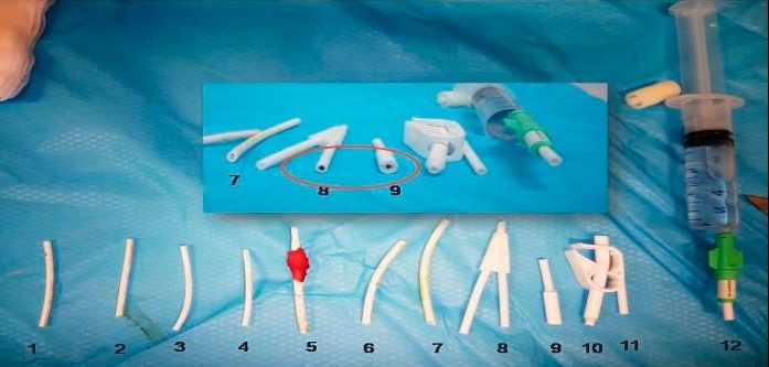
2008)) until a clot was aspirated into the CVC lumen. In Changes in infusion pressure can lead to thrombus
other cases, complete occlusions were observed between formation resulting from reflux (Hadaway, 2005). In our
infusion pumps or in the final stage of bolus drug admi- study, an aspiration clot was never observed in the first
nistration as well. The negative pressure applied between DHAP regime, reinforcing the idea that the extraluminal
infusion pumps and bolus drug administration could clot was produced in this phase, then aspirated and iden-
suggest a higher risk of retrograde flow along the external tified in subsequent CT phases. After the first DHAP
and internal lumen surface. (Figure 1) regime, negative pressure applied through the CVC
lumen during blood withdrawal could remove the exter-
Vein vessel nal CVC surface fibrin tail and aspirate it into the lumen.
Displacement of CVC
undergoing pressure The clot was aspirated through the CVC lumen until the
Hickman® bifurcation (Figure 3), as the difference bet-
Infusion Pump Or
Agressive flushing
ween lumen sizes is an important factor to position the
clot in this region. Indeed, the narrower lumen increa-
Mural Thrombus
sed the reflux distance observed, which could explain the
Possible vessel damage higher frequency of complete occlusion observed in the
Vein vessel 0.6 mm CVC lumen.
Fibrin sheat
Catheter tip clot
Period between Pumps
or Syringe pluger Blood
rebounds (retrograde reflux)
Figure 1. Risk of a retrograde flow along the external and
internal lumen surface.
ON 42 > ANO XIV ∙ JAN-JUN 2021 19References
• Zakhour, R., Chaftari, A. M., & Raad, I. I. (2016). Catheter-
related infections in patients with haematological
malignancies: novel preventive and therapeutic strategies.
The Lancet Infectious Diseases, 16(11), e241-e250.
• Joint Commission, Joint Commission Resources, Inc, &
Joint Commission International. (2012). Preventing central
line-associated bloodstream infections: a global challenge,
a global perspective. Joint Commission Resources.
• Baskin JL, Pui CH, Reiss U, Wilimas JA, Metzger ML, Ribeiro
RC, et al, (2009). Management of occlusion and thrombosis
associated with long-term indwelling central venous
catheters. The Lancet, 374(9684), 159-169.
• Callister D, Limchaiyawat P, Eells SJ, Miller LG. (2015). Risk
Figure 3. Aspiration clot into the CVC lumen. Factors for Central Line–Associated Bloodstream Infections
in the Era of Prevention Bundles. Infection Control &
Hospital Epidemiology, 36(2), 214-216
When infusion pump flow rates are increased, the nega- • Cesaro, S., Corrò, R., Pelosin, A., Gamba, P., Zadra, N.,
tive pressure applied through the CVC lumen between Fusaro, F., ... & Cavaliere, M. (2004). A prospective survey
on incidence and outcome of Broviac/Hickman catheter-
pumps could aspirate the clot through the catheter tip related complications in pediatric patients affected
by hematological and oncological diseases. Annals of
into the lumen (Hadaway, 2005).The inappropriate drug Hematology, 83(3), 183-188.
bolus administration by aggressive flushing through the • Smith, S. N., Moureau, N., Vaughn, V. M., Boldenow, T.,
administration system could produce the same effect as Kaatz, S., Grant, P. J., ... & Chopra, V. (2017). Patterns
and predictors of peripherally inserted central catheter
well (syringe plunger rebounds and draws blood back), occlusion: the 3P-O study. Journal of Vascular and
Interventional Radiology, 28(5), 749-756.
(Hadaway, 2005). In both cases, an aspiration clot resul-
• Napalkov, P., Felici, D. M., Chu, L. K., Jacobs, J. R.,
ting in catheter tip occlusion could be observed. (Figure &Begelman, S. M. (2013). Incidence of catheter-related
complications in patients with central venous or
4) hemodialysis catheters: a health care claims database
analysis. BMC Cardiovascular Disorders, 13(1), 86.
• Kitchens, C. S., Konkle, B. A., & Kessler, C. M. (2013).
Consultative Hemostasis and Thrombosis: Expert Consult-
Online and Print. Elsevier Health Sciences.
• Moore, R. A., Adel, N., Riedel, E., Bhutani, M., Feldman,
D. R., Tabbara, N. E., ... & Hassoun, H. (2011). High
incidence of thromboembolic events in patients treated
with cisplatin-based chemotherapy: a large retrospective
analysis. Journal of Clinical Oncology, 29(25), 3466.
• Gunawansa, N., Sudusinghe, D. H., & Wijayaratne,
D. R. (2018). Hemodialysis catheter-related central
venous thrombosis: clinical approach to evaluation and
management. Annals of Vascular Surgery, 51, 298-305.
• Beard, J. D., Gaines, P. A., & Loftus, I. (Eds.). (2013).
Figure 4. Thrombus reflux resulting in a catheter tip occlusion Vascular and Endovascular Surgery E-Book: Companion to
undergoing continuum infusion. Specialist Surgical Practice. Elsevier Health Sciences.
• Hadaway, L. C. (2005). Reopen the pipeline for IV therapy.
Nursing 2005, 35(8), 54-61.
Conclusions • Chopra, V., Flanders, S. A., Saint, S., Woller, S. C., O’Grady,
N. P., Safdar, N., ... & Pittiruti, M. (2015). The Michigan
Aggressive infusion pump flow rates can increase occlu- Appropriateness Guide for Intravenous Catheters (MAGIC):
sion risk resulting from thrombus reflux into the CVC results from a multispecialty panel using the RAND/UCLA
appropriateness method. Annals of Internal Medicine,
lumen and aspiration clot. 163(6_Supplement), S1-S40.
• Lim, M. Y., Al-Kali, A., Ashrani, A. A., Begna, K. H.,
Elliott, M. A., Hogan, W. J., ...& Patnaik, M. S. (2013).
Acknowledgments Comparison of complication rates of Hickman® catheters
versus peripherally inserted central catheters in patients
The authors wish to thank Dr Gillian Ray-Barruel for with acute myeloid leukemia undergoing induction
editing assistance. chemotherapy. Leukemia& Lymphoma, 54(6), 1263-1267.
• Cummings-Winfield, C., & Mushani, T. (2008). Restoring
patency to central venous access devices. Clinical Journal of
Oncology Nursing, 12(6), 925.
20 ON 42 > [JAN-JUN 2021]You can also read