A robotic respiration phantom with patient data synchronization for medical tomography - IOPscience
←
→
Page content transcription
If your browser does not render page correctly, please read the page content below

Journal of Physics: Conference Series

PAPER • OPEN ACCESS

A robotic respiration phantom with patient data synchronization for
medical tomography
To cite this article: T Szabaa et al 2021 J. Phys.: Conf. Ser. 1782 012037

View the article online for updates and enhancements.

                                This content was downloaded from IP address 46.4.80.155 on 06/05/2021 at 12:04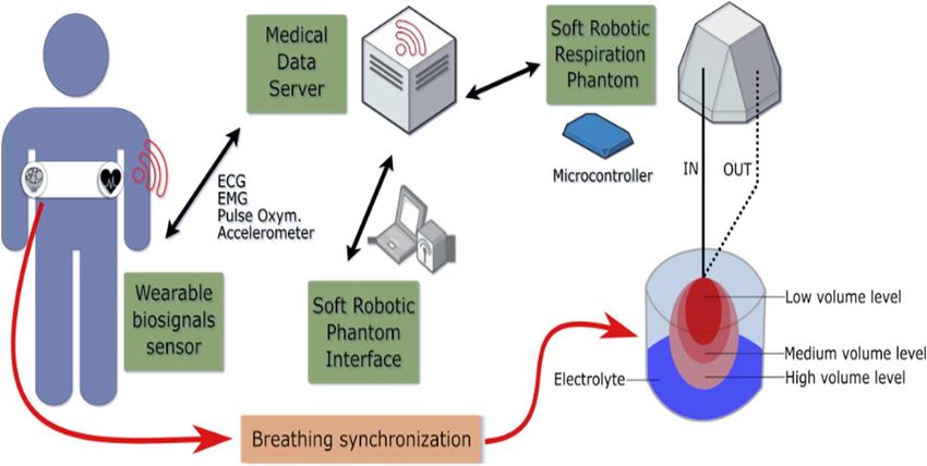
PTZE2020                                                                                                        IOP Publishing
Journal of Physics: Conference Series                         1782 (2021) 012037          doi:10.1088/1742-6596/1782/1/012037

A robotic respiration phantom with patient data
synchronization for medical tomography

                     T Szabała1, T Rymarczyk1,2 and A Vejar1,2
                     1
                         Research and Development Center, Netrix S.A., Lublin, Poland
                     2
                         University of Economics and Innovation, Lublin, Poland

                      e-mail: tomasz.rymarczyk@netrix.com.pl, andres.vejar@netrix.com.pl

                     Abstract. In order to benchmark on-line algorithms for electrical tomography we have designed
                     a dynamic soft robotic phantom system. The robotic phantom will be synchronized with real-
                     time measurements of a patient and it will support on-line tomographic algorithms during
                     dynamic conditions. The system would allow to embody the kinematics in the tomographic
                     inversion, for instance when using model predictive control to trigger the data acquisition at the
                     beginning and at the end of the breathing process.

1. Introduction
With the possibility to choose multiple algorithms for tomographic inversion and optimization [1-14]
and in order to achieve a good performance, there is an extreme increment of complexity when the
scenario is dynamic. With the combination of hardware, software and patient dependent variables,
testing the health state of the lungs in dynamic conditions might seem as a difficult task. For example,
by the use of time-dependent partial differential equations, the increment of model formulation
complexity is enormous. Simplification of static techniques while testing the state of patient during
research, could provide invalid results. That is the reason of the use of robotic phantom in medicine, e.g.
to assure accurate dosage on radiotherapy [15].

                                           Figure 1. Dynamic Phantom platform

              Content from this work may be used under the terms of the Creative Commons Attribution 3.0 licence. Any further distribution
              of this work must maintain attribution to the author(s) and the title of the work, journal citation and DOI.
Published under licence by IOP Publishing Ltd                          1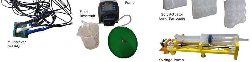
PTZE2020                                                                                   IOP Publishing
Journal of Physics: Conference Series          1782 (2021) 012037    doi:10.1088/1742-6596/1782/1/012037

   We have designed a robotic pump–lung system, presented in Figure 1 in which an elastic balloon is
actuated to represent the lungs, as a first approach. As a second approach for more appropriate
biomechanics, we use a soft-robotic lung surrogate. With these systems, we can recreate the alternating
conditions of the human lungs. The phantom is submerged in an electrolyte tank, and it is analyzed by
electrical excitation on the tank-electrolyte interface for further generation of electrical tomography
imaging.

2. System Design
The diagram of Figure 1 briefly describes how the system works. The real-time information, delivered
by wearable biosignals sensor flows to the Medical Data Server. Sensors are installed onto a thoracic
belt and they are operated by a small microcontroller. The sensors used are accelerometer, ECG, EMG
and pulse using and optelectronic sensor. They continuously send data, which is mined and encoded into
commands for the Robotic Respiration System. The Breathing synchronization process convert the
biosignals to the actuator signals transmitted by the Microcontroller into the Robotic Respiration
System. Robotic Respiration System is placed in a special tank, filled with electrolyte for the study of
electrical tomographic imaging. Because of multiple conditions tested with the system, doing research
on a human patient is unpractical. The phantom dynamically imitate the behavior of human lungs to
explore the behavior of algorithms and data acquisition system with respect to respiration biomechanics.

3. Soft Robotic Respiration Phantom
Soft-robotics [16] is a subset of robotics characterized by the use of actuators systems that qualitatively
resembles bio-mechanical systems. That is represented by the compliance degree of the material [17],
and their consequently fuzzy border between the concepts of mechanism and the materials that compose
the mechanism.
    This characteristics are very important for biomedical applications [18] and appropriate for a
respiration phantom, given that it needs to replicate the human respiratory process in a manner as similar
to reality as possible. In this study we use as a first approach a latex balloon as actuator system, where
the fluid transfer allows to change dynamically the shape of the balloon representing lung shape
variation. The fluid transfer is provided by a pump subsystem, that is driven by a microcontroller which
allows to dynamically pump-in and pump-out fluid into the balloon system. In the pilot study we use a
controlled peristaltic pump to drive the actuation in the robotic phantom. It can generate a pressure
pattern that allows the oscillatory behavior of the balloon, following the volume dynamics of Figure 3
but it is far to be representative of the real respiration, giving the switching behaviour of the pump. To
get another level of biomimicry as a second approach we replicate the work of [19] on surrogate lungs
to use this design as a soft robotic actuator. Our production process is presented in Figure 2. To reduce
the impulsive effect of the peristaltic pump, a customized syringe-based pump, in the style of [20], was
designed and constructed using additive manufacturing.

                                Figure 2. Soft Actuator Production Process

                                                     2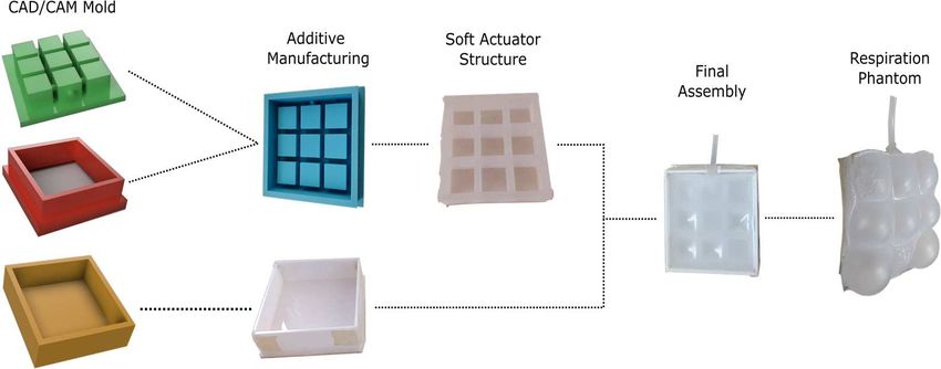
PTZE2020                                                                                   IOP Publishing
Journal of Physics: Conference Series          1782 (2021) 012037    doi:10.1088/1742-6596/1782/1/012037

                             Figure 3. EIT tank system for Dynamic Phantom

   The Figure 3 describes the tomographic tank system. Main reservoir is a 10L food-grade bucket. On
the surface of the bucket, we placed 32 graphite electrodes made of HB-type 33 mm diameter graphite
leads. Commonly in use in electrochemistry [21] graphite electrodes are proven to be effective in
biosensing applications [22]. The graphite electrodes are placed in 3D printed holders and they are
connected with small cooper plates and soldered to the wires. The balloon can variate its shape given a
reference signal transmitted to the pump automatically using a microcontroller that control speed steps,
pump switch direction. In this manner, the effect of the 3 points peristaltic actuation per cycle can be
controlled.

4. Wearable Biosignal Sensors
Considering modern MCU-based systems, we can collect large amounts of data from multiple sensors.
To obtain the redundant breathing data and other patients bio signals, we equipped the belt with multiple
sensor units. To monitor the chest movement during breath, we have used accelerometer and gyroscope.
We have installed the sensors on an elastic belt, in the middle of a chest. To monitor patients hearth, we
have used an opto-sensor to track the pulse signal. It gives us a simple hearth activity information to
analyze the joint effect of breathing pattern and blood pulse pattern.

Figure 4. Breathing activity (top left) and blood pulse opto-sensor (bottom left) signals. Right:
Gyroscope signal representing movements on the normal direction from the chest surface during five
breathing cycles

5. EIT Reconstruction
The tomography acquisition with the device [23], with an emitter with waveform reconfigurable
capabilities [24]. The used tomographer was architectured using a FPGA device [25,26]. To synchronize
the respiration changes into the robotic phantom, a map of the volume variation of the soft-actuator to
the real-time measured chest activity of Figure 4 can be estimated. In this section we simulate the

                                                     3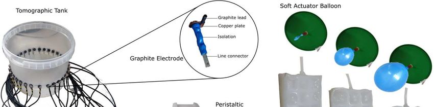
PTZE2020 IOP Publishing
Journal of Physics: Conference Series 1782 (2021) 012037 doi:10.1088/1742-6596/1782/1/012037
tomographic tank system of Figure 3, under the volume variations provided by the breathing activity
tracking. This volume variations are translated to controlled inputs to the soft-robotic system actuator,
modifying the conductivity properties of the system. The possibility of exploration of tomographic
reconstruction algorithms under dynamics changes in the observed system is one of the main goals of
this project.
In this section we present the numerical models and simulation of the tank system subject to
dynamic mechanical perturbation of the soft robotic actuator, to emulate the respiration activity.
For simplicity in design, a 32 electrode tomographic system with fixed-type electrodes is structured
as follows:
• There are four types of electrodes: positive excitation (set A), negative excitation (set B), positive
sensing (set M), negative sensing (set N).
• Every physical electrode can be of only one type.
• The electrodes types are assigned to the physical electrodes in counter-clockwise order following
the ABMN pattern.
For the ei electrodes with i = 1..32 we have: (e1 = a1,e2 = b1,e3 = m1,e4 = n1),
(e5 = a2,e6 = b2,e7 = m2,e8 = n2), ..., (e29 = a8,e30 = b8,e31 = m8,e32 = n8), with aj ∈ A,bj ∈ B,mj ∈ M,nj ∈ N,
and j = 1..8.
This settings allows for |A × B × M × N| = 84 = 4096 unique measurement configurations.
For testing the forward problem solver and the reconstruction of the simulated data, we study different
states in the evolution of a respiration cycle.
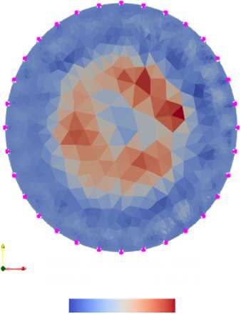
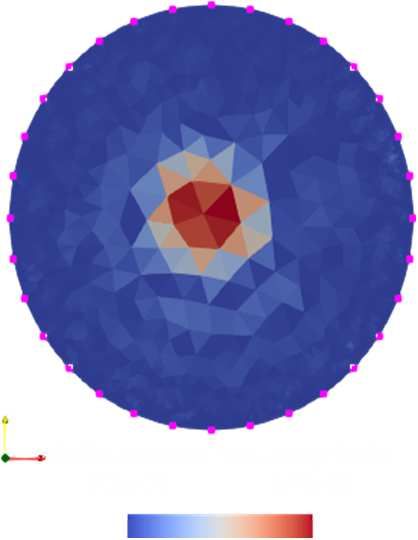
Figure 5. Main elements of the reconstruction of a small inclusion. Left: noised
permittivity distribution of the empty system used as a background. Center: base
system with inclusion. Right: real part of reconstructed delta conductivity
Reconstruction is based in the Gauss-Newton method, with a regularization term following
Tikhonov. The algorithm of reconstruction that we use adapts the pyEIT library [27] to be compatible
with the fixed electrode tomographic scheme presented. The reconstruction for the surface data of is
presented in Figure 5 where three main elements are displayed. The first element is the permittivity
distribution of the empty system, selected as a background value polluted with random noise. The second
element is the base system were the inclusion was incorporated, note that the background noise is still
the same than in the first element, but the difference of magnitude in the permittivity of the inclusion
makes appear the background as almost homogeneous. The third element presents the magnitude of the
reconstructed delta conductivity. The location of the electrodes on the surface are marked with pink
circles, with the first electrode e1 located at 0 degrees (center-left) and continuing there in counter-
clockwise fashion.
A comparison of tomographic reconstruction for different robotic respiration phantom states is
presented in Figure 6. The five columns represent the dynamics of inspiration, from minimum volume
in column 1 to maximum volume in column 5. The corresponding respiratory states are presented in row
1. As we can appreciate from the results, the reconstructed system output is coherent with the variations
of the internal structure of the system.
4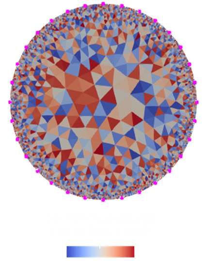
PTZE2020                                                                                  IOP Publishing
Journal of Physics: Conference Series          1782 (2021) 012037   doi:10.1088/1742-6596/1782/1/012037

Figure 6. Comparison of reconstruction from phantom respiration state. Top row: respiratory state.
Bottom row: delta conductivity reconstructed

6. Conclusions and Further Work
Using a soft robotic actuator driven by fluid transport we can emulate the movement of lungs. Different
levels of biomimicry can be achieved using combination of flexible materials, multiscale structures and
controlled actuators for fluid transport. As a proof-of-concept we use an automatized peristaltic pump,
with a microcontroller interface to connect the system to a medical data server. The server provide the
reference signal to the microcontroller, that is encoded to drive the peristaltic pump into the desired
respiration behaviour. In this way the system can be operated using the in-vivo data provided by a
wearable biosignal belt or to test arbitrary respiratory behaviours using the soft robotic phantom
interface. The peristaltic pump allows us to prove the concept in a rapid manner. To eliminate the
discrete jumps produced by the control of the pump we build a continuous inflow outflow pump based
on cylinder and piston system with similar functionality to current mechanical ventilators. After a
successful test of the system using a simple latex balloon actuator, we recreate a soft robotic surrogate
lung with multiple air chambers. More advanced models can consider soft robotic actuators for the
diaphragm and the intercostal muscles, for a robust execution of the biomechanics of respiration. The
current ongoing research in the system is to explore electric tomography data acquisition techniques, in
particular to select appropriate modulation for the emitted waveforms for analyzing human tissues in
dynamic state and evaluate the performance of the dynamic tomographic imaging algorithms. Our
efforts are directed to provide modern solutions to medicine to support the diagnostic of cardio
respiratory diseases.

References
[1]    Rymarczyk T., Characterization of the shape of unknown objects by inverse numerical methods,
       Przegląd Elektrotechniczny, 88 (2012), No. 7b, 138-140
[2]    Rymarczyk T., Kłosowski G., Tchórzewski P., Cieplak T., Kozłowski E.: Area monitoring using
       the ERT method with multisensor electrodes, Przegląd Elektrotechniczny, 95 (2019), No. 1,
       153-156
[3]    Kłosowski G., Rymarczyk T., Kania K., Świć A., Cieplak T., Maintenance of industrial reactors
       based on deep learning driven ultrasound tomography, Eksploatacja i Niezawodnosc –
       Maintenance and Reliability; 22 (2020), No 1, 138–147
[4]    Kłosowski G., Rymarczyk T., Wójcik D., Skowron S., Adamkiewicz P., The Use of Time-
       Frequency Moments as Inputs of LSTM Network for ECG Signal Classification, Electronics, 9
       (2020), No. 9, 1452

                                                    5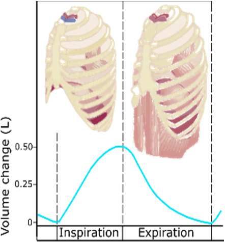
PTZE2020                                                                                IOP Publishing
Journal of Physics: Conference Series        1782 (2021) 012037   doi:10.1088/1742-6596/1782/1/012037

[5]     Kłosowski G., Rymarczyk T., Cieplak T., Niderla K., Skowron Ł., Quality Assessment of the
        Neural Algorithms on the Example of EIT-UST Hybrid Tomography, Sensors, 20 (2020), No.
        11, 3324
[6]     Romanowski, A. Big Data-Driven Contextual Processing Methods for Electrical Capacitance
        Tomography. IEEE Trans. Ind. Informatics, 15 (2019), 1609–1618
[7]     Kryszyn J., Smolik W., Toolbox for 3d modelling and image reconstruction in electrical
        capacitance tomography, Informatyka, Automatyka, Pomiary w Gospodarce i Ochronie
        Środowiska (IAPGOŚ), 2017, (1), 137-145
[8]     Dušek J., Hladký D., Mikulka J., Electrical Impedance Tomography Methods and Algorithms
        Processed with a GPU, In PIERS Proceedings, 2017, 1710-1714
[9]     Rymarczyk T., Using electrical impedance tomography to monitoring flood banks, International
        Journal of Applied Electromagnetics and Mechanics, 45 (2014), 489–494
[10]    Filipowicz S.F., Rymarczyk T., The Shape Reconstruction of Unknown Objects for Inverse
        Problems, Przegląd Elektrotechniczny, 88 (2012), No 3A, 55-57
[11]    Koulountzios P., Rymarczyk T., Soleimani M., A quantitative ultrasonic travel-time
        tomography system for investigation of liquid compounds elaborations in industrial processes,
        Sensors, 19 (2019), No. 23, 5117
[12]    Szczesny, A.; Korzeniewska, E. Selection of the method for the earthing resistance
        measurement. Przegląd Elektrotechniczny, 94 (2018), 178–181
[13]    Fraczyk, A.; Kucharski, J. Surface temperature control of a rotating cylinder heated by moving
        inductors. Appl. Therm. Eng., 125 (2017), 767–779
[14]    Kozłowski E., Mazurkiewicz D., Żabiński T., Prucnal S., Sęp J., Assessment model of cutting
        tool condition for real-time supervision system, Eksploatacja i Niezawodnosc – Maintenance
        and Reliability, 21 (2019), No. 4, 679–685
[15]    Nioutsikou E, Symonds-Tayler J R N, Bedford J L and Webb S 2006 Physics in Medicine and
        Biology 51 3359–3374 ISSN 0031-9155 publisher: IOP Publishing
[16]    Polygerinos P, Correll N, Morin S A, Mosadegh B, Onal C D, Petersen K, Cianchetti M, Tolley
        M T and Shepherd R F 2017 Advanced Engineering Materials 19 1700016
[17]    Manti M, Cacucciolo V and Cianchetti M 2016 IEEE Robotics & Automation Magazine 23 93–
        106 URL https://doi.org/10.1109/mra.2016.2582718
[18]    Cianchetti M, Laschi C, Menciassi A and Dario P 2018 Nature Reviews Materials 3 143–153
        URL https://doi.org/10.1038/s41578-018-0022-y
[19]    Ranunkel O, Güder F and Arora H 2019 ACS Applied Bio Materials 2 1490–1497
[20]    Samokhin A 2020 Journal of Analytical Chemistry 75 416–421
[21]    Sharma S, Jain R, Raja A N et al. 2019 Journal of The Electrochemical Society 167 037501
[22]    Torrinha Á, Amorim C G, Montenegro M C and Araújo A N 2018 Talanta 190 235–247
[23]    Rymarczyk T and Vejar A 2019 2019 Applications of Electromagnetics in Modern Engineering
        and Medicine (PTZE) pp 198–202
[24]    Rymarczyk T and Vejar A 2020 Przegląd Elektrotechniczny 2020 164–167 ISSN 0033-2097
[25]    Rymarczyk T., Nita P., Vejar A., Woś M., Stefaniak B., Adamkiewicz P.: Wearable mobile
        measuring device based on electrical tomography, Przegląd Elektrotechniczny, 95 (219), No. 4,
        211-214
[26]    Rymarczyk T, Nita P, Vejar A, Stefaniak B and Sikora J 2019 Przegląd Elektrotechniczny 1
        135–138 URL https://doi.org/10.15199/48.2019.06.24
[27]    Liu B, Yang B, Xu C, Xia J, Dai M, Ji Z, You F, Dong X, Shi X and Fu F 2018 SoftwareX 7
        304–308

                                                   6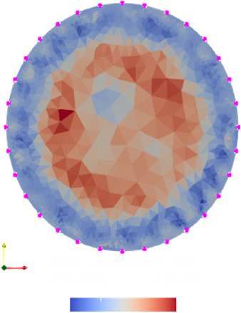
You can also read