Insulin-like growth factor-1 inhibits apoptosis of rat gastric smooth muscle cells under high glucose condition via adenosine ...
←
→
Page content transcription
If your browser does not render page correctly, please read the page content below
FOLIA HISTOCHEMICA                                                        ORIGINAL PAPER
ET CYTOBIOLOGICA
Vol. 60, No. 1, 2022
pp. 74–88

Insulin-like growth factor-1 inhibits apoptosis of rat
gastric smooth muscle cells under high glucose
condition via adenosine monophosphate-activated
protein kinase (AMPK) pathway
Xiang-zi Zhang1, 2, Yan Sun1, Mo-han Zhang1*, Zheng Jin1*
1
 Yanbian University College of Medicine, Yanji 133000, China
2
 Department of Stomatology, Affiliated Hospital of Yanbian University,Yanji 133000, China

Abstract
Introduction. Diabetic gastroparesis (DGP) is a common chronic complication of diabetes characterized by decreased
gastric motility, and an effective number of gastric smooth muscle cells (GSMCs) ensures gastric motility. A previous
study documented that apoptosis was present in gastric smooth muscles in rats with DGP and adenosine monophos-
phate-activated protein kinase (AMPK) was an important factor of apoptosis of rat GSMCs cultured under high
glucose conditions. This study aimed to explore the effect of insulin-like growth factor-1 (IGF-1) on apoptosis of high
glucose cultured rat GSMCs after silencing of AMPK and elucidate the underlying mechanism.
Material and methods. A total of 120 rats were divided into normal control (NC, n = 20), diabetic gastropa-
resis (DGP, n = 50) and DGP + IGF-1 (n = 50) groups. After establishing the rat model of DGP, rats in the
DGP+IGF-1 group received an intraperitoneal injection of IGF-1 at a dose of 1.5 μg/kg/d for 10 weeks. The
level of AMPK activity, liver kinase B1 (LKB1) activity, and calcium/calmodulin-dependent protein kinase b
(CaMKKb) expression in rat gastric smooth muscle tissues was detected by Western blot analysis. Apoptosis
in rat gastric smooth muscle tissues was detected by TUNEL assay. We also cultured rat GSMCs in vitro under
high glucose (HG) condition (35 mM), incubated cells with IGF-1, and silenced AMPK with siRNA. The cells
were divided into HG, HG + IGF-1, HG + siRNA, and HG + siRNA + IGF-1 groups. The apoptosis rates of
rat GSMCs after silencing AMPK were detected by TUNEL assay and flow cytometry, and apoptosis-related
protein expression in rat GSMCs was detected by Western blot.
Results. IGF-1 decreased LKB1 activity, CaMKKb expression, AMPK activity, and inhibited apoptosis in rat
gastric smooth muscle tissues. Compared with rat GSMCs cultured in vitro under HG conditions, apoptosis rates
were reduced after treatment with IGF-1 and AMPK silencing (both p < 0.01). Apoptosis rates were higher in
the HG + siRNA group compared with the HG + IGF-1 group (p < 0.05). IGF-1 down-regulated the expres-
sion of calcium/calmodulin-dependent kinase II (CaMKII) and p53, up-regulated the expression of p21, PLC-b3,
PI3K p110 Ser1070, and the activities of Akt, p70S6K, mTORC1, and mTORC2. IGF-1 also up-regulated Bcl-2
expression and down-regulated the expression of BAX and Caspase-3.
Conclusions. IGF-1 can inhibit the apoptosis of rat GSMCs under high glucose conditions, its mechanism may
be related to the regulation of expression and activity of p53, PI3K, TSC-2, Akt, mTOR, 4E-BP1, p70S6K, p21,
CaMKII, and PLC-b3 in rat GSMCs acting through AMPK pathway. (Folia Histochemica et Cytobiologica 2022,
Vol. 60, No. 1, 74–88)

Key words: rat; gastric smooth muscle cells; IGF-1; high glucose; apoptosis; AMPK signaling; siRNA

Correspondence adresse: Mo-han Zhang,
Yanbian University College of Medicine, Yanji 133000, China
e-mail: mhzhang@ybu.edu.cn
Zheng Jin,
Yanbian University College of Medicine, Yanji 133000, China
e-mail: jinzheng@ybu.edu.cn

This article is available in open access under Creative Common Attribution-Non-Commercial-No Derivatives 4.0 International (CC BY-NC-ND 4.0) license, allowing to
download articles and share them with others as long as they credit the authors and the publisher, but without permission to change them in any way or use them commercially.

©Polish Society for Histochemistry and Cytochemistry
Folia Histochem Cytobiol. 2022                                                                  www.journals.viamedica.pl/folia_histochemica_cytobiologica
10.5603/FHC.a2022.0005
ISSN 0239-8508, e-ISSN 1897-5631
IGF-1 inhibits apoptosis of gastric smooth muscle cells                                                                 75

Introduction                                                glucose conditions and the underlying mechanisms
                                                            have yet not been reported.
Diabetic gastroparesis (DGP) is manifested by gastric           In the present study, we established a rat model
dysmotility [1]. The specific pathogenesis is not yet       of DGP, observed the effect of IGF-1 on the sponta-
known, but long-term high blood glucose levels are          neous contraction of gastric smooth muscle, as well
the cause of DGP. Although the contraction of gastric       as its effect on AMPK activation and apoptosis in
smooth muscle cells (GSMCs) in the body is regulated        rat gastric smooth muscle tissues. We also cultured
by neural and humoral factors [2], the related changes      rat GSMCs under high glucose conditions, observed
in GSMCs themselves are also factors that should            the apoptosis of rat GSMCs after silencing AMPK,
not be ignored. Apoptosis refers to the autonomous          explored whether IGF-1 regulates apoptosis through
programmed death of cells that is controlled by genes       AMPK, detected the phosphorylation changes in the
in order to maintain the stability of the internal en-      AMPK signaling pathway by using AMPK Signaling
vironment, which has become a research hotspot in           Phospho-Antibody Array, identified apoptosis-related
various biological fields [3]. Apoptosis is an important    proteins. The purpose of this study is to elucidate the
factor affecting the number of functional cells and an      possible mechanism of action of IGF-1 in DGP and to
increased apoptosis rate can reflect a greater propor-      provide a scientific theoretical, and experimental basis
tion of apoptotic cells. A previous study documented        for the exploration of new treatment options for DGP.
the existence of spontaneous contraction disorders
and apoptosis in gastric smooth muscles in DGP rats         Materials and methods
[4], and the authors suggested that high glucose serum
concentration can induce apoptosis of GSMCs and             Materials. The following reagents were purchased: strep-
participate in the occurrence of gastric dysmotility.       tozotocin (STZ) (No. S0130, Sigma, St. Louis, MO, USA),
Finding effective interventions to suppress apopto-         IGF-1 (No. REGFP-0901, Cyagen Biosciences, Inc., Santa
sis of rat GSMCs cultured under high glucose (HG)           Clara, CA, USA), TUNEL apoptosis detection kit (No.
conditions is undoubtedly positive for the prevention       4030ES50, Yeasen Biotechnology Co., Ltd., Shanghai,
of DGP and other complications of diabetes.                 China), hematoxylin and eosin (HE) staining kit (Cat. No.
    Under HG conditions (glucose concentration in           G1120, Solarbio Science and Technology Co., Ltd., Beijing,
cell culture medium > 30 mM), cell apoptosis can            China), Apoptosis detection kit (No. 556547, BD Bio-
be regulated by a variety of cytokines and signaling        sciences, Heidelberg, Germany), Rat GSMCs (NO. RAT-
pathways [5, 6]. Adenosine monophosphate-activated          iCell-d005, iCell, Shanghai, China), primary smooth muscle
protein kinase (AMPK) is a heterotrimer composed            cell low serum culture system (No. PriMed-iCell-004, iCell
of a catalytic subunit (a) and two regulatory subunits      Bioscience, Shanghai, China), lentivirus-expressed small
(b and g) [7]. Phosphorylation of AMPK is required          interfering RNA (siRNA, AMPKa1+a2, Genepharma,
for AMPK activity. Phosphorylation at T172 of the           Suzhou, China), the protein antibody array (No. PAM174,
a-subunit is a key way to regulate AMPK activity. Our       Full Moon BioSystems, Sunnyvale, CA, USA), phospho
previous animal experiment [4] found that AMPK              (p)-AMPK Thr172 antibody (No. ab133448, Abcam, Cam-
activity first decreased and then increased in rat          bridge, UK), AMPK antibody (No. ab131512, Abcam),
GSMCs cultured under HG condition, moreover, in             liver kinase B1 (LKB1) antibody (No. 3047, Cell Signaling
vitro experiment confirmed that AMPK promoted the           Technology, Danvers, MA, USA), phospho (p)-LKB1 Ser428
apoptosis of rat GSMCs cultured in HG concentration         antibody (No. 3482, Cell Signaling Technology), calcium/
at the early stages, the effect was not obvious; but with   /calmodulin-dependent protein kinase b (CaMKKb) anti-
prolonged exposure to high glucose, AMPK was the            body (No. ab96531, Abcam), calcium/calmodulin-depend-
pro-apoptotic factor. Thus, the results indicated that      ent kinase II (CaMKII) antibody (No. ab52476, Abcam),
high glucose concentration can induce apoptosis by          p53 antibody (No. 32532, Cell Signaling Technology),
regulating AMPK activity [4]. Insulin-like growth           p21 antibody (No. ab80633, Abcam), phospholipase C-b3
factor (IGF-1) is a polypeptide similar in structure        (PLC-b3) antibody (No. 14247, Cell Signaling Technology),
to insulin. IGF-1 acts in endocrine, autocrine, and         phospho (p)-phosphoinositide 3-kinase (PI3K) p110 Ser1070
paracrine modes, and plays an important role in cell        antibody (No. bs-6417R, BIOSS Biological Technology. Co.
proliferation and differentiation [8]. IGF-1 has an         Ltd, Beijing, China), protein kinase B (Akt) antibody (No.
inhibitory effect on apoptosis of endothelial cells         9272, Cell Signaling Technology), phospho (p)-Akt Ser473
after spinal cord injury [9]. Moreover, IGF-1 can also      antibody (No. 4060, Cell Signaling Technology), mecha-
regulate the biological activity of AMPK [10, 11].          nistic target of rapamycin (mTOR) antibody (No. 2792,
However, the effects of IGF-1 on AMPK activity and          Cell Signaling Technology), phospho (p)-mTOR Ser2448
AMPK-mediated apoptosis of GSMCs under high                 antibody (SAB4504476, Sigma), phospho (p)-mTOR Ser2481

©Polish Society for Histochemistry and Cytochemistry
Folia Histochem Cytobiol. 2022                                   www.journals.viamedica.pl/folia_histochemica_cytobiologica
10.5603/FHC.a2022.0005
ISSN 0239-8508, e-ISSN 1897-5631
76                                                                                                         Xiang-zi Zhang et al.

antibody (SAB4301526, Sigma,), eukaryotic translation            stained with HE to observe the changes of gastric smooth
initiation factor 4E (eIF4E)-binding protein 1 (4E-BP1)          muscle tissues of rats in each group.
antibody (No. 9452, Cell Signaling Technology), p70 ribo-
somal protein S6 kinase (p70S6K) antibody (No. 9202, Cell        Detection of changes in the spontaneous contraction of iso-
Signaling Technology), phospho (p)-p70S6K Thr389 antibody        lated gastric smooth muscle. Eight rats each from the NC,
(No. 9205, Cell Signaling Technology), B-cell lymphoma 2         DGP, and DGP + IGF-1 groups were randomly selected
(Bcl-2) antibody (No. ab196495, Abcam, Cambridge, UK),           and euthanized after a 24 h fast. The whole stomach of rats
Bcl-2-associated X protein (Bax) antibody (No. ab32503,          was taken and cut along the lesser curvature, the contents
Abcam), Caspase-3 antibody (No. ab4051, Abcam), b-actin          of the stomach were washed out in oxygen-saturated Kreb’s
antibody (A5316, Sigma).                                         solution at 4°C. Circular muscle strips of the antrum (2 mm
                                                                 × 12 mm) were dissected. Strip was attached to an isomet-
Animals, the diabetic gastroparesis model, and experimen-        ric tension transducer in Kreb’s solution, the solution was
tal groups. One hundred and twenty male adult Sprague            maintained at 37°C and bubbled continuously with a mix-
Dawley rats, weighing 200 ± 20 g, were provided by the           ture of 95% O2 and 5% CO2. Upon the experiment, strips
Experimental Animal Center of Yanbian University.                were preloaded with 0.25 g and incubated for 40 min. After
All animal experimental procedures were approved by              stabilization of spontaneous contractions of the strips, the
the Ethics Committee of Yanbian University College of            smooth muscle contraction was recorded simultaneously
Medicine.                                                        using a four-channel physiological signal recording system
     0.5% STZ solution was prepared in 0.1 mol/L citrate         (RM6240, Chengdu Instrument Factory, Chengdu, China).
buffer (pH 4.0). Rats were housed in a room with a tem-          Care was taken to prevent the adhesion of the strips.
perature of 20°C, relative humidity of 60%, and a 12-hr
light: dark cycle.                                               Detection of the expression of p-AMPK Thr172, AMPK,
     They had free access to food and water and were adapted     p-LKB1 Ser428, LKB1, CaMKKβ in rat gastric smooth
to the environment for 1 week. Rats were fasted for 12 h         muscle tissues by Western blot analysis. Eight rats each from
before weighing and administration of a single intraperi-        the DGP and DGP + IGF-1 groups were randomly selected
toneal injection of 65 mg/kg 0.5% STZ. The animals had           and euthanized after a 24 h fast, then the whole stomach was
free access to water throughout the experiment. Seven            excised. Gastric smooth muscle tissues were placed in a 1.5 mL
days after injection, blood samples were collected via the       centrifuge tube. 100 ml of ice-cold RIPA lysis buffer containing
tail vein. Blood glucose concentration was measured by           1 mM PMSF was added for 10 mg of tissue. Then the samples
OneTouch Verio Reflect™ system (LifeScan, Wayne, PA,             were homogenized on ice using a handheld homogenizer (MT-
USA). Blood glucose levels > 350 mg/dl (19.45 mM) indi-          30k, Hangzhou Mioyi Instrument Co., Ltd., Hangzhou, China)
cated the successful establishment of STZ-induced diabetes.      and centrifuged at 12,000 g for 5 min at 4°C. Total proteins
According to the results from our previous study, DGP was        were then extracted and protein concentrations were determined
developed at 6 weeks after the establishment of the diabetic     using a BCA protein assay kit (cat. no. PC0020, Beijing Solar-
rat model [4, 12].                                               bio Science & Technology Co., Ltd., Beijing, China). Proteins
     One hundred rats with DGP were selected and divided         were boiled for 2 min, and 40 μg of proteins were loaded into
into DGP and DGP + IGF-1 groups with 50 rats per group,          each well. After separation of the proteins by electrophoresis
and 20 untreated, normal rats were chosen as normal control      in 10% polyacrylamide gel, the proteins were transferred to
(NC) group. Rats in the DGP + IGF-1 group received an            polyvinylidene difluoride membrane. The membranes were
i.p. injection of IGF-1 at a dose of 1.5 μg/kg/d for 10 weeks,   blocked with 5% skimmed milk powder in TBS-T buffer solu-
and rats in the DGP and NC groups received an i.p. injection     tion. After the blocked membranes were washed, they were
of the same dose of normal saline for 10 weeks.                  incubated with primary antibodies, p-AMPK Thr172 (1:1,000),
                                                                 AMPK (1:1,000), p-LKB1 Ser428 (1:1,000), LKB1 (1:1,000),
Detection of morphological changes in gastric smooth             CaMKKβ (1:1,000), and β-actin (1:500) at 4°C overnight.
muscle tissues by HE staining. Eight rats each from the NC       After washing the membranes, they were incubated with HRP
and DGP and DGP + IGF-1 groups were randomly selected            conjugated goat anti-rabbit IgG secondary antibody for 1 h at
and euthanized after a 24 h fast. Then, the whole stomach        RT. Then the membranes were washed and exposed, and β-actin
was rapidly excised, the gastric smooth muscle tissues were      was used as the internal reference.
fixed in Bouin’s fluid (25 ml of 40% formaldehyde, 75 ml
of saturated aqueous picric acid, 5 ml of glacial acetic acid)   Detection of apoptosis in gastric smooth muscle tissues by
for 12 hours, dehydrated, and infiltrated with paraffin wax.     TUNEL assay. Eight rats each from the DGP and DGP +
The infiltrated tissues were then embedded into paraffin         IGF-1 groups were randomly selected and euthanized after
blocks. Paraffin blocks were cut into 7 μm-thick sections at     a 24 h fast. Then the whole stomach was rapidly excised,
room temperature (RT). After dewaxing, the section was           the gastric smooth muscle tissues were fixed, dehydrated,

©Polish Society for Histochemistry and Cytochemistry
Folia Histochem Cytobiol. 2022
10.5603/FHC.a2022.0005                                                 www.journals.viamedica.pl/folia_histochemica_cytobiologica
ISSN 0239-8508, e-ISSN 1897-5631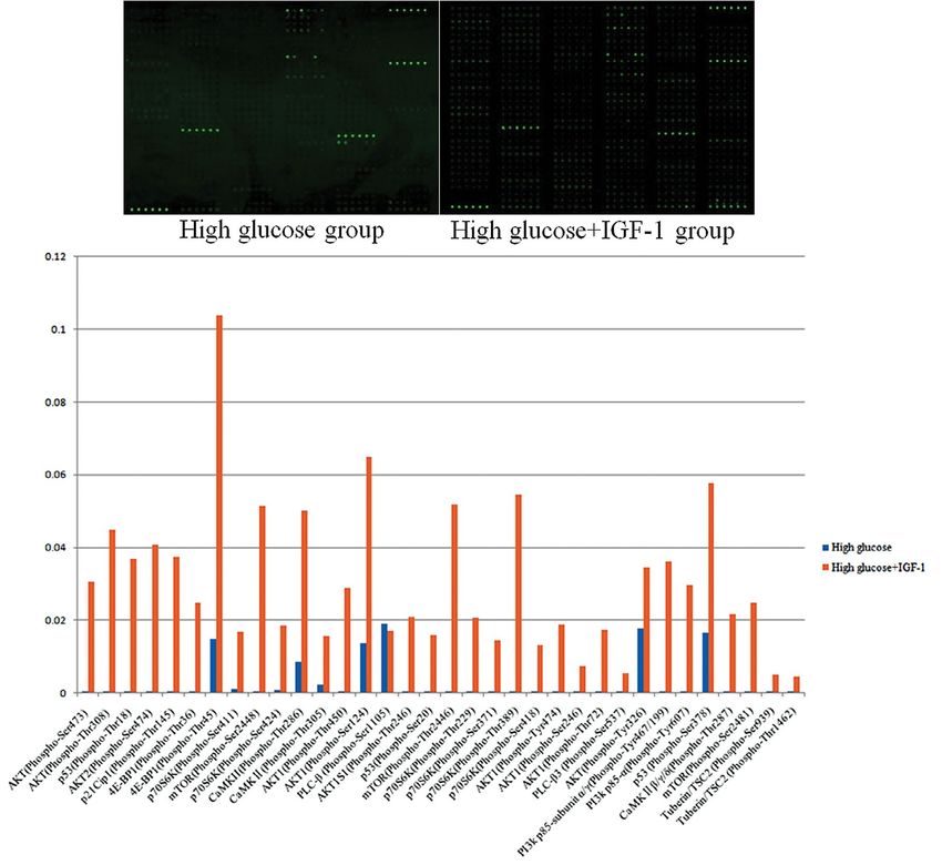
IGF-1 inhibits apoptosis of gastric smooth muscle cells                                                                        77

infiltrated with paraffin wax, and embedded in a paraffin         IGF-1 groups. Proteins were extracted according to the
block. Paraffin block was cut into 7 μm-thick sections at RT.     kit instructions. Apoptosis-related proteins in the AMPK
After dewaxing, apoptosis was detected by using TUNEL             pathway were identified.
Apoptosis Detection Kit according to the manufacturer’s
instructions. Five fields at the magnification of 200× were       Detection of apoptosis-related protein expression in rat
randomly selected from each section and apoptotic cells           GSMCs by Western blot analysis. Rat GSMCs in the
were observed under a fluorescence microscope (Cytation           logarithmic growth phase were obtained, divided into HG
5 Cell Imaging Microplate Detection System, BioTek In-            and HG + IGF-1 groups, and cultured for 48 h. The pro-
struments, Inc., Winooski, VT, USA), green fluorescence           cedures of Western blot were the same as those mentioned
in the nucleus indicated TUNEL-positive apoptotic cells.          above. Antibodies were CaMKII (1:1,000), p53 (1:1,000),
The fluorescence intensity was quantitatively analyzed using      PLC-b3 (1:1,000), p21 (1:1,000), PI3K p110 Ser1070 (1:500),
Image-Pro Plus 6.0 (Media Cybernetics, Inc., Rockville,           Akt (1:1,000), p-Akt Ser473 (1:1,000), 4E-BP1 (1:1,000),
MD, USA).                                                         p70S6K(1:1,000), p-p70S6K Thr389 (1:1,000), p-mTOR
                                                                  Ser2448 (1:500), p-mTOR Ser2481 (1:500), mTOR (1:1,000),
Silencing of AMPK with siRNA. The deep-frozen rat gastric         Bcl-2 (1:1,000), BAX(1:1,000), Caspase-3(1:1,000), b-actin
smooth muscle cells purchased from iCell Bioscience Inc           (1:500). The experiments were repeated at least 3 times.
(Shanghai, China) were recovered and cultured in primary
smooth muscle cell low serum (10%) culture system (iCell          Statistical analysis. Statistical analyses were performed
Bioscience) and passaged 2–3 times. Rat GSMCs in the log-         with SPSS 21.0 software (IBM Co., Armonk, NY, USA).
arithmic growth phase were obtained, digested with trypsin        All figures were drawn using GraphPad Prism5 software
(2.5%), and resuspended in primary smooth muscle cell low         (GraphPad Inc., San Diego, CA, USA). Data are expressed
serum culture system. 1×10⁶ of cells were added into a 24-well    as mean ± standard error of the mean (SEM). Differences
plate and incubated at 37°C overnight. The solution containing    between groups were compared using a t-test and two-way
siRNA-expressing lentiviral vectors (AMPKα1 + α2) was mixed       analysis of variance (ANOVA). p < 0.05 was considered to
and diluted with the primary smooth muscle cell low serum         indicate a significant difference, p < 0.01 was considered to
culture system in a 1:2 ratio, the total volume was 500 µl. The   indicate a highly significant difference.
old medium was aspirated from the plate, lentiviral dilutions
were added, and after incubation at 37°C for 24 h, the medium     Results
was aspirated and replaced with 1 mL of fresh medium. After
incubation at 37°C for 72 h, the cells were observed and photo-   Comparison of blood glucose concentrations
graphed under a fluorescence inverted microscope. Cells were      and body weight between groups
collected for subsequent experiments when transfection effi-      The body weight of rats in the DPG group was lower
ciency reached > 70%. Transfection efficiency was determined      than in normal control (NC) rats (174.8 ± 3.26 g vs.
by monitoring the expression of green fluorescent protein using   219.0 ± 5.18 g, respectively, p < 0.01), and the blood
fluorescence microscopy.                                          glucose concentration of rats in the DPG group was
                                                                  higher than in the NC group (28.46 ± 1.47 mM vs.
Detection of the apoptosis rates of rat GSMCs after silenc-       7.01 ± 0.28 mM, respectively, p < 0.01).
ing AMPK by flow cytometry. After recovering the primary              After treatment with IGF-1, the body weight
rat GSMC line, cells were passaged 2–3 times. Cell density        of rats in the DGP+IGF-1 group was higher than
was adjusted to 1 × 10⁶/ml. Then cells were divided into          the DGP group (197.4 ± 3.17 g vs. 174.8 ± 3.26 g,
HG (35 mmol/L) and HG + IGF-1 (100 ng/ml) groups.                 p < 0.01), and lower than the NC group (197.4 ± 3.17 g
The IGF-1 concentration of 100 ng/ml was determined by            vs. 219.0 ± 5.18 g, p < 0.01). The blood glucose
prior dose-response experiments. After silencing AMPK in          concentrations of rats in the DGP + IGF-1 group
rat GSMCs, cells were passaged 1 to 2 times, and the cell         were lower than the DGP group (10.26 ± 0.69 mM
density was adjusted to 1 × 10⁶/ml. Then cells were divid-        vs. 28.46 ± 1.47 mM, p < 0.01), and higher than the
ed into HG + siRNA and HG + siRNA+IGF-1 groups.                   NC group (10.26 ± 0.69 mM vs. 7.01 ± 0.28 mM,
Flow cytometry was performed at 48 h of culture to detect         p < 0.05, Fig. 1).
the apoptosis rates of rat GSMCs. The experiments were
repeated at least 3 times.                                        Morphological changes of gastric smooth muscle
                                                                  tissues in each group
Detection of the changes in the AMPK pathway by using             HE staining showed that in the NC group, gastric
protein antibody arrays. After culturing cells for 48 hours,      smooth muscles were arranged regularly and tightly,
the protein antibody array was performed to detect the            with moderate intercellular gaps, and did not present
changes in the AMPK pathway in the HG and HG +                    vacuolar degeneration. In the DGP group, the cyto-

©Polish Society for Histochemistry and Cytochemistry
Folia Histochem Cytobiol. 2022                                          www.journals.viamedica.pl/folia_histochemica_cytobiologica
10.5603/FHC.a2022.0005
ISSN 0239-8508, e-ISSN 1897-5631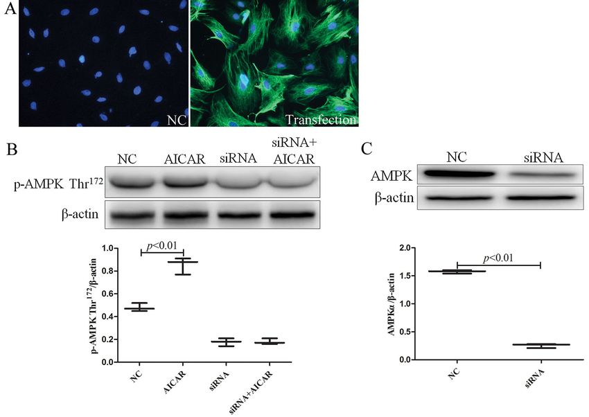
78                                                                                                        Xiang-zi Zhang et al.

Figure 1. The effect of IGF-1 (1.5 μg/kg/d, i.p., 10 weeks) on body weight and blood glucose levels of streptozotocin-induced
diabetic gastroparesis (DGP) rats. NC — control rats; DGP + IGF-1 — DGP rats that received IGF-1. Data are expressed
as mean ± SEM, n = 8 per group.

Figure 2. The effect of IGF-1 (1.5 μg/kg/d, i.p., 10 weeks) on the morphological changes of gastric smooth muscle tissues of
DGP rats. Magnification: 200×. N = 8 per group. Abbreviations as in the description of Fig. 1.

plasm of the gastric smooth muscle was lightly stained          and higher as compared with the DGP group (0.62 ±
and translucent, vacuolar degeneration was observed.            0.05 vs. 0.41 ± 0.02, p < 0.01, Fig. 3).
In the DGP + IGF-1 group, gastric smooth muscles
were arranged regularly with fewer vacuoles (Fig. 2).           Changes in the levels of LKB1, AMPK activities,
                                                                and CaMKKb expression in rat gastric smooth
Changes in the spontaneous contraction of gastric               muscle tissues between groups
smooth muscle between groups                                    Ratio of phospho-AMPK Thr172/AMPK and phos-
The frequency and amplitude of spontaneous contrac-             pho-LKB1 Ser 428/LKB1 were both higher in the
tion of gastric smooth muscles were both lower in the           DGP group than that in the DGP + IGF-1 group
DGP group than that in the NC group (13.82 ± 0.46               (0.92 ± 0.06 vs. 0.77 ± 0.03, p < 0.05; 0.83 ± 0.05
per sec vs. 29.52 ± 1.06 per sec, p < 0.05; 0.41 ± 0.02         vs. 0.61 ± 0.04, p < 0.05, respectively). CaMKKb ex-
vs. 0.90 ± 0.05, frequency and amplitude, respectively,         pression was increased in the DGP group than that in
p < 0.01). After treatment with IGF-1, the frequency            the DGP + IGF-1 group (0.68 ± 0.04 vs. 0.50 ± 0.03,
of spontaneous contractions in the DGP + IGF-1 was              p < 0.01, Fig. 4).
lower than that in the NC group (20.08 ± 0.85/sec vs.
29.52 ± 1.06/sec, p < 0.01), and was higher than in             Changes in apoptosis in rat gastric smooth
the DGP group (20.08 ± 0.85/sec vs. 13.82 ± 0.46/sec,           muscle tissues between groups
p < 0.01). The amplitude of spontaneous contractions            The nuclei of TUNEL-positive cells exhibited green
in DGP + IGF-1 group was reduced as compared with               fluorescence, the results showed that significantly
the NC group (0.62 ± 0.05 vs. 0.90 ± 0.05, p < 0.01),           more TUNEL-positive nuclei were found in the DGP

©Polish Society for Histochemistry and Cytochemistry
Folia Histochem Cytobiol. 2022
10.5603/FHC.a2022.0005                                                www.journals.viamedica.pl/folia_histochemica_cytobiologica
ISSN 0239-8508, e-ISSN 1897-5631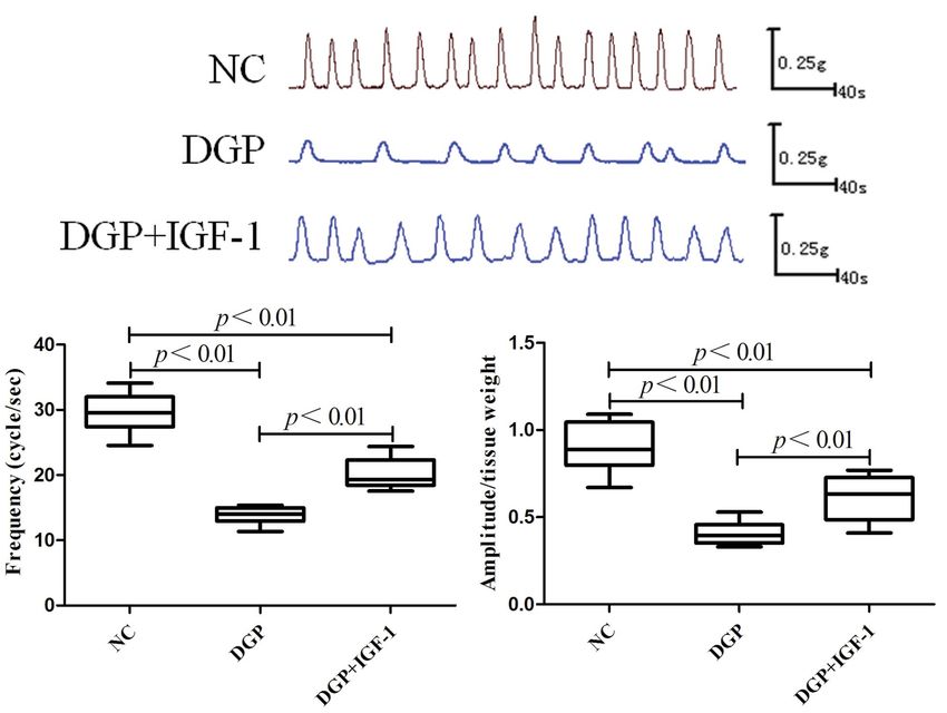
IGF-1 inhibits apoptosis of gastric smooth muscle cells 79
Figure 3. The effect of IGF-1 (1.5 μg/kg/d, i.p., 10 weeks) on the frequency and amplitude of spontaneous contractions of
gastric smooth muscle DGP rats. Data are expressed as mean ± SEM, n = 8 per group. Abbreviations as in the description
of Fig. 1.
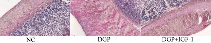
group versus the DGP + IGF-1 group (90.05 ± 1.77 Effects of IGF-1 on the apoptosis of rat GSMCs
vs. 68.72 ± 2.44, p < 0.01, Fig. 5). after silencing AMPK
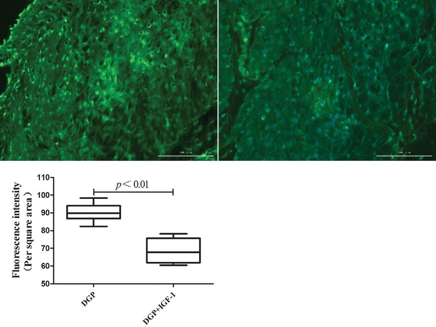
GSMCs were incubated with or without IGF-1 (100 ng/
Effects of silencing of AMPK with siRNA /ml). Apoptosis rates were higher in the GSMCs cul-
The lentiviral transfection efficiency for cells was tured under HG condition compared with the HG +
more than 70% (Fig. 6A). After adding 5-aminoimida- IGF-1 group (7.96 ± 0.04% vs. 5.71 ± 0.13%, p < 0.01)
zole-4-carboxamide1-b-D-ribofuranoside (AICAR), as well as the HG + siRNA group (7.96 ± 0.04 vs. 6.59
the expression of phospho-AMPK Thr172 was higher ± 0.16, p < 0.01). Apoptosis rates were higher in the
in the NC+AICAR group compared with the NC HG + siRNA group compared with the HG + IGF-1
group (0.85 ± 0.04 vs. 0.48 ± 0.02; p < 0.01), and group (6.59 ± 0.16 vs. 5.71 ± 0.13, p < 0.05). There were
phospho-AMPK Thr172 expression was not signifi- no statistically significant differences between the HG
cantly changed after transfection with AMPK a1/ + siRNA + IGF-1 and HG + IGF-1 groups (Fig. 7).
/a2 siRNA (Fig. 6B). After the cells were transfected
with lentivirus-expressed siRNA (AMPKa1+a2), Effect of IGF-1 on changes in AMPK signaling
the AMPKa expression was higher in the NC group pathway in rat GSMCs cultured under high
compared with the siRNA group (1.57 ± 0.02 vs. 0.25 glucose conditions
± 0.02; p < 0.01), indicating that AMPKa expression GSMCs were incubated with or without IGF-1 (100
was significantly inhibited (Fig. 6C). ng/ml). All target proteins were normalized to internal
©Polish Society for Histochemistry and Cytochemistry
Folia Histochem Cytobiol. 2022 www.journals.viamedica.pl/folia_histochemica_cytobiologica
10.5603/FHC.a2022.0005
ISSN 0239-8508, e-ISSN 1897-5631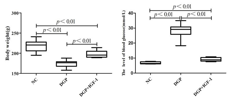
80                                                                                                         Xiang-zi Zhang et al.

Figure 4. The effect of IGF-1 (1.5 μg/kg/d, i.p, 10 weeks) on the activities of LKB1, AMPK, and the expression of CaMKKb
in the gastric smooth muscle of DGP rats. Data are expressed as mean ± SEM, n = 8 per group. Abbreviations as in the
description of Fig. 1.

Figure 5. Effect of IGF-1 (1.5 μg/kg/d, i.p., 10 weeks) on the apoptosis in gastric smooth muscle tissues of DGP rats. Data are
expressed as mean ± SEM, n = 8 per group. The nuclei of apoptotic cells exhibited green TUNEL. Magnification: 200×.
Abbreviations as in the description of Fig. 1.

©Polish Society for Histochemistry and Cytochemistry
Folia Histochem Cytobiol. 2022
10.5603/FHC.a2022.0005                                                 www.journals.viamedica.pl/folia_histochemica_cytobiologica
ISSN 0239-8508, e-ISSN 1897-5631
IGF-1 inhibits apoptosis of gastric smooth muscle cells                                                                       81

Figure 6. Effect of silencing of AMPK with small interfering RNA in rat gastric smooth muscle cells. A. Transfection efficiency
of lentiviral after 72 h of transfection (magnification: 200×). B. Changes in AMPK Thr172 phosphorylation after silencing
AMPK and adding 5-aminoimid-azole-4-carboxamide1-b-D-ribofuranoside (AICAR). C. Expression of AMPKa in NC
(control) and siRNA groups after silencing AMPK. The experiment was repeated three times independently.

Figure 7. Effects of IGF-1 (100 ng/ml) on apoptosis of high glucose (35 mM)-cultured rat gastric smooth muscle cells after
silencing of AMPK measured by flow cytometry as described in Methods. The experiment was repeated three times inde-
pendently.

©Polish Society for Histochemistry and Cytochemistry
Folia Histochem Cytobiol. 2022                                         www.journals.viamedica.pl/folia_histochemica_cytobiologica
10.5603/FHC.a2022.0005
ISSN 0239-8508, e-ISSN 1897-5631
82                                                                                                     Xiang-zi Zhang et al.

 Figure 8. Effect of IGF-1 (100 ng/ml) on changes in the AMPK signaling pathway in rat gastric smooth muscle cells cultured
 under high glucose concentration (35 mM). The experiment was repeated three times independently.

reference b-actin (i.e. dividing the signal intensity of      Ser473/Akt ratio were decreased in the HG group
the target protein with that of b-actin). Fold change         compared with the HG + IGF-1 group (0.55 ± 0.10
≥ 1.5 was used as inclusion criteria for differentially       vs. 0.93 ± 0.07, p < 0.05; 1.03 ± 0.04 vs. 1.43 ± 0.12,
expressed proteins. A total of 34 differentially phos-        p < 0.05, 0.57 ± 0.11 vs. 0.92 ± 0.02, p < 0.05; 0.37 ±
phorylated sites were identified, including 10 apopto-        0.01 vs. 1.30 ± 0.25, p < 0.05, respectively, Fig. 9A).
sis-related proteins, p53, PI3K, Akt, tuberous sclerosis          4E-BP1 and Bcl-2 expression and the ratio of
complex 2 (TSC-2), mTOR, 4E-BP1, p70S6K, p21,                 p-p70S6K Thr389/p70S6K and p-mTOR Ser2448/mTOR
CaMKII, and PLC-b3 (Fig. 8).                                  and p-mTOR Ser2481/mTOR were decreased in the
                                                              HG group compared with the HG + IGF-1 group
Effect of IGF-1 on the expression of apoptosis-               (0.72 ± 0.09 vs. 1.06 ± 0.06, p < 0.05; 0.33 ± 0.03
-related proteins in rat GSMCs cultured under                 vs. 0.43 ± 0.02, p < 0.05, 0.65 ± 0.08 vs. 1.17 ± 0.12,
high glucose conditions                                       p < 0.05; 0.59 ± 0.05 vs. 0.97 ± 0.04, p < 0.01; 0.48
P53 and CaMKII expression were increased in the HG            ± 0.01 vs. 1.02 ± 0.02, p < 0.01, respectively), while
group compared with the HG + IGF-1 group (1.05 ±              BAX and caspase-3 expression were increased in the
0.02 vs. 0.88 ± 0.03, p < 0.01; 0.92 ± 0.08 vs. 0.57 ±        HG group compared with the HG + IGF-1 group
0.02, p < 0.05, respectively), while p21 and PLC-b3 and       (0.41 ± 0.01 vs. 0.34 ± 0.01, p < 0.05; 0.25 ± 0.01 vs.
PI3K p110 Ser1070 expression and the ratio of p-Akt           0.17 ± 0.01, p < 0.01, respectively, Fig. 9B).

©Polish Society for Histochemistry and Cytochemistry
Folia Histochem Cytobiol. 2022
10.5603/FHC.a2022.0005                                             www.journals.viamedica.pl/folia_histochemica_cytobiologica
ISSN 0239-8508, e-ISSN 1897-5631
IGF-1 inhibits apoptosis of gastric smooth muscle cells 83
1.5 1.8 p < 0.05 1.2
p < 0.05 1.6 p < 0.01
CaMK II/b-actin
Camk II
PLC-b3/b-actin
1.1
1.0
p53/b-actin
1.4
1.0
PLC-b3 0.5
1.2
1.0 0.9
p53 0.0 0.8 0.8
High High glucose High High glucose High High glucose
p21 glucose + glucose + glucose +
IGF-1 IGF-1 IGF-1
PI3K p110 Ser1070
1.5 1.5
/b-actin
2.0 p < 0.05
p-Akt Ser1070 p < 0.05 p < 0.05
p-Akt Ser473/Akt
p21/b-actin
1.0 1.0 1.5
1070
Akt
PI3K p110 Ser
1.0
0.5 0.5
b-actin 0.5
IGF-1 – + 0.0 0.0 0.0
High High glucose High High glucose High High glucose
High glucose + + glucose + glucose + glucose +
IGF-1 IGF-1 IGF-1
1.5 1.5
/mTOR
/mTOR
p < 0.01 p < 0.01
1.0 1.0
2481
2448
p-mTOR Ser
p-mTOR Ser
0.5 0.5
p-mTOR Ser2448
0.0 0.0
p-mTOR Ser
2481
High High glucose High High glucose
glucose + glucose +
IGF-1 IGF-1
mTOR
1.5 1.5 p < 0.05
p-p70S6K Thr389/p70S6K
4E-BP1 p < 0.05
4E-BP1/b-actin
1.0 1.0
p-p70S6K Thr389
0.5 0.5
p70S6K
0.0 0.0
Bcl-2 High High glucose High High glucose
glucose + glucose +
BAX IGF-1 IGF-1
Caspase-3 0.50 p < 0.05 0.50 0.30
p < 0.01
Caspase-3/b-actin
0.45 0.45 p < 0.05 0.25
Bcl-2/b-actin
BAX/b-actin
b-actin 0.40
0.40 0.20
0.35
IGF-1 – + 0.35 0.15
0.30
High glucose + + 0.25 0.30 0.10
High High glucose High High glucose High High glucose
glucose + glucose + glucose +
IGF-1 IGF-1 IGF-1
Figure 9. Effect of IGF-1 (100 ng/ml) concentration on the expression of apoptosis-related proteins assessed by Western
blotting in rat gastric smooth muscle cells cultured under high glucose concentration (35 mM). The experiment was repeated
three times independently.
Discussion structure of rat gastric smooth muscle cells, inhibited
the apoptosis in gastric smooth muscle tissues, and
Gastric motility is an important determinant of gastric increased the amplitude and frequency of sponta-
emptying and an effective number of GSMCs ensures neous contractions of gastric smooth muscles in rats
gastric motility. Apoptosis is a common cellular pro- with DGP, indicating that inhibition of apoptosis has
cess that can reduce the number of cells. Our previous a significant effect on the recovery of gastric motility.
study [4] demonstrated that gastric motility disorders Our previous study [4] documented that AMPK is an
and apoptosis were present in gastric smooth muscles important factor of apoptosis of rat GSMCs cultured
in rats with DGPS. Because apoptosis is an important under high glucose conditions. In this study, we found
cause of gastric motility disorders, the inhibition of that IGF-1 inhibited AMPK activity, and the activity
apoptosis may play a positive role in ensuring gastric of its upstream activator LKB1, as well as CaMKKb
motility. In this study, we found that prolonged ad- expression. The biological role of CaMKKb is depend-
ministration of IGF-1 improved the morphological ent on intracellular Ca2+ levels. A previous study [13]
©Polish Society for Histochemistry and Cytochemistry
Folia Histochem Cytobiol. 2022 www.journals.viamedica.pl/folia_histochemica_cytobiologica
10.5603/FHC.a2022.0005
ISSN 0239-8508, e-ISSN 1897-563184 Xiang-zi Zhang et al.
found that IGF-1 can down-regulate the Ca2+ levels of the upstream protein of mTOR, small GTPase
in high glucose-induced rat GSMCs, suggesting that Rheb, thereby inhibiting the binding of Rheb to GTP,
IGF-1 down-regulated AMPK activity by inhibiting and down-regulating mTOR complex 1 (mTORC1)
the mode of AMPK activation. In this study, in order to activity [22, 23]. TSC-2 has multiple phosphorylation
investigate whether IGF-1 exerts an inhibitory effect on sites, activated Akt can phosphorylate TSC-2 at Ser939
apoptosis of high glucose-cultured rat GSMCs through and Thr1462, which in turn inhibits TSC-1/TSC-2 for-
the AMPK pathway, we silenced AMPK expression. mation and indirectly activates mTORC1 [24]. Acti-
Our findings showed that under high glucose condi- vated AMPK can phosphorylate TSC-2 at Ser1345 and
tions, IGF-1 inhibited in vitro apoptosis through the Ser1227, which directly activates TSC-2 and promotes
AMPK pathway. In order to explore the underlying TSC-1/TSC-2 formation, thereby inhibiting mTORC1
mechanism, we observed the effect of IGF-1 on chang- activity. Thus, the biological functions of mTOR are
es in the AMPK signaling pathway by protein antibody different when the TSC-2 is phosphorylated at differ-
arrays, and out of 34 different phosphorylated sites, ent sites [25]. In this study we demonstrated that in
10 involved apoptosis-related proteins. rat GSMCs cultured under high glucose conditions,
PI3K has serine/threonine (Ser/Thr) kinase activ- IGF-1 can increase the phosphorylation levels of
ity, can mediate a wide range of biological functions, TSC-2 at Ser939 and Thr1462 via Akt through inhibiting
including cell endocytosis, exocytosis, and vesicle AMPK, thereby suppressing the formation of TSC-1/
formation, as well as cell proliferation, differentiation, /TSC-2 complex, and inhibiting cell apoptosis through
apoptosis, and glucose transport [14]. PI3K is com- activating mTORC1.
prised of a regulatory subunit (p85) and a catalytic The mTOR is an atypical serine/threonine kinase
subunit (p110). The helical domain of the regulatory that exists in the form of mTORC1 and mTORC2
subunit p85a/p85b can bind p110, leading to the complexes [26]. mTORC1 and mTORC2 can sense
phosphorylation and activation of p110, followed by different nutritional and environmental factors, and
recruitment of PI3K to the plasma membrane. The participate in the regulation of energy metabolism
phosphorylation and activation of p110 can also be and cell apoptosis [26]. mTOR is co-regulated by
mediated by G protein-coupled receptors through Akt and AMPK and mTOR activity is indicated
binding of Ras/Rac to p110 [15]. A previous study [16] by its phosphorylation. A study found that mTOR
showed that AMPK can act as an upstream factor of regulated apoptosis of neuroblastoma cells mainly
PI3K to regulate its biological functions in a variety of through regulation of its downstream targets p70S6K
cells. We demonstrated in this report that under high and 4E- BP1 [27]. Multiple phosphorylation sites are
glucose conditions, IGF-1 can up-regulate the phos- present on mTOR, Ser2448 and Ser2481 are the phospho-
phorylation levels of PI3K p85 by inhibiting AMPK rylation sites of mTORC1 and mTORC2, respectively
activity, which in turn relieves the inhibition of PI3K [28]. The results of our study suggest that IGF-1 exerts
p110. thereby activating PI3K p110 phosphorylation. a regulatory effect on mTOR phosphorylation and
This may lead to the up-regulation of the biological expression by inhibiting AMPK, and/or Akt. In this
activities of PI3K, and inhibition of apoptosis. way, IGF-1 may activate mTOR, as well as mTORC1
Akt is a serine/threonine-specific protein kinase and mTORC2, respectively, and inhibit cell apoptosis
and its activity can be regulated by various upstream through regulation of p70S6K and 4E-BP1. Interestingly,
factors, including AMPK and PI3K [17, 18]. Akt ac- Sutton and Coran showed in a rat model of cocaine-in-
tivation involves the phosphorylation at Thr308, Ser473, duced nucleus accumbens sensitization and reward study
which is reflected by the ratio of p-AktSer473 to Akt. data suggesting that it reduced the phosphorylation of
Activated Akt can cause caspase3 inactivation and mTOR at Ser2448 by phosphorylating mTOR at Thr2446
inhibit apoptosis [19]. The results of our study showed and inhibited the mTORC1 activity, findings contrary
for the first time that under high glucose conditions, to our results [29]. The differences in the in vitro model,
IGF-1 can up-regulate the phosphorylation levels of cell type, and culture conditions may be responsible for
Akt at ten identified sites and stabilize Akt expression the discrepancy between theirs and our results.
by inhibiting AMPK, which is also involved in inhib- P70S6K is a downstream substrate of mTOR and
iting apoptosis by increasing Akt activity. can be activated by PI3K-Akt and protein kinase C
TSC-2 is a downstream target of AMPK and also a pathways. p70S6K is involved in the regulation of
downstream target of Akt [20, 21]. The biological role transcription and translation, cell proliferation and
of TSC-2 is mainly achieved through its downstream apoptosis [30]. p70S6K has several phosphorylation
mammalian target of rapamycin (mTOR). TSC-2 can sites, p70S6K phosphorylation at Ser411, Ser424, Thr421,
form a heterodimeric complex with TSC-1 (TSC-1/ Ser418, and Ser371 leads to the release p70S6K from
/TSC-2), TSC-1/TSC-2 complex can inhibit the activity C-terminal self-inhibition and facilitate p70S6K acti-
©Polish Society for Histochemistry and Cytochemistry
Folia Histochem Cytobiol. 2022
10.5603/FHC.a2022.0005 www.journals.viamedica.pl/folia_histochemica_cytobiologica
ISSN 0239-8508, e-ISSN 1897-5631IGF-1 inhibits apoptosis of gastric smooth muscle cells 85
vation [31]. p70S6K activation requires the phospho- under high glucose conditions, IGF-1 can increase
rylation of p70S6K, the phosphorylation of p70S6K at the phosphorylation of p53 at the 3 identified sites
Thr389 and Thr229 are crucial for p70S6K activity, the and decrease p53 expression through inhibiting the
ratio of p-p70S6K Thr389/p70S6K can reflect p70S6K AMPK pathway, thereby regulating the biological
activity [32, 33]. Activated p70S6K can phosphorylate activities of Bcl-2, Bax, and Caspase-3, and inhibiting
Bad at Ser136 and cause its dissociation from Bcl-2 cell apoptosis.
and Bcl-xL, which exerts an antiapoptotic effect [30]. P21 has GTPase activity and is involved in regulat-
In this study, results from protein antibody arrays ing cell transmembrane signaling, cell proliferation,
showed that under high glucose conditions, IGF-1 differentiation, and apoptosis [40]. Studies [41, 42]
up-regulated in rat GSMCs p70S6K phosphorylation have revealed that p21 exerts an inhibitory effect on
at six sites whereas Western blot analysis showed that apoptosis, and this effect is dependent on p53. The p21
IGF-1 stabilized p70S6K expression, indicating that protein can also exert a pro-apoptotic effect which is
IGF-1 can up-regulate p70S6K phosphorylation and not dependent on p53 [40]. The p21 expression can be
stabilize its expression through inhibiting AMPK, regulated by upstream factors such as AMPK and Akt
thereby maximizing the activity of p70S6K, and inhib- [43, 44]. In this study, results from protein antibody
iting apoptosis of high glucose-cultured rat GSMCs. arrays and western blot analysis indicate that IGF-1
4E-BP1 is a small-molecular-weight protein that exerts a regulatory effect on p21 phosphorylation
can bind to eIF4E, thereby inhibiting translation and expression via Akt and p53 pathways through
initiation. The phosphorylation level of 4E-BP1 is inhibiting AMPK, thereby inhibiting the apoptosis
closely related to its biological function, high levels of rat GSMCs.
of phosphorylated 4EBP1 promote the dissociation of Ca2+ can induce endoplasmic reticulum stress
eIF4E from 4E-BP1, and low levels of phosphorylated and initiate ER stress-induced apoptosis signaling
4EBP1enhances the binding of 4E-BP1 to eIF4E [34]. pathway [45]. PLCb/IP3/Ca2+ is a basic pathway in
A previous study [35] showed that increasing the level the PLC pathway [46]. IP3 generation can stimulate
of phosphorylated 4E-BP1 had an inhibitory effect on Ca2+ influx, the release of endoplasmic reticulum and
the apoptosis of rat neural cells. The reason may be sarcoplasmic reticulum, and increase the cytoplasmic
that eIF4E promotes the translation of anti-apoptotic Ca2+ levels. PLC-b3 is one of the PLC subtypes, and
proteins after dissociating 4E-BP1 from eIF4E [35]. is mainly present in the gastrointestinal tract [47].
4E-BP1 has many phosphorylation sites, phospho- PLC-b Ser1105 is also known as the inhibitory type of
rylation of 4E-BP1 at Thr36, Thr45, Ser65, Thr70 are PLC, the increase in PLC-b Ser1105 expression can in-
associated with the mTOR pathway. Phosphorylation hibit PLC-b3 activity and expression, phosphorylation
of 4E-BP1 at Thr37, Thr46, Ser65, Thr70 promotes its of PLC-b3 at Ser537 can promote PLC-b3 activity and
dissociation from eIF4E [36, 37]. In our current study, expression [48]. In this study, the obtained results
results from protein antibody arrays and Western blot suggested that IGF-1 can up-regulate the phosphoryl-
analysis showed that IGF-1 up-regulated the 4E-BP1 ation of PLC-b3 (Ser537), stabilize its phosphorylation
expression in rat GSMCs. The results suggested that at Ser1105, increase PLC-b3 expression by inhibiting
IGF-1 can increase the phosphorylation levels of 4E- AMPK, and can decrease intracellular Ca2+ levels
BP1 (Thr36/45), stabilize its phosphorylation at Ser65 and inhibiting cell apoptosis.
via mTOR through inhibiting AMPK, thereby main- Calcium-calmodulin-dependent protein kinase II
taining EIF4E/4E-BP dissociation. Our results differ (CaMKII) is a member of the Ca2+/calmodulin-de-
slightly from the findings of the above-mentioned pendent protein kinase family. CaMKII phosphoryl-
previous studies [36, 37]. The discrepancy between ation is required for CaMKII activity [49]. Increased
our results and previous reports may be also due to CaMKII activity can cause intracellular Ca2+ accu-
the use of different cell types and culture conditions. mulation and induce apoptosis of gastric cancer cells
P53 can regulate apoptosis through its downstream [50]. Increased levels of phosphorylated CaMK and
targets, Bcl-2, Bax, and Caspase-3 [38]. The p53 pro- decreased levels of non-phosphorylated CaMKII
tein has many phosphorylation sites, phosphorylation cause decreased CaMKII activity [51]. The results of
of P53 at Ser15, Ser18, Ser20, Ser315, Ser378, Ser366 can our study showed that IGF-1 down-regulated CaM-
promote DNA transcription [39]. In this study, re- KII expression in rat GSMCs, suggesting that IGF-1
sults from protein antibody arrays showed that in rat could increase the phosphorylation of CaMKII at the
GSMCs cultured under high glucose conditionIGF-1 identified sites and down-regulate CaMKII expression
up-regulated p53 phosphorylation whereas Western through inhibiting AMPK, thereby inhibiting CaMKII
blot analysis showed that IGF-1 down-regulated wild activity, decreasing the intracellular Ca2+ levels and
type p53 expression. These results indicated that suppressing apoptosis.
©Polish Society for Histochemistry and Cytochemistry
Folia Histochem Cytobiol. 2022 www.journals.viamedica.pl/folia_histochemica_cytobiologica
10.5603/FHC.a2022.0005
ISSN 0239-8508, e-ISSN 1897-563186 Xiang-zi Zhang et al.
Authors’ contributions
IGF-1
Mo-han Zhang and Zheng Jin designed the study,
LKB1 CaMKKb supervised the data collection, and reviewed the draft
of the manuscript; Xiang-zi Zhang wrote the draft of
AMPK
the manuscript, analyzed the data, interpreted the
CaMK II PI3K
Akt TSC-1/TSC-2
data; Yan Sun analyzed the data, interpreted the data
PLC-b3
and reviewed the draft of the manuscript. All authors
Ca2+
mTORC1 mTOR mTORC2 have read and approved the manuscript.
p53
4E-BP1
p21 Competing interests
4E-BP1/eIF4E
p70S6K
The authors state that there are no conflicts of interest
Caspase-3 eIF4E to disclose.
& Bcl-2
BAX
References
Apoptosis 1. Zhao J, Frøkjaer JB, Drewes AM, et al. Upper gastrointes-
tinal sensory-motor dysfunction in diabetes mellitus. World
J Gastroenterol. 2006; 12(18): 2846–2857, doi: 10.3748/wjg.
Figure 10. Possible mechanism of IGF-1 effects acting via v12.i18.2846, indexed in Pubmed: 16718808.
2. Ramzan Z, Duffy F, Gomez J, et al. Continuous glucose mon-
AMPK pathway on inhibiting gastric smooth muscle cell
itoring in gastroparesis. Dig Dis Sci. 2011; 56(9): 2646–2655,
apoptosis under high glucose condition in STZ-induced doi: 10.1007/s10620-011-1810-z, indexed in Pubmed: 21735078.
diabetic rat. 3. Hrdinka M, Yabal M. Inhibitor of apoptosis proteins in human
health and disease. Genes Immun. 2019; 20(8): 641–650, doi:
10.1038/s41435-019-0078-8, indexed in Pubmed: 31110240.
4. Zhang MH, Fang XS, Guo JY, et al. Effects of AMPK on
In summary, the results of our study suggest that apoptosis and energy metabolism of gastric smooth muscle
IGF-1 can inhibit apoptosis in the gastric smooth cells in rats with diabetic gastroparesis. Cell Biochem Bio-
phys. 2019; 77(2): 165–177, doi: 10.1007/s12013-019-00870-9,
muscle tissues of DGP rats, which may contribute to indexed in Pubmed: 30968342.
the recovery of gastric motility. Moreover, IGF-1 can 5. Pang L, Yang K, Zhang Z. High-glucose environment accel-
also inhibit apoptosis of rat GSMCs cultured under erates annulus fibrosus cell apoptosis by regulating endoplas-
high glucose conditions. The inhibitory effect of IGF-1 mic reticulum stress. Biosci Rep. 2020; 40(7), doi: 10.1042/
BSR20200262, indexed in Pubmed: 32515472.
on apoptosis is mainly achieved by regulating the ex- 6. Yang M, Lin Y, Wang Y, et al. High-glucose induces cardiac
pression or activity of p53, PI3K, TSC-2, Akt, mTOR, myocytes apoptosis through Foxo1/GRK2 signaling pathway.
4E-BP1, p70S6K, p21, CaMKII, and PLC-b3 through Biochem Biophys Res Commun. 2019; 513(1): 154–158, doi:
the AMPK pathway as shown in Fig. 10. 10.1016/j.bbrc.2019.03.193, indexed in Pubmed: 30952428.
7. Meng S, Cao J, He Q, et al. Metformin activates AMP-acti-
vated protein kinase by promoting formation of the abg het-
Funding erotrimeric complex. J Biol Chem. 2015; 290(6): 3793–3802,
doi: 10.1074/jbc.M114.604421, indexed in Pubmed: 25538235.
This work was supported by the National Natural Sci- 8. Nambam B, Schatz D. Growth hormone and insulin-like
growth factor-I axis in type 1 diabetes. Growth Horm IGF
ence Foundation of China (grant number 82060154). Res. 2018; 38: 49–52, doi: 10.1016/j.ghir.2017.12.005, indexed
in Pubmed: 29249623.
Ethics approval 9. Li H, Kong R, Wan B, et al. Initiation of PI3K/AKT pathway
by IGF-1 decreases spinal cord injury-induced endothelial ap-
optosis and microvascular damage. Life Sci. 2020; 263: 118572,
Ethical approval was obtained from the Ethics Com- doi: 10.1016/j.lfs.2020.118572, indexed in Pubmed: 33065147.
mittee of Yanbian University College of Medicine. 10. Luo Li, Lu AM, Wang Y, et al. Chronic resistance training
activates autophagy and reduces apoptosis of muscle cells by
Statement of informed consent modulating IGF-1 and its receptors, Akt/mTOR and Akt/
FOXO3a signaling in aged rats. Exp Gerontol. 2013; 48(4):
427–436, doi: 10.1016/j.exger.2013.02.009, indexed in Pu-
Not applicable. bmed: 23419688.
11. Vanamala J, Reddivari L, Radhakrishnan S, et al. Resvera-
Availability of data and materials trol suppresses IGF-1 induced human colon cancer cell pro-
liferation and elevates apoptosis via suppression of IGF-1R/
Wnt and activation of p53 signaling pathways. BMC Cancer.
All data generated or analyzed during this study are 2010; 10: 238, doi: 10.1186/1471-2407-10-238, indexed in Pu-
included in this published article. bmed: 20504360.
©Polish Society for Histochemistry and Cytochemistry
Folia Histochem Cytobiol. 2022
10.5603/FHC.a2022.0005 www.journals.viamedica.pl/folia_histochemica_cytobiologica
ISSN 0239-8508, e-ISSN 1897-5631IGF-1 inhibits apoptosis of gastric smooth muscle cells 87
12. Zhang MH, Jiang JZ, Cai YL, et al. Significance of dynamic doi: 10.1080/13813455.2016.1275701, indexed in Pubmed:
changes in gastric smooth muscle cell apoptosis, PI3K-AKT- 28084108.
mTOR and AMPK-mTOR signaling in a rat model of dia- 26. Saxton RA, Sabatini DM. mTOR signaling in growth, metab-
betic gastroparesis. Mol Med Rep. 2017; 16(2): 1530–1536, olism, and disease. Cell. 2017; 168(6): 960–976, doi: 10.1016/j.
doi: 10.3892/mmr.2017.6764, indexed in Pubmed: 28627597. cell.2017.02.004, indexed in Pubmed: 28283069.
13. Fang XS, Zhang MH, Zhang XZ, et al. Insulin-like growth 27. Sangaunchom P, Dharmasaroja P. Caffeine potentiates
factor-1 inhibits the apoptosis of rat gastric smooth mus- ethanol-induced neurotoxicity through mTOR/p70S6K/4E-
cle cells cultured under high glucose condition through BP1 Inhibition in SH-SY5Y Cells. Int J Toxicol. 2020; 39(2):
PI3K-Akt-PKC-Ca2+ pathway. Biotechnology & Bi- 131–140, doi: 10.1177/1091581819900150, indexed in Pu-
otechnological Equipment. 2019; 33(1): 456–464, doi: bmed: 31955628.
10.1080/13102818.2019.1585206. 28. Mukaida S, Evans BA, Bengtsson T, et al. Adrenoceptors pro-
14. Miricescu D, Totan A, Stanescu-Spinu II, et al. PI3K/AKT/ mote glucose uptake into adipocytes and muscle by an insu-
mTOR signaling pathway in breast cancer: from molecular lin-independent signaling pathway involving mechanistic target
landscape to clinical aspects. Int J Mol Sci. 2020; 22(1), doi: of rapamycin complex 2. Pharmacol Res. 2017; 116: 87–92,
10.3390/ijms22010173, indexed in Pubmed: 33375317. doi: 10.1016/j.phrs.2016.12.022, indexed in Pubmed: 28025104.
15. Martini M, De Santis MC, Braccini L, et al. PI3K/AKT signa- 29. Sutton LP, Caron MG. Essential role of D1R in the regula-
ling pathway and cancer: an updated review. Ann Med. 2014; tion of mTOR complex1 signaling induced by cocaine. Neu-
46(6): 372–383, doi: 10.3109/07853890.2014.912836, indexed ropharmacology. 2015; 99: 610–619, doi: 10.1016/j.neurop-
in Pubmed: 24897931. harm.2015.08.024, indexed in Pubmed: 26314207.
16. Yan J, Wang C, Jin Y, et al. Catalpol ameliorates hepat- 30. Zhang XH, Chen SY, Tang L, et al. Myricetin induces apop-
ic insulin resistance in type 2 diabetes through acting on tosis in HepG2 cells through Akt/p70S6K/bad signaling and
AMPK/NOX4/PI3K/AKT pathway. Pharmacol Res. 2018; mitochondrial apoptotic pathway. Anticancer Agents Med
130: 466–480, doi: 10.1016/j.phrs.2017.12.026, indexed in Pu- Chem. 2013; 13(10): 1575–1581, doi: 10.2174/187152061366
bmed: 29284152. 6131125123059, indexed in Pubmed: 23438827.
17. Liu Y, Deng J, Fan D. Ginsenoside Rk3 ameliorates high- 31. Ragan TJ, Ross DB, Keshwani MM, et al. Expression, purifi-
fat-diet/streptozocin induced type 2 diabetes mellitus in mice cation, and characterization of a structurally disordered and
via the AMPK/Akt signaling pathway. Food Funct. 2019; functional C-terminal autoinhibitory domain (AID) of the 70
10(5): 2538–2551, doi: 10.1039/c9fo00095j, indexed in Pu- kDa 40S ribosomal protein S6 kinase-1 (S6K1). Protein Expr
bmed: 30993294. Purif. 2008; 57(2): 271–279, doi: 10.1016/j.pep.2007.09.014,
18. Ersahin T, Tuncbag N, Cetin-Atalay R. The PI3K/AKT/ indexed in Pubmed: 17980619.
mTOR interactive pathway. Mol Biosyst. 2015; 11(7): 1946– 32. Keshwani MM, von Daake S, Newton AC, et al. Hydropho-
–1954, doi: 10.1039/c5mb00101c, indexed in Pubmed: 25924008. bic motif phosphorylation is not required for activation loop
19. Shariati M, Meric-Bernstam F. Targeting AKT for cancer phosphorylation of p70 ribosomal protein S6 kinase 1 (S6K1).
therapy. Expert Opin Investig Drugs. 2019; 28(11): 977–988, J Biol Chem. 2011; 286(26): 23552–23558, doi: 10.1074/jbc.
doi: 10.1080/13543784.2019.1676726, indexed in Pubmed: M111.258004, indexed in Pubmed: 21561857.
31594388. 33. Magnuson B, Ekim B, Fingar DC. Regulation and function
20. Yin H, Zhao L, Li S, et al. Impaired cellular energy metab- of ribosomal protein S6 kinase (S6K) within mTOR signal-
olism contributes to duck-enteritis-virus-induced autophagy ling networks. Biochem J. 2012; 441(1): 1–21, doi: 10.1042/
via the AMPK-TSC2-MTOR signaling pathway. Front Cell BJ20110892, indexed in Pubmed: 22168436.
Infect Microbiol. 2017; 7: 423, doi: 10.3389/fcimb.2017.00423, 34. Cheng CY, Kao ST, Lee YC. Ferulic acid ameliorates cerebral
indexed in Pubmed: 29018776. infarction by activating Akt/mTOR/4E‑BP1/Bcl‑2 anti‑apop-
21. McCampbell AS, Mittelstadt ML, Dere R, et al. Loss of p27 totic signaling in the penumbral cortex following permanent
associated with risk for endometrial carcinoma arising in the cerebral ischemia in rats. Mol Med Rep. 2019; 19(2): 792–804,
setting of obesity. Curr Mol Med. 2016; 16(3): 252–265, doi: doi: 10.3892/mmr.2018.9737, indexed in Pubmed: 30569126.
10.2174/1566524016666160225153307, indexed in Pubmed: 35. Cheng CY, Kao ST, Lee YC. Ferulic acid ameliorates cerebral
26917264. infarction by activating Akt/mTOR/4E‑BP1/Bcl‑2 anti‑apop-
22. Bonucci M, Kuperwasser N, Barbe S, et al. mTOR and S6K1 totic signaling in the penumbral cortex following permanent
drive polycystic kidney by the control of Afadin-dependent cerebral ischemia in rats. Mol Med Rep. 2019; 19(2): 792–804,
oriented cell division. Nat Commun. 2020; 11(1): 3200, doi: doi: 10.3892/mmr.2018.9737, indexed in Pubmed: 30569126.
10.1038/s41467-020-16978-z, indexed in Pubmed: 32581239. 36. Qin X, Jiang B, Zhang Y. 4E-BP1, a multifactor regulated
23. Rozas NS, Redell JB, Hill JL, et al. Genetic activation of multifunctional protein. Cell Cycle. 2016; 15(6): 781–786,
mTORC1 signaling worsens neurocognitive outcome after doi: 10.1080/15384101.2016.1151581, indexed in Pubmed:
traumatic brain injury. J Neurotrauma. 2015; 32(2): 149–158, 26901143.
doi: 10.1089/neu.2014.3469, indexed in Pubmed: 25025304. 37. So L, Lee J, Palafox M, et al. The 4E-BP-eIF4E axis promotes
24. Al-Attar R, Childers CL, Nguyen VuC, et al. Differential pro- rapamycin-sensitive growth and proliferation in lymphocytes.
tein phosphorylation is responsible for hypoxia-induced regu- Sci Signal. 2016; 9(430): ra57, doi: 10.1126/scisignal.aad8463,
lation of the Akt/mTOR pathway in naked mole rats. Comp indexed in Pubmed: 27245614.
Biochem Physiol A Mol Integr Physiol. 2020; 242: 110653, doi: 38. Lieschke E, Wang Z, Kelly GL, et al. Discussion of some
10.1016/j.cbpa.2020.110653, indexed in Pubmed: 31926299. ‘knowns’ and some ‘unknowns’ about the tumour suppressor
25. Al Dera H, Eleawa SM, Al-Hashem FH, et al. Enhanced p53. J Mol Cell Biol. 2019; 11(3): 212–223, doi: 10.1093/jmcb/
hepatic insulin signaling in the livers of high altitude na- mjy077, indexed in Pubmed: 30496435.
tive rats under basal conditions and in the livers of low 39. Liu Y, Tavana O, Gu W. p53 modifications: exquisite decorations
altitude native rats under insulin stimulation: a mechanis- of the powerful guardian. J Mol Cell Biol. 2019; 11(7): 564–577,
tic study. Arch Physiol Biochem. 2017; 123(3): 145–158, doi: 10.1093/jmcb/mjz060, indexed in Pubmed: 31282934.
©Polish Society for Histochemistry and Cytochemistry
Folia Histochem Cytobiol. 2022 www.journals.viamedica.pl/folia_histochemica_cytobiologica
10.5603/FHC.a2022.0005
ISSN 0239-8508, e-ISSN 1897-563188 Xiang-zi Zhang et al.
40. Karimian A, Ahmadi Y, Yousefi B. Multiple functions of p21 46. Finkelstein M, Etkovitz N, Breitbart H. Ca signaling in
in cell cycle, apoptosis and transcriptional regulation after mammalian spermatozoa. Mol Cell Endocrinol. 2020; 516:
DNA damage. DNA Repair (Amst). 2016; 42: 63–71, doi: 110953, doi: 10.1016/j.mce.2020.110953, indexed in Pubmed:
10.1016/j.dnarep.2016.04.008, indexed in Pubmed: 27156098. 32712383.
41. Yu D, Liu Q, Qiao Bo, et al. Exposure to acrylamide inhib- 47. Makhlouf G, Murthy K. Signal transduction in gastrointes-
its uterine decidualization via suppression of cyclin D3/p21 tinal smooth muscle. Cell Signal. 1997; 9(3-4): 269–276, doi:
and apoptosis in mice. J Hazard Mater. 2020; 388: 121785, 10.1016/s0898-6568(96)00180-5.
doi: 10.1016/j.jhazmat.2019.121785, indexed in Pubmed: 48. Nalli AD, Kumar DP, Al-Shboul O, et al. Regulation of
31818667. Gbgi-dependent PLC-b3 activity in smooth muscle: inhibito-
42. Kaluzki I, Hailemariam-Jahn T, Doll M, et al. Dimethylfu- ry phosphorylation of PLC-b3 by PKA and PKG and stim-
marate inhibits colorectal carcinoma cell proliferation: evi- ulatory phosphorylation of Gai-GTPase-activating protein
dence for cell cycle arrest, apoptosis and autophagy. Cells. RGS2 by PKG. Cell Biochem Biophys. 2014; 70(2): 867–880,
2019; 8(11), doi: 10.3390/cells8111329, indexed in Pubmed: doi: 10.1007/s12013-014-9992-6, indexed in Pubmed: 24777815.
31661890. 49. Simon B, Huart AS, Temmerman K, et al. Molecular
43. He P, Li Z, Xu F, et al. AMPK activity contributes to G2 mechanisms of protein kinase regulation by calcium/calm-
Arrest and DNA damage decrease via p53/p21 pathways in odulin. Bioorg Med Chem. 2015; 23(12): 2749–2760, doi:
oxidatively damaged mouse zygotes. Front Cell Dev Biol. 10.1016/j.bmc.2015.04.051, indexed in Pubmed: 25963826.
2020; 8: 539485, doi: 10.3389/fcell.2020.539485, indexed in 50. Banerjee C, Khatri P, Raman R, et al. Role of calmod-
Pubmed: 33015052. ulin-calmodulin kinase II, cAMP/protein kinase A and
44. Yang D, Zhang Qi, Ma Y, et al. Augmenting the therapeutic ERK 1/2 on Aeromonas hydrophila-induced apoptosis
efficacy of adenosine against pancreatic cancer by switching of head kidney macrophages. PLoS Pathog. 2014; 10(4):
the Akt/p21-dependent senescence to apoptosis. EBioMed- e1004018, doi: 10.1371/journal.ppat.1004018, indexed in
icine. 2019; 47: 114–127, doi: 10.1016/j.ebiom.2019.08.068, Pubmed: 24763432.
indexed in Pubmed: 31495718. 51. Jayanthi LD, Wilson JJ, Montalvo J, et al. Differential reg-
45. McConkey D, Orrenius S. The role of calcium in the regu- ulation of mammalian brain-specific proline transporter by
lation of apoptosis. Biochem Biophys Res Commun. 1997; calcium and calcium-dependent protein kinases. Br J Phar-
239(2): 357–366, doi: 10.1006/bbrc.1997.7409, indexed in macol. 2000; 129(3): 465–470, doi: 10.1038/sj.bjp.0703071,
Pubmed: 9344835. indexed in Pubmed: 10711344.
Submitted: 17 July, 2021
Accepted after reviews: 27 January, 2022
Available as AoP: 14 February, 2022
©Polish Society for Histochemistry and Cytochemistry
Folia Histochem Cytobiol. 2022
10.5603/FHC.a2022.0005 www.journals.viamedica.pl/folia_histochemica_cytobiologica
ISSN 0239-8508, e-ISSN 1897-5631You can also read