International Journal of Veterinary Science
←
→
Page content transcription
If your browser does not render page correctly, please read the page content below
P-ISSN: 2304-3075; E-ISSN: 2305-4360

                       International Journal of Veterinary Science
                                               www.ijvets.com; editor@ijvets.com

    Research Article                                                             https://doi.org/10.47278/journal.ijvs/2020.020

Comparative Anatomical, Light and Scanning Electron Microscopical Studies
Between Intromittent and Non-Intromittent Typed Phallus of Domestic Goose
(Anser anser domestica) and Turkey (Meleagris gallopavo domestica)
Samah H El- Bably1* and Shaymaa Hussien2
1
 Department of Anatomy and Embryology, Faculty of Veterinary Medicine, Cairo University, Giza, Egypt
2
 Department of Histology and cytology, Faculty of Veterinary Medicine, Cairo University, Giza, Egypt
*Corresponding author: drsamah_elbably@yahoo.com, Alternative email: samah.hussien@cu.edu.eg

Article History: 20-073            Received: 22-Mar-20                Revised: 04-Jun-20           Accepted: 17-Jun-20

    AB S T RA C T
    The work applied on the phallus of the adult goose and turkey to give knowledge for the phallus functional morphology
    and the mechanism of copulation of these domestic birds. It helped the surgical interfering of the wild geese and the
    artificial insemination in the turkey for commercial production. The phallus of the goose and the phallic bodies of the
    turkey were demonstrated by anatomical, histological, histochemical and scanning electron microscopy to compare the
    micromorphological features. The goose has an intromittent type phallus. It consisted of inner glandular part and outer
    cutaneous one. The former lined by mucous secretory cells, while the later cover externally by stratified squamous non-
    keratinized epithelium. The turkey phallus was a non-intromittent type. It composed of a median phallic furrow on the
    crest at the ventral vent lip and flanked on either side by lateral phallic bodies. The later lined by stratified squamous
    non-keratinized and supported by longitudinal oriented skeletal muscle which circular in the furrow between two phallic
    bodies. Additionally, lymphatic aggregation was observed in phallus of two birds. This study helped in comparative
    studies and surgical operations.
    Key words: Phallus, Copulatory organ, Goose, Turkey, Scan electron microscope.
                                                                                                  ©2020 IJVS - All Rights Reserved

                      INTRODUCTION                                    third one involved a complete absence of the phallus as in
                                                                      pigeon (Briskie and Montgomeric 1997; Chen 2005;
      Several studies applied by many authors on the male             Brennan et al. 2008; Herrera et al. 2013).
reproductive system of the fowl (Tingari 1971; Amer and
Shahin 1975). The avian male external genitalia showed a                          MATERIALS AND METHODS
great anatomical variation in size and shape even between
closely related species. All birds reproduced by internal             Animal ethics
fertilization except the phallus of some species of birds                  All animal related procedures in the study were
could be capable of intromission, so the phallus had a                approved ethically by Cairo University institutional animal
variety of forms according to its anatomical structures               care and use committee (CU-IACUC). Approval document
(Hosken and Stockley 2004).                                           number is CU-11-F-32-19. The study was applied on
      The avian phallus is divided into three categories, the         seemingly healthy adult ten male native geese and turkeys,
first was the intromittent type that is divided into two types        weighting 4.5-6kg, 3.5-5kg respectively; they were
according to presence or absence of a blind tubular cavity            obtained from the chicken farmhouses around Cairo,
within the phallus. The blind cavity absents in kiwis and             Egypt.
ostrich, while it is present in duck and goose, this type of
phallus is entered the female cloaca during coitus. The               Gross anatomical study
second was the non-intromittent type, which was a reduced                  The phallus was seen in situ at the ventral lip of the
type but not lost, as in the fowl and turkey and some song            vent in the turkey, while laid in a phallic pouch close to
birds, this type did not enter the female cloaca but deposited        cloaca in goose. By using 50 cm syringe filled with warm
the semen outside on the female external genitalia. The               saline solution, its nozzle was introduced into the phallic

Cite This Article as: El-Bably SH and Hussien S, 2021. Comparative anatomical, light and scanning electron
microscopical studies between intromittent and non-intromittent typed phallus of domestic goose (Anser anser domestica)
and turkey (Meleagris gallopavo domestica). International Journal of Veterinary Science 10(1): 19-24.
https://doi.org/10.47278/journal.ijvs/2020.020

                                                                 19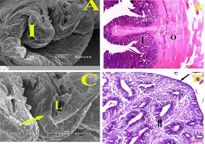
Int J Vet Sci, 2021, 10(1): 19-24.
pouch and injected with 10% normal saline solution, the sac (sacci cutanei phalli) (Fig. 1B/3), and the small thin one
phallus could be everted outside the pouch, passed into called glandular sac (sacci glandularis phalli) (Fig. 1B/4).
caudal direction, through the vent, the length could be At erected state the phallus was a very long structure
measured during erection by using a ruler, also could be composed of base, body and apex, its length was about 8-
demonstrated by the morphology and different parts (Reilly 10 cm. The shaft of the phallus (Fig. 1B/5), was demarcated
2001). Sony digital camera 24mp, 18x was used for by a phallic sulcus (sulcus phalli) which was a spirally
photographed the existing results. The nomenclature longitudinal groove, placed between two tubes of the
recommended in this study was given by Nomina phallus, extended from the ejaculatory fossa to the apex of
Anatomica Avium. the phallus called sulcus spermaticus (Fig. 1B/ 6). Also, the
body was characterized by presence of phallic ridges
General histological study spread all over the phallus, arranged in a numerous row,
After complete anesthesia, the birds were slaughtered these ridges were pointed and directed backward towards
and the phallus of both birds was immediately dissected out the base called rugae phalli (Fig. 2/1). The base of the
and sectioned into small pieces. The specimens were fixed phallus (Fig. 3A) supported by a cartilaginous body (corpus
in neutral buffered formalin. They were dehydrated in cartilaginous) (Fig. 3B/4). The suspensory ligament of the
ascending degree of alcohols, cleared in benzene finally phallus is called suspensorium phalli (Fig. 3C/6). The apex
embedded in paraplast. Serial and step serial sections of 5- of the phallus (apex phalli erecti) was the terminal part of
6μm thick were obtained and stained with Hematoxylin and the phallus (Fig. 3D/7), it had the opening of glandular sac
Eosin (H&E), Masson’s trichrome (Bancroft and Gamble (ostium sacci glandularis phalli) that allowed the seminal
2008). fluid to be come out from the phallus (Fig. 3D/8).
Histochemical study Goose histological finding
The specimens were obtained, fixed and processed as Histologically, the phallus of goose consisted of the
listed in general histological study and Periodic Acid Schiff inner glandular part and outer cutaneous layer. The inner
technique (PAS) stained for neutral mucopolysaccharides glandular part was lined with mucous secretary cells and
detection Carson (1990). contained mucous gland (Figs. 4A, 4B and 4C) whose
revealed PAS positive reaction (Fig. 4D) and surrounded
Scanning electron microscope by outer cutaneous layer of collagen fibers (Fig. 5A); the
Small specimens taken from phallus of geese and latter layer was composed of two distinct parts: an inner
turkeys were washed by 0.1M Na-cacodylate buffer. They
were fixed in a mixture of 2.5% paraformaldehyde and
2.5% glutaraldehyde in 0.1M Na-cacodylate buffer, pH 7.3
for 4 hours at 4°C, thereafter, they were washed in the same
buffer used and post-fixed in 1% osmic acid in 0.1 M Na-
cacodylate buffer for further 2h at room temperature. The
samples were dehydrated and critical point dried with a
polaron apparatus. Finally, they were coated with gold and
observed with JEOL scanning electron microscope (JSM-
5400 LV) at KV 10 Polák et al. (1983). All specimens were
prepared and analyzed in electron microscope Unit, of
applied centre for entomonematodes, Faculty of
Agriculture, Cairo University, Egypt.
RESULTS
The phallus was the organ of copulation in the male’s Fig. 1: A photograph showing the length and parts of erected
birds, could be categorized into two types, true intromittent pallus of the male goose. A: Length of the flaccid phallus. B:
phallus, which was introduced into the female cloaca Length of the fully erected phallus. 1) anticlock wise coils, 2)
during coitus and non-intromittent phallus was not entered phallic flexure, 3) cutaneous sac, 4) glandular sac, 5) body, 6)
the cloaca but it expelled the sperms onto the external spermatic sulcus, 7) apex, 8) base and 9) vent.
female genitalia directly.
Goose anatomical finding
The phallus was an intermittent type (phallus
protrudens). It was a spiral-coiled organ. At the rest state,
the phallus of goose was invested within a double
peritoneal membrane called phallic pouch, slightly sited
left to median plane close to the cloaca. It was about 2 and
half coils, organized in an anticlock wise direction (Fig.
1A/1) and these coils were arranged as a double U-shaped
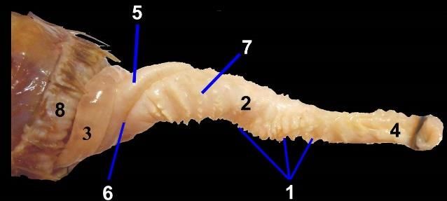
tube that were formed of a thick, large part twisted by a Fig. 2: A photograph showing a fully erected goose phallus
small, thin tube, in between the phallic flexure (Flexura consisted of 1) phallic ridges, 2) body, 3) base, 4) apex, 5)
phalli) (Fig. 1B/2). The large thick part called cutaneous glandular sac, 6) phallic flexure, 7) cutaneous sac and 8) vent.
20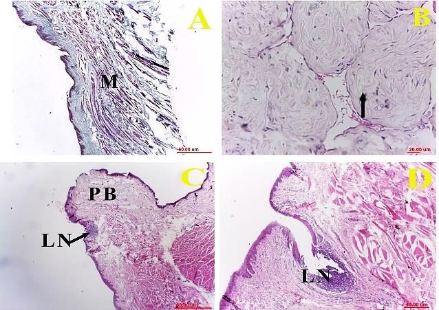
Int J Vet Sci, 2021, 10(1): 19-24.
Fig. 3: A photograph showing the different parts of the
phallus in the male goose. A: Base, B: Cartilaginous body,
C: Phallic pouch and D: Apex and opening of glandular
sac. Other parts are 1) glandular sac, 2) cutaneous sac, 3)
vent, 4) hyaline cartilage, 5) phallic pouch, 6) suspensory
ligament, 7) apex and 8) opening of glandular sac.
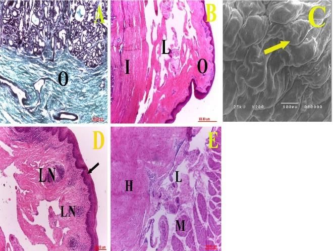
Fig. 4: A transverse section of goose
phallus, A: Scanning electron
microscopy showing inner glandular
part (I) (Bar=500µm); B: A
photomicrograph showing inner
glandular part (I) and outer
collagenous part (O), H&E stain, X40;
C: Scanning electron microscopy
showing mucous secretory cells
(arrow) and lymphatic capillaries (L)
(Bar=100µm), and D: A
photomicrograph showing inner
glandular part lined with PAS positive
mucous secretory cells (arrow) and
mucous gland (double arrows). PAS
stain (X400).
Fig. 5: A transverse section of goose
phallus, A: A photomicrograph showing
outer cutaneous layer of collagen fibers
(O), Masson’s trichrome stain (X100);
B: A photomicrograph showing an inner
layer of loose circumferential
connective tissue (I) & an outer dense
disorganized one (O) in between the two
layers was large lymphatic lumen (L),
H&E stain (X40); C: Scanning electron
microscopy showing the surface
epithelium of its cutaneous part lined by
stratified squamous non keratinized
epithelium (arrow) (Bar=100µm); D: A
photomicrograph showing a numerous
lymph nodules (LN) observed under the
stratified squamous non keratinized
epithelium (arrow), and E: A
photomicrograph of goose phallus base
showing hyaline cartilage (H),
lymphatic capillaries (L) and skeletal
muscle (M), H&E stain, X100.
21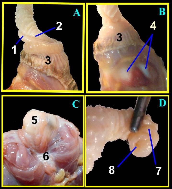
Int J Vet Sci, 2021, 10(1): 19-24.

                                                                              keratinized epithelium (Fig. 7B) and characterized by
                                                                              neither tubular nor glandular portion detected. This
                                                                              cutaneous portion was arranged as two plugged portions,
                                                                              lateral phallic bodies (Fig. 7A), connected by furrow.
                                                                              Longitudinal oriented skeletal muscle was observed in the
                                                                              phallic body while circular orientation in the furrow (Figs.
                                                                              7C and 8A). Highly vascularized and innervated dense
                                                                              irregular connective tissue mainly collagen fibres with
                                                                              peripheral nerve ending, lamellar corpuscle, were
                                                                              observed beneath the stratified squamous non keratinized
                                                                              epithelium (Figs. 7C and 8B). Lymphatic nodules were
                                                                              detected beneath the epithelium in the phallic bodies as
                                                                              well as at commissure between the phallic bodies and the
                                                                              furrow (Figs. 8C and 8D).
Fig. 6: A photograph showing the phallus in the male Turkey.
Various parts are 1) lateral phallic bodies, 2) median furrow, 3)                                    DISCUSSION
opening of the vertical fissure, 4) dorsal lip, 5) vent and 6) ventral
lip.
                                                                                   The present study revealed that, the phallus of the
layer of loose circumferential and an outer dense                             goose was an intromittent type, it was a spiral coiled
disorganized layer in between the two layers was large                        structure invested within a double peritoneal membrane
lymphatic lumen (Fig. 5B), as well as lymphatic                               (Phallic pouch), similar to King (1981), Kevin (2000),
capillaries were observed in the connective tissue beneath                    Brennan et al. (2009) and El Gindy et al. (2016) in duck
the glandular portion (Fig. 4C). The surface epithelium of                    and King and McLelland (1984), Brennan and Prum (2012)
its cutaneous part was lined by stratified squamous non                       in Ostrish and kiwis and Rajendranath et al. (2013) in emu,
keratinized epithelium Fig. (5C). Numerous lymph                              while, the phallus of the turkey was an non intromittent
nodules were observed under the epithelium (Fig. 5D).                         type and lied on the crest of the ventral lip of the vent, these
The base of the phallus was supported by hyaline                              results similar to the results in the domestic fowl that were
cartilage, lymphatic capillaries and skeletal muscle (Fig.                    confirmed by Bull et al. (2007).
5E).                                                                               The coils of phallus were about 2 and half coils, had a
                                                                              anticlock wise direction, but El Gindy et al. (2016) in
Turkey anatomical findings
                                                                              balady duck recorded 3 and half coils. When dissected it
     The male copulatory organ was appeared in the
                                                                              was seen as a U-shaped tube, attached to the cloaca at both
ventral wall of the vent; it was non intromittent type. It
                                                                              ends that simulated Kevin (2000) in Argentine duck and El
was composed of two lateral phallic bodies on each side
at the ventral wall of the vent (Fig. 6/1), separated by a                    Gindy et al. (2016) in balady duck and. This tube is formed
median furrow or groove (Fig. 6/2). Two lateral phallic                       of two parts; thick and thin tubes in between phallic flexure,
bodies were appeared as a short, rounded structure, which                     the thick part is called cutaneous sac while, the thin one is
was enlarged and engorged with fluid; the fluid passed                        called glandular sac similar to the findings of King and
into the median furrow.                                                       McLelland (1984), Baumel et al. (1993) and El Gindy et al.
                                                                              (2016) in duck, but in this study, the two tubes in the goose
Turkey histological findings                                                  were a very characteristic than the same tubes in the duck
    The phallus of turkey was formed of cutaneous portion                     as the cutaneous tube was a very larger than the glandular
only (Fig. 7A) lined with stratified squamous non                             one.

                                                                                                     Fig. 7: A transverse section of turkey
                                                                                                     phallus.     A:     Scanning      electron
                                                                                                     microscopy showing the cutaneous
                                                                                                     portion     in    the     phallic    body
                                                                                                     (Bar=500µm); B: Scanning electron
                                                                                                     microscopy showing the cutaneous
                                                                                                     portion lined by stratified squamous non
                                                                                                     keratinized (Bar=10µm) and C: A
                                                                                                     photomicrograph showing longitudinal
                                                                                                     oriented skeletal muscle (M) and
                                                                                                     lamellar corpuscle (arrow) in the phallic
                                                                                                     body, Masson’s trichrome stain, Bar=
                                                                                                     60µm.

                                                                         22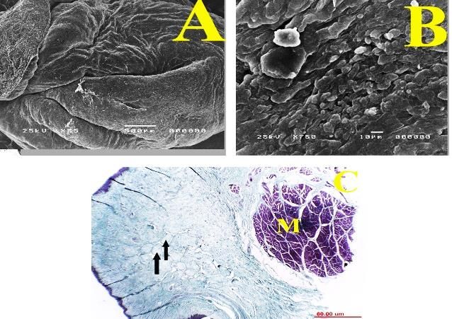
Int J Vet Sci, 2021, 10(1): 19-24.

                                                                                           Fig. 8: A transverse section of turkey
                                                                                           phallus. A: A photomicrograph showing
                                                                                           circular oriented skeletal muscle (M) in
                                                                                           the furrow, Masson’s trichrome stain,
                                                                                           X40; B: A photomicrograph showing
                                                                                           lamellar corpuscle (arrow) in the phallic
                                                                                           body, H&E stain, X400; C: A
                                                                                           photomicrograph showing lymphatic
                                                                                           nodule (LN) in the phallic body (PB),
                                                                                           H&E stain, X40 and D: A
                                                                                           photomicrograph showing lymphatic
                                                                                           nodule at the commissure between the
                                                                                           phallic body and the furrow (LN), H&E
                                                                                           stain, X100.

     In the turkey, the phallus was formed of two lateral            Laysan ducks that was recorded by Herrera et al. (2014),
phallic bodies on each side at the ventral wall of the vent.         this coloration was not recorded in the goose in this study
Two lateral phallic bodies were appeared as a short,                 and also was not recorded by El Gindy et al. (2016) in
rounded structure, separated by a median furrow, these               Balady duck. The external surface of the phallus was
results were confirmed by Bull et al. (2007) in fowl but the         characterized by presence of ridges that covering the wall
author reported presence of a median phallic body between            of the phallus, as reported by Brennan et al. (2009) in
the two phallic bodies, which was not found in the turkey            Mallarad duck, El Gindy et al. (2016) in balady duck but
in this study and also stated that, the deposited semen              this study added that these phallic ridges were pointed,
became outside the female cloaca, on the everted vagina              distributed all over the phallus in a several rows.
during copulation process, then the spermatozoa                      Meanwhile Kevin (2000) in Argentine duck reported
transported into the oviduct. In goose, the large cutaneous          spines directed backwards towards the base of the phallus.
sac of phallus was characterized by a spirally longitudinal               The penis of goose had the inner glandular part lined
phallic sulcus that was extended from the base to the apex,          with secretory cells surrounded by outer cutaneous layer of
also called sulcus spermaticus due to the sperms                     collagen fibers; the latter layer was composed of two
transported through this groove. The results are in line with        distinct parts: an inner layer of lose circumferential and an
the previous studies of King (1981), Duan (2000), El Gindy           outer dense disorganized layer in between the two layers
et al. (2016) in balady duck, Wang et al. (2008) in goose,           was large lymphatic lumen. These results agreed with
while Yong and Zhanjun (2011) in ostrich recorded a penile           Brennan et al. (2009) in mallard duck, Brennan and Prum
groove at the back of the penis which was a closed and like          (2012) in emus, El Gindy et al. (2016) in Balady duck,
a tube when the penis erected.                                       while Brennan and Prum (2012) in ostrich recorded that,
     In this study, the fully erected phallus of goose was           the penis lacked a blind tubular cavity and formed of
consisted of a large left and a small right tube twisted             fibrous bodies composed of dense, largely disorganized
around each other, which formed a bulbous base, body and             collagen matrix except in the areas surrounded the narrow
terminated by the apex of the phallus, as recorded by King           lymphatic spaces.
and McLelland (1984) in duck. These regions were found                    In line with the findings of King and McLelland
in the phallus of all types of Anseriforms but the divisions         (1984), Brennan et al. (2009) in duck and El Gindy et al.
of these regions differed according to each author, as               (2016) in Balady duck, the base of phallus was supported
Brennan et al. (2009) in Muscovy duck divided it into basal          by cartilaginous body which was formed of hyaline
and apical regions, Baumel et al. (1993) in duck                     cartilage and its cutaneous part.
demonstrated four components, phallic base, phallic body,                 In the present study lymphocytic aggregations noticed
phallic sac and phallic pouch. While in the turkey, the              in the phallus of goose and turkey. Lymphocytic
phallus was composed of a median phallic groove flanked              aggregation had developed a number of different
on either side by lateral phallic bodies. Regarding the total        immunological strategies including cell mediated one.
length of the phallus, it was ranged from 8-10cm during              These lymphocytes might add a more protective condition
fully erection in goose, while, reached 22cm in Argentine            for the sperms. In the current study, PAS reaction was
duck (Kevin 2000), 20cm in ostrich (Elias et al. 2007;               detected in the surface epithelium and the gland. This
Brennan and Prum 2012), 25-30cm in ostrich (Yong and                 revealed presence of neutral mucoplysaccharides (Zaher et
Zhanjun 2011) who stated that this penis was very strong,            al. 2012).
while the penis of the turkey as in chickens was not strong,
3 cm in rhea (Góes et al. 2010), 8.5 cm in Laysan ducks and          Conclusion
5.3cm in Mandarin ducks (Herrera et al. 2014) and ranged                 The study gives a complete knowledge for phallus of
from 13-15cm in Balady duck (El Gindy et al. 2016). The              geese and turkey that will help in the surgical operations of
phallus had some black coloration on its outer surface in            the wild geese, and the artificial insemination in the turkey,
Mandarin ducks and also lacked this coloration in the                which is an obligatory method for commercial production.

                                                                23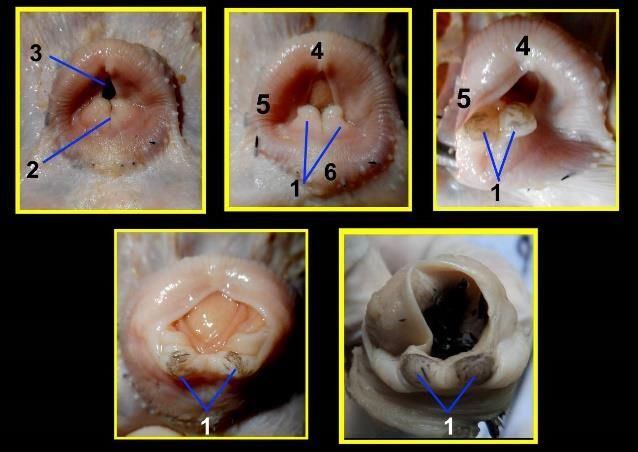
Int J Vet Sci, 2021, 10(1): 19-24.

Author contributions                                                            (Struthio camelus). Anatomia, Histologia, Embryologia, 36:
    All authors contributed to the reagents/materials/                          255-262.
analysis tools, collected the material, analyzed the data and              Góes PAA, Cavalcante AKS, Nichi M, Perez EG de A, Barnabe
wrote the manuscript.                                                           RC and Barnabe VH, 2010. Reproductive characteristics of
                                                                                captive greater rhea (Rhea americana) males reared in the
                                                                                state of Sao Paulo Brazil. Brazil Journal of Poultry Science,
                       REFERENCES                                               12: 57-62. https://doi.org/10.1590/S1516-635X2010000100
                                                                                009
Amer FI and Shahin MA, 1975. The post-hatching development                 Herrera AM, Brennan PLR and Cohn MJ, 2014. Development of
     of the gonads in the fowl, Gallus domesticus. Annals of                    avian external genitalia: interspecific differences and sexual
     Zoology 11: 1-25.                                                          differentiation of the male and female Phallus. Sexual
Bancroft JD and Gamble M, 2008. Theory and practice of                          Development, 9: 43-52.
     histological techniques. 6th Ed. Churchill Livingston, New            Herrera AM, Shuster SG, Peritton CL and John MJ, 2013.
     York, USA.                                                                 Developmental basis of Phallus reduction during bird
Baumel JJ, King SA, and Breasile JE, 1993. Nomina Anatomica                     evolution.      Current      Biology,      23:     1065-1075.
     Avium. Published by the Nuttall Ornithological Club. No:                   https://doi.org/10.1016/j.cub.2013.04.062
     23, Cambridge, Massachusets.                                          Hosken DJ and Stockley P, 2004. Sexual selection and genital
Brennan PLR, Birkhead TR, Zyskowski K, van der Waag J and                       evolution. Trends in Ecology & Evolution 19: 87-93.
     Prum RO, 2008. Independent evolutionary reductions of the             Kevin GMC, 2000. The 20-cm spiny penis of the Argentine Lake
     phallus in basalbirds. Journal of Avian Biology 39: 487-492.               Duck (Oxyura vittata). The Auk 117: 820-825.
     https://doi.org/10.1111/j.0908-8857.2008.04610.x                      King AS, 1981. Phallus. In form and function in birds. Academic
Brennan PLR, Clark CJ and Prum RO, 2009. Explosive eversion                     Press, New York, USA, pp: 107-148.
     and functional morphology of the duck penis supports sexual           King AS and McLelland J, 1984. Birds, Their Structure and
     conflict in waterfowl genitalia. Proceedings of the Royal                  Function. 2nd Ed, Bailliere Tindall, England: pp: 145-165.
     Society B, Biological Sciences 277: 1309-1314.                        Polák Š, Brozman M and Jakubovský J, 1983. Concerning
     https://doi.org/10.1098/rspb.2009.2139                                     problem of applying raster electron microscopy in studying
Brennan PLR and Prum RO, 2012. The erection mechanism of                        the ortho- and pathomorphology of the human spleen.
     ratite penis. Journal of Zoology 286: 140-144.                             Bratislava Lekarske Listy 80: 267-278.
     https://doi/org/10.1111/j.1469-7998.2011.00858.x                      Rajendranath N, S Chandrasekhara RT, Kumar PD, Raghavendar
Briskie JV and Montgomerie R, 1997. Sexual selection and the intro-             K and Kumar V, 2013. Gross anatomical studies on the
     mittent organ of birds. Journal of Avian Biology 28: 73-86.                apparatus of the Emu (Dromaius novaehollandiae). Indian
Bull ML, Martins MRF, Cesario MD, Padovani CR, Mendes AA,                       Journal of Veterinary Anatomy 25: 81-82.
     2007. Anatomical study on domestic fowl (Gallus                       Reilly JS, 2001. Euthanasia of animals used for scientific
     domesticus) reproductive system. International Journal of                  purposes; section 13: Birds (Class Aves), ANZCCART, 2nd
     Morphology 25: 709-716.                                                    Ed. pp: 79-82.
Carson FL, 1990. Histotechnology: A Self-Insructional Text.                Tingari MD, 1971. On the structure of the epididymal region and
     American Society of Clinical Pathologists. Chicago, pp: 294.               ductus deferens of the domestic fowl (Gallus domesticus).
Chen YT, 2005. Matching methods and the relevant management                     Journal of Anatomy, 109: 423-435.
     skills of pigeon breeders. China Poultry, 27: 26-28.                  Yong Z and Zhanjun R, 2011. Anatomic study on the main male
Duan X, 2000. The main points of artificial insemination. China                 reproductive organs of Ostrich. Global Journal of Health
     Poultry, 22: 17-18.                                                        Science 3: 2011.
EL Gindy EM, Hassan AM, El-Bably SH, Shaker NA and                         Wang, Wenqiang and Huaqi Z, 2008. Artificial insemination
     Hussien SH, 2016. Morphological studies on the male                        technology and the application in Goose. China Poultry, 30:
     copulatory organ of Balady Duck (Anas boscas domestius)                    37-38.
     with special reference to its blood supply. Giza Veterinary           Zaher M, El_Ghareeb AW, Hamdi H and and AbuAmod F,
     Medical Journal.                                                           2012. Histochemical adaptation of avian alimentary canal to
Elias MZJ, Aire TA and Soley JT, 2007. Macroscopic features of the              their food habits. I: Coturnix coturnix. Life Science Journal
     arterial supply to the reproductive system of the male Ostrich             9: 253-275.

                                                                      24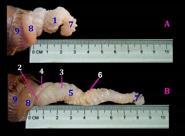
You can also read