Isolated saccular aneurysm of the external jugular vein
←
→
Page content transcription
If your browser does not render page correctly, please read the page content below
Clinical Case Report
Isolated saccular aneurysm of the external jugular vein
Hari Janardanan Pillai1 , Nilanjan Roy1 , Pankaj Purushotam Rao1 ,
Khushdeep Kaur Shergill2 , Divya Shelly2 , Basil Badarudeen1
How to cite: Pillai HJ, Roy N, Rao PP, Shergill KK, Shelly D, Badarudeen B. Isolated saccular aneurysm of the external jugular
vein. Autops Case Rep [Internet]. 2021;11:e2020188. https://doi.org/10.4322/acr.2020.188
ABSTRACT
Venous aneurysm of the head and neck is a rare clinical entity due to its asymptomatic nature and tendency of clinicians
to report only surgical results. Whereas the primary aneurysm of internal jugular vein (IJV) in children is being increasingly
recognized, secondary aneurysms of veins of the head and neck in adults, notably the external jugular vein (EJV) aneurysm
remains only in anecdotal case reports. We present the case of a 63-year-old previously healthy woman who presented with
a gradually progressive right lateral neck swelling over the last 18 months. Following the evaluation, she was diagnosed
as a case of isolated spontaneous right-sided EJV aneurysm and was managed by surgical excision of the aneurysm.
Keywords
Aneurysm; Jugular Veins; Venous Thrombosis.
CASE REPORT
A 63-year-old, previously healthy lady, presented lesion with no internal vascularity concerning the
with complaints of insidious-onset swelling over right EJV. Magnetic Resonance Venogram (MRV)
the right side of her neck since18 months, which reported a focal outpouching arising from the
gradually progressed from the initial size of 2 cm to anterolateral aspect of right EJV immediately cranial
5 cm at presentation. She also noted an increase in to its confluence with right subclavian vein measuring
the size of the swelling during straining and coughing. 3.3 x 3.4 x 3.7 cm with an intraluminal thrombus
Though the swelling was initially painless, she (Figure 2). The remaining vasculature of the neck
experienced a vague, dull aching pain in the swelling
appeared normal. She was diagnosed as a case of
in the last couple of months, which forced her to seek
an isolated saccular aneurysm of the right EJV. Given
medical evaluation. There was no other contributory
the recent onset pain and progressive increase in size,
history of trauma or surgical intervention in the
she underwent exploration of the right side of the
neck. On evaluation, she had a non-tender, soft,
neck, which revealed a 4x4 cm saccular aneurysm of
and non‑pulsatile swelling in her right supraclavicular
region, measuring 5 cm x 4cm (Figure 1A, B). the proximal right EJV with intraluminal thrombus
The swelling was reducible and enlarged in size (Figure 1C and D).
with the Valsalva maneuver. The ultrasonography Proximal and distal control of EJV were obtained,
with color Doppler of her neck revealed a 3.5 cm and the aneurysm was excised with ligation of
1
Armed Forces Medical College, Department of Surgery, Pune, Maharashtra, India
2
Indian Navy Hospital Ship Asvini, Mumbai, Maharashtra, India
Copyright: © 2020 The Authors. This is an Open Access article distributed under the terms of the Creative
Commons Attribution License, which permits unrestricted use, distribution, and reproduction in any medium,
provided the original work is properly cited.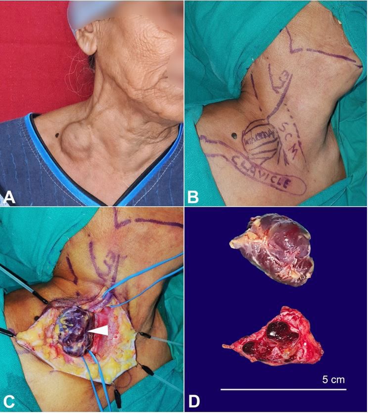
Isolated saccular aneurysm of the external jugular vein
Figure 1. A – Gross examination of the neck region with a 5-cm swelling over right supraclavicular region;
B – preoperative marking of the patient after draping in the OR, SCM – sternocleidomastoid muscle, Clavicle – Right
Clavicle, EJV - Right EJV; C – intraoperative image. Note the saccular aneurysm along the looping of the right EJV
(arrowhead); D – Gross view of the excised specimen of aneurysm (upper image) and the cut surface of the opened
specimen showing intraluminal thrombus (lower image).
the right EJV. The patient had an uneventful section of the aneurysm showed an abnormally dilated
postoperative recovery and was discharged on venous lumen with thrombus, surrounded externally
postoperative day 2. by a thinned tunica media (H&E stain x 100). Elastic
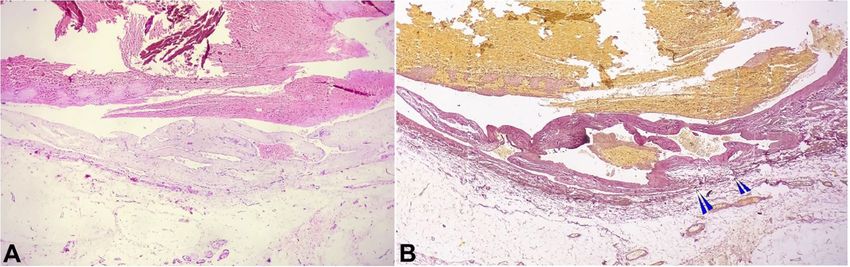
Histopathology of the specimen was consistent Van Gieson (EVG) stain revealed a complete loss
with a true aneurysm of the vein. The transverse of elastic fibers in the vessel wall (Figure 3). Her follow
2-5 Autops Case Rep (São Paulo). 2021; 11:e2020188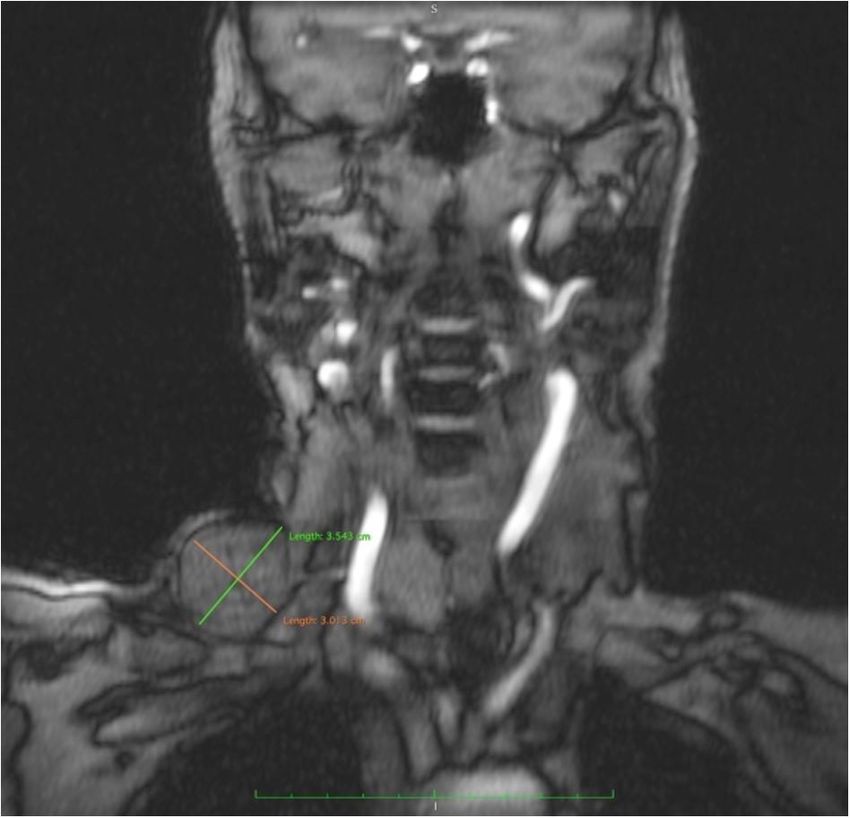
Pillai HJ, Roy N, Rao PP, Shergill KK, Shelly D, Badarudeen B

up visit at one month revealed a well-healed scar in        or acquired. Congenital aneurysms are fusiform
the neck with no complaints.                                dilatations of the vein, commonly involving IJV, and
                                                            are supposed to be a consequence of the weakness
                                                            of the elastic layers and muscle cells.2,3,6 The acquired
DISCUSSION
                                                            aneurysms are typically saccular, occur spontaneously
     Venous aneurysms in the head and neck are              or secondary to trauma, surgical intervention or
rarely encountered in clinical practice owing to the        diseases involving veins like endophlebosclerosis and
low-pressure system of the superior vena cava. 1-4          endophlebohypertrophy.7,8
The majority of them involve the internal jugular                 EJV aneurysm, by far, is exceedingly rare, and
vein (IJV), followed by the EJV and the anterior veins in   its exact incidence is still not known.9 EJV aneurysm
decreasing frequency.5 They can either be congenital        is commonly found in elderly males, on the right
                                                            side. The risk factors for their development include
                                                            trauma, thoracic outlet syndrome, tumors, hormonal
                                                            therapy, or increased pressure in the vena cava
                                                            system. 3-5 They are commonly saccular and rarely
                                                            fusiform.3 The majority of the patients present with an
                                                            asymptomatic, soft, non- tender compressible swelling
                                                            in the lateral aspect of the neck, which classically
                                                            shows an increase in size on straining or with Valsalva
                                                            maneuver in the absence of any bruit. Some patients
                                                            may complain of dull aching or, more commonly, a
                                                            “discomfort” or feeling of tightness over the swelling.10
                                                            The differential diagnosis includes (i) laryngocele,
                                                            (ii) cavernous hemangioma, (iii) pharyngeal pouch,
                                                            (iv) external laryngeal diverticulum, (v) superior
                                                            mediastinal cyst, and (vi) cervical arterial, venous
                                                            aneurysms or veno‑lymphatic malformations. 10,11
                                                            Noninvasive diagnostic imaging modality like doppler
                                                            ultrasonography of the neck veins confirms the
Figure 2. MR venogram of the neck (T1 weighted              diagnosis. 12 Further, CT angiography with digital
image) showing focal outpouching arising from the           subtraction angiography and MR venography can
anterolateral aspect of the right EJV, immediately          help to delineate the anatomical extent of the
cranial to its confluence with the right subclavian vein,   aneurysm, presence of feeder vessels, intraluminal
measuring 3.5 × 3.01 cm.                                    thrombus besides aiding in pre-operative planning,9,10,13

Figure 3. Photomicrograph of the surgical specimen showing the complete loss of the elastic fibers in the vessel wall
(multiple arrowheads); A – (H&E, 100X) and B – (EVG stain, 100X).

Autops Case Rep (São Paulo). 2021; 11:e2020188                                                                              3-5
Isolated saccular aneurysm of the external jugular vein
EJV aneurysms rarely give rise to complications like REFERENCES
thrombophlebitis, rupture, thrombus formation
secondary to trauma, or pulmonary embolism.14,15 1. Başbuğ HS, Bitargil M, Karakurt A, Özışık K. External
jugular vein aneurysm in a young woman: an uncommon
The indications for treatment are primarily cause of neck mass. International Journal of the
cosmetic and rarely due to complications of the Cardiovascular Academy. 2016;2(1):16-8. http://dx.doi.
aneurysm per se.16 Clinical progression of EJV aneurysm org/10.1016/j.ijcac.2015.12.005.
is insidious and protracted without significant 2. Ekim H, Ozen S. Primary venous aneurysm of the external
morphological transformations. Asymptomatic patients jugular vein. East J Med. 2002;7(1):24-5.
can be reassured and conservatively managed with
3. Kim SW, Chang JW, Lee S. Unusual presentation of
watchful waiting.2,9,14,16,17 The management modalities a cervical mass revealed as external jugular venous
for an aesthetically disfiguring and complicated aneurysm. Vasc Specialist Int. 2016;32(4):205-7. http://
aneurysm include surgical excision or endovascular coil dx.doi.org/10.5758/vsi.2016.32.4.205. PMid:28042563.
embolization.17 The treatment of choice for saccular 4. Drakonaki EE, Symvoulakis EK, Fachouridi A, Kounalakis D,
aneurysms of the EJV is complete surgical excision Tsafantakis E. External jugular vein aneurysm presenting as a
without venous reconstruction through a longitudinal, cervical mass. Int J Otolaryngol. 2011;2011:485293. http://
lateral neck incision under general or local anesthesia dx.doi.org/10.1155/2011/485293. PMid:21716689.
with minimal intraoperative and postoperative 5. Siani A, Flaishman I, Schioppa A, Zaccaria A, Baldassarre
complications. Intravenous coil embolization, along E. Jugular venous phlebectasia: uncommon in children,
anecdotal in adults. Am J Surg. 2008;195(3):419-20.
with percutaneous injection of sclerosant foam, has
http://dx.doi.org/10.1016/j.amjsurg.2007.01.041.
recently emerged as a minimally invasive alternative.9,13 PMid:18308045.
Though there is no evidence, at present, to prove
6. Porcellini M, Selvetella L, Bernardo B, Del Guercio L,
its clinical efficacy compared to standard surgical
Baldassarre M. Aneurysms of the external jugular vein.
excision, it offers a superior cosmetic outcome with G Chir. 1996;17(5):238-41. PMid:8755223.
lesser hospital stay at the expense of incurring higher
7. Anzidei MSG, Fraioli F, Catalano C. Multi-detector ct
treatment costs.
imaging: principles, head, neck and vascular systems.
Boca Raton: CRC Press; 2014.
CONCLUSION 8. Mohanty D, Jain BK, Garg PK, Tandon A. External jugular
venous aneurysm: a clinical curiosity. J Nat Sci Biol Med.
Aneurysms of the EJV can occur spontaneously or 2013;4(1):223-5. http://dx.doi.org/10.4103/0976-
as a sequel to trauma, surgical intervention, or diseases 9668.107296. PMid:23633867.
affecting the vein walls. It can be asymptomatic or 9. Pandey NN, Sinha M, Deshpande A, Kumar S.
can present with complications like thrombosis or External jugular vein aneurysm: successful endovascular
inflammation. Clinical examination augmented by management of an exceedingly rare entity. BMJ Case
Rep. 2020;13(2):e233572. http://dx.doi.org/10.1136/
non-invasive color doppler ultrasonography confirms bcr-2019-233572. PMid:32041750.
the diagnosis. Management depends on the symptoms
10. Verma RK, Kaushal D, Panda NK. External jugular vein
complex of the patient. Whereas asymptomatic
aneurysm with thrombus presenting as painful neck mass:
patients can be offered watchful waiting, symptomatic a case report. Oman Med J. 2013;28(4):278-80. http://
patients have to be managed with either surgical dx.doi.org/10.5001/omj.2013.77. PMid:23904923.
excision or endovascular embolization with minimal
11. Al-Shaikhi A, Kay S, Laberge JM. External jugular
postoperative morbidity. venous aneurysm: an unusual cause of a neck mass
in a young child. J Pediatr Surg. 2003;38(10):1557-9.
http://dx.doi.org/10.1016/S0022-3468(03)00526-8.
ACKNOWLEDGEMENTS PMid:14577090.
12. Lucatelli P, Tommasino G, Guaccio G, Benvenuti A, Ricci
Dr. Sagar Iyer for the compilation of
C. External jugular vein spontaneous aneurysm, diagnosis,
intra‑operative photographs and Dr. Isha Sharma and treatment with video. Ann Vasc Surg. 2017;41:282.
for photomicrograph of histopathology e11-e13.
4-5 Autops Case Rep (São Paulo). 2021; 11:e2020188Pillai HJ, Roy N, Rao PP, Shergill KK, Shelly D, Badarudeen B
13. Rajadurai A, Abdul Aziz A, Mat Daud NA, Abdul Wahab AF, a source of thrombotic complications. Int Angiol.
Muda AS. Embolisation of external jugular vein aneurysm: 2010;29(3):284-5. PMid:20502418.
a case report. Malays J Med Sci. 2017;24(6):107-
12. http://dx.doi.org/10.21315/mjms2017.24.6.14. 16. Calligaro KD, Ahmad S, Dandora R, et al. Venous
PMid:29379394. aneurysms: surgical indications and review of the
literature. Surgery. 1995;117(1):1-6. http://dx.doi.
14. Lee HY, Cho SH, Ko TY, et al. Saccular aneurysm of the org/10.1016/S0039-6060(05)80222-3. PMid:7809821.
external jugular vein: a case report. Korean J Thorac
Cardiovasc Surg. 2014;47(2):171-3. http://dx.doi. 17. Parashi HS, Rawekar KH, Joshi MM, Namdev HS,
org/10.5090/kjtcs.2014.47.2.171. PMid:24782973. Jadhao MR, Bhosle KN. Saccular aneurysm of external
jugular vein with partial thrombosis. Asian Cardiovasc
15. Ioannou CV, Kostas T, Tsetis D, Georgakarakos E, Gionis Thorac Ann. 2018; 26( 8) : 625- 7. http://dx.doi.
M, Katsamouris AN. External jugular vein aneurysm: org/10.1177/0218492316686477. PMid:30335501.
This study carried out at the Armed Forces Medical College, Pune, Maharashtra, India.
Authors’ contributions: Nilanjan Roy and Hari Janardanan Pillai did surgery of patient. Hari Janardanan Pillai
and Khushdeep Kaur Shergill wrote the manuscript. Basil Badarudeen collated the photographs and proofread
the manuscript. Pankaj Purushotam Rao and Divya Shelly did the literature review and critical review of the
manuscript. All authors read and approved the final manuscript.
Ethics statement: The authors retain informed consent signed by the patient authorizing pictures and data
publication. The manuscript is by the Institutional Ethics committee rules.
Conflict of interest: None
Financial support: None
Submitted on: May 4th, 2020
Accepted on: June 1st, 2020
Correspondence
Hari Janardanan Pillai
Armed Forces Medical College, Department of Surgery
Wanowrie, Pin code 411040, Pune, India
Phone: +91 (70) 3039-9113
drharijp@gmail.com
Autops Case Rep (São Paulo). 2021; 11:e2020188 5-5You can also read