Joint Preparation and Ray Shortening in Arthroscopic Versus Open First Metatarsophalangeal Fusion: A Cadaver Study - Cureus
←
→
Page content transcription
If your browser does not render page correctly, please read the page content below
Open Access Original
                                          Article                                                DOI: 10.7759/cureus.9633

                                          Joint Preparation and Ray Shortening in
                                          Arthroscopic Versus Open First
                                          Metatarsophalangeal Fusion: A Cadaver
                                          Study
                                          Haley McKissack 1 , Bradley Alexander 1 , Gean C. Viner 1 , Eildar Abyar 1 , Nicholas A. Andrews 1
                                          , Ashish Shah 1

                                          1. Orthopaedic Surgery, University of Alabama at Birmingham, Birmingham, USA

                                          Corresponding author: Ashish Shah, ashishshah@uabmc.edu

                                          Abstract
                                          Purpose
                                          This study compares the amount of joint preparation and first ray shortening following first
                                          metatarsophalangeal (MTP) joint fusion utilizing open conical reaming versus arthroscopic
                                          technique.

                                          Methods
                                          Ten below-knee cadaver specimens were randomly assigned to undergo either open or
                                          arthroscopic first MTP fusion. Following fixation, first ray length measurements were obtained
                                          from pre-operative and post-operative radiographs and were used to determine first ray
                                          shortening. Additionally, the ratio of first ray length to second ray length was calculated both
                                          pre-operatively and post-operatively and compared between the two approaches. All ankles
                                          were then completely dissected, and prepared surface areas were demarcated. ImageJ photo
                                          analysis software (National Institutes of Health, Bethesda, MD, USA) was used to calculate the
                                          percentage of prepared and unprepared cartilage of each articular surface of each specimen.

                                          Results
                                          Overall, the open approach resulted in 99.3% ± 1.6% joint surface preparation, whereas the
                                          arthroscopic approach yielded 92.9% ± 7.2% (p = 0.089). On average, the head of the first
                                          metatarsal was significantly more prepared with the use of the open approach (99.5% ± 1.1%)
                                          than with the arthroscopic approach (96.6% ± 1.5%) (p = 0.008). However, with respect to the
                                          base of the phalanx, the average difference in preparation between the arthroscopic approach
                                          and the open approach was not statistically significant (90.0% ± 12.8% vs. 99.0% ± 2.2%; p =
Received 07/09/2020
                                          0.160). The average amount of first ray shortening in the arthroscopic approach was 2.2 ± 1.8
Review began 07/20/2020
Review ended 07/27/2020
                                          mm compared to 2.1 ± 3.2 mm in the open approach (p = 0.934). The average change in the first
Published 08/09/2020                      to second ray length ratio was 0.02 for both approaches (p = 0.891).

© Copyright 2020
McKissack et al. This is an open          Conclusion
access article distributed under the
terms of the Creative Commons             Arthroscopic first MTP fusion can be used to achieve joint preparation comparable to open
Attribution License CC-BY 4.0., which     technique while maintaining first ray length.
permits unrestricted use, distribution,
and reproduction in any medium,
provided the original author and
source are credited.                      Categories: Orthopedics

                                          How to cite this article
                                          Mckissack H, Alexander B, Viner G C, et al. (August 09, 2020) Joint Preparation and Ray Shortening in
                                          Arthroscopic Versus Open First Metatarsophalangeal Fusion: A Cadaver Study. Cureus 12(8): e9633. DOI
                                          10.7759/cureus.9633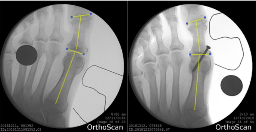
Keywords: mtp fusion, arthroscopic, joint preparation, ray shortening

                                   Introduction
                                   Open and arthroscopic first metatarsophalangeal (MTP) joint fusions are effective treatments
                                   for a variety of conditions such as rheumatoid arthritis, hallux rigidus, and severe hallux
                                   valgus [1]. Multiple surgical techniques have been described in the literature with regard to
                                   bone preparation and joint fixation, with varying degrees of success [2-6]. Regardless of
                                   technique, the important aspects of fusion are adequate preparation of the joint, maintaining
                                   the soft tissue envelope, and stable fixation. Singh et al. compared first MTP joint fusion
                                   utilizing flat cut or conical reaming technique for bone preparation with regard to the amount
                                   of first ray shortening that occurs and found no significant difference between both
                                   techniques [4]. These two techniques require an open approach to access the joint for surface
                                   preparation. In contrast, the arthroscopic technique is a minimally invasive procedure that
                                   allows joint visualization and surface preparation with less soft tissue damage. The first study
                                   describing first MTP arthroscopy was published in 1972, but due to a number of factors,
                                   arthroscopic treatments for first MTP joint have not fully spread [7,8]. This could be attributed
                                   to the learning curve that comes with arthroscopic surgery and the proven success of the open
                                   approach in first MTP fusion.

                                   One potential complication of first MTP fusion is first ray shortening, which can lead to
                                   symptomatic forefoot disorders such as transfer metatarsalgia of the lesser toes [6,7,9]. The
                                   second metatarsal seems to be especially vulnerable to developing transfer metatarsalgia due to
                                   it being usually longer than the other metatarsals and fixed between three cuneiforms, making
                                   it relatively immobile [10]. Patients can develop altered gait mechanics that manifest in the
                                   way of decreased ankle plantarflexion at toe-off and decreased step gait [11]. Currently, there is
                                   no consensus regarding the amount of first ray shortening that is acceptable [9]. To our
                                   knowledge, no study has compared the difference in joint preparation or the amount of first ray
                                   shortening following first MTP joint fusion utilizing open versus arthroscopic technique. The
                                   purpose of this study was to compare first MTP joint fusion utilizing open conical reaming
                                   versus arthroscopic technique for joint preparation with regard to the amount of first ray
                                   shortening that occurs. We hypothesize that arthroscopic MTP fusion will be as effective as the
                                   open technique, without any increase in first ray shortening.

                                   Materials And Methods
                                   Specimen preparation
                                   Ten fresh-frozen, unmatched below-knee cadaver leg specimens were utilized. Each specimen
                                   was inspected visually and radiographically to ensure the absence of any gross musculoskeletal
                                   pathology. Due to the nature of these specimens, we were not able to review or compare past
                                   medical records. There were four male donors and six female donors. Mean specimen age at
                                   death was 66.2 ± 15.3 years (range: 36-86 years). All specimens were stored at -20°C and then
                                   thawed at room temperature for 24 hours prior to testing.

                                   Open technique
                                   All specimens were prepared by a single foot and ankle fellowship-trained orthopedic surgeon.
                                   The first MTP joint was approached through a standard dorsal longitudinal incision. Soft tissue
                                   was dissected and extensor hallucis longus was freed and carefully retracted. The dorsal aspect
                                   of the joint capsule was incised and elevated subperiosteally over the first metatarsal and
                                   proximal phalanx to allow exposure and preparation of the joint. A 1.6-mm Kirschner (K)-wire
                                   was inserted into the metatarsal head, and a conical reamer (Arthrex Inc., Naples, FL, USA) was
                                   slid over the guidewire to expose the subchondral bone. The proximal phalanx was then
                                   prepared in a similar fashion. The joint was then placed in 15 degrees of dorsiflexion and 10

2020 McKissack et al. Cureus 12(8): e9633. DOI 10.7759/cureus.9633                                                               2 of 9
degrees of valgus using a 1.6-mm K-wire and a compression screw placed from distal-medial to
proximal-lateral in a standard lag fashion. Anterior-posterior and lateral fluoroscopic images
were obtained following fixation.
Arthroscopic technique
All specimens were prepared by a single foot and ankle fellowship-trained orthopedic surgeon.
The great toe was distracted with traction over a pulley attached to the surgical table. A 30-
degree, 2.4-mm arthroscope (Arthrex Inc.) was used for visualization. A two-portal approach
was utilized in five feet. The dorsomedial portal site was marked at the medial aspect of the
first MTP joint line, and the dorsolateral portal site was marked on the lateral aspect of the
extensor hallucis longus tendon at the level of the first MTP joint line. An 18-gauge needle was
inserted at the marked site, and the skin incision was widened utilizing a hemostat. The
arthroscope was inserted into one portal, and a 2.4-mm arthroscopic shaver (Arthrex Inc.) was
inserted into the other. The shaver was used to remove the articular cartilage and expose the
subchondral bone. The dorsolateral and dorsomedial portals were used interchangeably for
working and viewing until both joint surfaces were adequately prepared. Following joint
preparation, the MTP joint was then fixed in 15 degrees of dorsiflexion and 10 degrees of
valgus using a 1.6-mm K-wire. The MTP joint was then fixed with a compression screw placed
from distal-medial to proximal-lateral in a standard lag fashion. Anterior-posterior and lateral
fluoroscopic images were obtained following fixation.
First ray length measurement
Pre-operative and post-operative lengths of the first metatarsal were measured from anterior-
posterior radiographs using ImageJ (Wayne Rasband, National Institutes of Health, Bethesda,
MD) (Figure 1). The length of the first ray was measured from the base of the first metatarsal to
the joint line of the head of the first proximal phalanx, as described by Singh et al [4]. The line
of measurement was drawn from the midpoint of the joint line of the first metatarsal base to
the midpoint of the joint line of the proximal phalanx head. This technique is demonstrated in
Figure 1. Similarly, length of the second ray was measured from the base of the second
metatarsal to the distal end of the head of the second proximal phalanx. Change in pre-
operative and post-operative length of the first ray was calculated for each specimen. The
average change in first ray length was compared between specimens prepared using the
arthroscopic approach and those prepared using the open approach. Additionally, the ratio of
first ray length to second ray length was calculated both pre-operatively and post-operatively
and compared between the two approaches.
2020 McKissack et al. Cureus 12(8): e9633. DOI 10.7759/cureus.9633 3 of 9FIGURE 1: First Ray Plain Film
Pre- and post-operative measurement of the first ray length using ImageJ software analysis of
fluoroscopic images.
Joint preparation measurement
Following completion of arthrodesis and after fluoroscopic images were obtained, the joint was
dissected to expose the articular surface of each side of the MTP joint. High-resolution pictorial
images of both joint surfaces were obtained. Estimated fusion contact area was assessed and
outlined, and the total fusion contact surface area and the amount of unprepared cartilage on
the proximal phalanx and distal metatarsal head were measured using ImageJ software
(Figure 2) [12-14]. These measurements were used to calculate the percentages of prepared and
unprepared cartilage of each articular surface for each specimen.
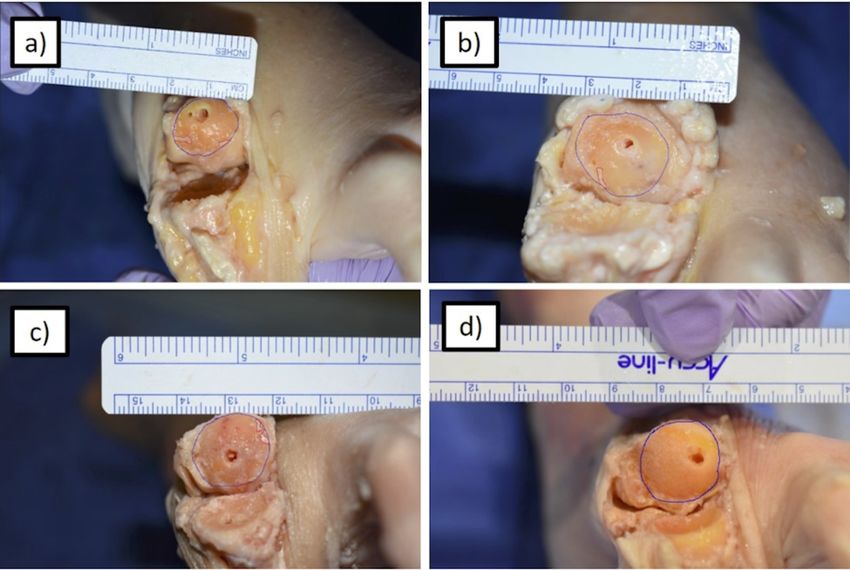
FIGURE 2: MT Head Preparation
Measurement of total prepared fusion contact surface area using ImageJ software for (a) the most
incomplete arthroscopically prepared MT head, (b) the most complete arthroscopically prepared MT
head, (c) the most incomplete open technique prepared MT head, and (d) the most complete open
technique prepared MT head. The blue circle represents the estimated fusion contact surface area.
Red outlines represent areas of remaining articular cartilage.
MT, metatarsal
Statistical analysis
Data were collected and analyzed using descriptive statistics in Microsoft Excel (Microsoft
Corp., Redmond, WA, USA) and SPSS Statistics (IBM, Armonk, NY, USA). All outcome measures
were compared using paired t-tests to examine the difference between the arthroscopic and
2020 McKissack et al. Cureus 12(8): e9633. DOI 10.7759/cureus.9633 4 of 9open MTP fusion technique groups (α ≤ 0.05). An a priori power analysis was performed to
determine what power could be achieved based on the number of specimens available for this
investigation. Based on the clinical experience of board-certified foot and ankle orthopedic
surgeons, it was determined that a 10% difference in joint preparation and 5-mm difference in
ray shortening would be clinically significant. Four specimens were in each group for ray
shortening analysis yielding a beta of 0.26, and five specimens were in each group for joint
preparation analysis yielding a beta of 0.72.
Results
Prepared surface area
The percent of joint preparation for each cadaver is detailed in Table 1. On average, the head of
the first metatarsal was significantly more prepared with the use of the open approach (99.5% ±
1.1%) than with the arthroscopic approach (96.6% ± 1.5%) (p = 0.008). However, with respect to
the base of the phalanx, the average difference in preparation between the open approach and
the arthroscopic approach was not statistically significant (99.0% ± 1.6% vs. 90.0% ± 12.8%; p =
0.159). When assessing the average total prepared surface area of the entire joint with both
articular surfaces combined, the open approach yielded an average of 99.3% ± 1.6%, whereas the
arthroscopic approach yielded an average of 92.9% ± 7.2. These differences were not
statistically significant (p = 0.089).
Cadaver Preparation Percent of Metatarsal Percent of Phalanx Percent of Total Joint Surface
No. Method Prepared Prepared Prepared
1 Scope 95.2 69.7 80.8
2 Scope 98.5 85.8 91.3
3 Scope 96.1 100.0 98.1
4 Scope 95.5 100.0 97.9
5 Scope 98.0 94.6 96.1
6 Open 100.0 100.0 100.0
7 Open 100.0 100.0 100.0
8 Open 97.6 95.0 96.4
9 Open 100.0 100.0 100.0
10 Open 100.0 100.0 100.0
TABLE 1: Percent of Joint Surface Prepared by Cadaver
First ray length
Radiographs for eight total specimens, four in the arthroscopic group and four in the open
group, were available for first ray length analysis (Table 2). Average decrease in length of the
first ray using the open approach was 2.1 ± 3.2 mm, whereas average decrease using the
arthroscopic approach was 2.2 ± 1.8 mm. The difference in the average change in length
2020 McKissack et al. Cureus 12(8): e9633. DOI 10.7759/cureus.9633 5 of 9between the two approaches was not statistically significant (p = 0.934). The average change in
the first to second ray length ratio was 0.02 for both approaches (p = 0.891). Of note, the length
of one specimen prepared using the open approach increased by 1 mm, presumably secondary
to correction of preoperative hallux valgus. We did not see any differences in change in ray
length based on the gender or age of the cadaver specimen.
Cadaver No. Preparation Method Ratio Difference Length Difference (mm)
1 Scope 0.04 4.267
2 Scope Radiograph unavailable
3 Scope 0.01 0.791
4 Scope 0.01 0.77
5 Scope 0.03 3.162
6 Open 0.06 6.37
7 Open Radiograph unavailable
8 Open -0.01 -1.01
9 Open 0.03 2.64
10 Open 0.00 0.36
TABLE 2: MTP Shortening by Cadaver
MTP, metatarsophalangeal
Discussion
First MTP fusion has been shown to improve the stability, propulsive power, and overall
function of the foot, with high patient satisfaction rates [15-17]. The success rate of first MTP
fusion was shown to be 90% in one study [18]. More recent studies have shown that revision
rates for first MTP fusion are between 0% and 11.7% [19]. One of the complications of first MTP
fusion is transfer metatarsalgia due to shortening of the first metatarsal. Jung et al.
demonstrated an increase in plantar pressure of the second to fifth metatarsals when
shortening of the first metatarsal occurs [9]. The increase in pressure was found to be
proportional to the increase in first ray shortening. Additionally, the first MTP plays an
important role in normal gait by bearing 40% to 60% of body weight, which increases two to
three times during running activities. Therefore, first ray length must be maintained during
joint preparation for MTP arthrodesis. Singh et al. investigated the amount of first ray
shortening in first MTP arthrodesis using flat cuts compared to conical reamers and concluded
that there was no significant difference in terms of shortening [4]. The results of our study
demonstrate no significant difference in the amount of first ray shortening between
arthroscopic and open joint preparation.
As with any fusion, outcomes of first MTP fusion are dependent on adequate preparation of the
joint, regardless of surgical technique. For first MTP fusion, joint preparation can be
accomplished through an open approach using a conical reamer or flat cutter or with an
2020 McKissack et al. Cureus 12(8): e9633. DOI 10.7759/cureus.9633 6 of 9arthroscopic technique using an arthroscopic shaver. To our knowledge, this is the first study to
compare joint preparation and first ray shortening between an open conical reaming technique
and arthroscopic technique. In our study, the difference in the amount of overall joint
preparation between the open and arthroscopic groups was not statistically significant.
Although the difference in the amount of total surface area prepared on the metatarsal head
between the two groups was statistically significant, it is unclear whether this difference is
clinically relevant. The percent of the metatarsal head that was prepared using arthroscopy in
our study was similar to the amount prepared in a study by Vaseenon and Phisitkul utilizing a
similar arthroscopic technique (92.9% vs. 93.31%) [20].
In general, arthroscopic technique is associated with decreased infection rate, scarring, and
bleeding, as well as improved cosmesis and faster recovery [21]. MTP arthroscopy was first
described by Watanabe in 1972 [8]. In recent years, the role of arthroscopy of the first ray has
expanded as it has been described in the treatment of various forefoot pathologies [22]. Ahn et
al. described a series of 59 patients with a variety of pathologies treated with first MTP
arthroscopy that demonstrated an improvement in outcomes scores, high patient satisfaction
with the procedure, and a low rate of complications [23]. Additionally, Lui reported on 121
cases of first MTP arthroscopies in patients with hallux valgus and found that of patients with
preoperative MTP joint pain, 90% had complete or significant relief of pain
postoperatively [24]. Similarly, arthroscopic ankle arthrodesis has been shown to result in
better long-term outcomes scores and shorter hospitalization [25].
Although arthroscopic management of forefoot pathologies has several advantages, it is not
without limitations. Arthroscopy of the forefoot is technically demanding and requires
adequate training and surgical experience. First MTP arthroscopy is most commonly performed
through two dorsally based portals, which lead to difficulty visualizing and treating dorsal
structures. Although our study utilized two portals, a three portal technique has been shown to
provide better exposure and allow more joint preparation when compared to two-portal
technique [20]. Similarly, newer techniques have been described utilizing four portals for
increased visualization of the MTP joint. Additionally, arthroscopy may not be possible in
patients with high angular deformity or those with severe arthritic changes due to osteophyte
formation and lack of joint space. Furthermore, traction is necessary for performing
arthroscopy in order to widen the joint space, which may result in nerve injury.
Valuable information can be gained from a study that uses a cadaveric model. However, there
are limitations on the number of specimens that can be acquired for these types of studies.
Despite the power being inadequate due to the logistical limitations of having access to more
specimens, the study is still the first of its kind to look at how surgical technique effects ray
shortening and joint preparation in MTP fusion, therefore still providing value to the field of
foot and ankle surgery. Second, the specimens used for this study did not include severely
arthritic joints, thus it is difficult to assess how effective arthroscopic joint preparation is in
patients with significant MTP joint pathology. Finally, we used anterior-posterior simulated
weight-bearing fluoroscopic images for measurements and it is possible that the axis of the
beam did not align with the anterior-posterior axis of the foot. We attempted to minimize this
error by measuring the length of the second metatarsal and including the ratio of first and
second ray lengths in the analysis. Finally, the accuracy of our measurement is limited by the
ImageJ software; however, the same software was used on all specimens, and the effect of this
error is felt to be minimal.
Conclusions
Arthroscopic first MTP fusion can be used to achieve comparable total joint preparation to open
technique despite increased metatarsal head preparation in the open group. The clinical
significance of increased metatarsal head preparation is currently unknown. First ray length
2020 McKissack et al. Cureus 12(8): e9633. DOI 10.7759/cureus.9633 7 of 9was maintained in both arthroscopic and open groups. Our study is the first to examine how
surgical technique affects ray shortening and joint preparation in MTP fusion. This cadaveric
study’s findings suggest that arthroscopic first MTP fusion is likely an effective surgical option
for surgeons with adequate arthroscopic experience. Further research is necessary to correlate
our findings clinically.
Additional Information
Disclosures
Human subjects: All authors have confirmed that this study did not involve human
participants or tissue. Animal subjects: All authors have confirmed that this study did not
involve animal subjects or tissue. Conflicts of interest: In compliance with the ICMJE uniform
disclosure form, all authors declare the following: Payment/services info: All authors have
declared that no financial support was received from any organization for the submitted work.
Financial relationships: All authors have declared that they have no financial relationships at
present or within the previous three years with any organizations that might have an interest in
the submitted work. Other relationships: All authors have declared that there are no other
relationships or activities that could appear to have influenced the submitted work.
References
1. Fitzgerald JAW, Wilkinson JM: Arthrodesis of the metatarsophalangeal joint of the great toe .
Clin Orthop Relat Res. 1986, 124:288-292. 10.1055/s-2008-1044562
2. Bei C, Gross CE, Adams S, Parekh SG: Dual plating with bone block arthrodesis of the first
metatarsophalangeal joint: a clinical retrospective review. Foot Ankle Surg. 2015, 21:235-239.
10.1016/j.fas.2015.01.004
3. Gross CE, Bei C, Gay T, Parekh SG: A short-term retrospective of first metatarsophalangeal
joint arthrodesis using a plate with PocketLock fixation. Foot Ankle Spec. 2015, 8:466-471.
10.1177/1938640015585968
4. Singh B, Draeger R, Del Gaizo DJ, Parekh SG: Changes in length of the first ray with two
different first MTP fusion techniques: a cadaveric study. Foot Ankle Int. 2008, 29:722-725.
10.3113/FAI.2008.0722
5. Sullivan C, Singh B, Parekh SG: Technique tip: using 1.7-mm screws in a 1/3 semitubular plate
to achieve additional fixation with a first MTP fusion. Foot Ankle Int. 2008, 29:518-520.
10.3113/FAI.2008.0518
6. Taylor D, Sage R, Pinzur M: Arthrodesis of the first metatarsophalangeal joint . Am J Orthop.
2004, 33:285-288.
7. Nakajima K: Arthroscopy of the first metatarsophalangeal joint . J Foot Ankle Surg. 2018,
57:357-363. 10.1053/j.jfas.2017.10.003
8. Watanabe M: Selfoc-Arthroscope, Watanabe No. 24 Arthroscope, Monograph. Teishin
Hospital, Tokyo; 1972.
9. Jung HG, Zaret DI, Parks BG, Schon LC: Effect of first metatarsal shortening and dorsiflexion
osteotomies on forefoot plantar pressure in a cadaver model. Foot Ankle Int. 2005, 26:748-
753. 10.1177/107110070502600913
10. Harris RI, Beath T: The short first metatarsal; its incidence and clinical significance . J Bone
Joint Surg Am. 1949, 31:553-565. 10.2106/00004623-194931030-00012
11. DeFrino PF, Brodsky JW, Polio FE, Crenshaw SJ, Beischer AD: First metatarsophalangeal
arthrodesis: a clinical, pedobarographic and gait analysis study. Foot Ankle Int. 2002, 23:496-
502. 10.1177/107110070202300605
12. Hartig SM: Basic image analysis and manipulation in imageJ. Curr Protoc Mol Biol. 2013,
102:14.15.1-14.15.12. 10.1002/0471142727.mb1415s102
13. Schindelin J, Arganda-Carreras I, Frise E, et al.: Fiji: an open-source platform for biological-
image analysis. Nat Methods. 2012, 9:676-682. 10.1038/nmeth.2019
14. Abyar E, McKissack HM, Pinto MC, Littlefield ZL, Moraes LV, Stefani K, Shah A: Subtalar
fusion preparation: what are we really doing? A cadaver study. Foot Ankle Spec. 2019, 13:201-
206. 10.1177/1938640019846970
2020 McKissack et al. Cureus 12(8): e9633. DOI 10.7759/cureus.9633 8 of 915. Brodsky JW, Passmore RN, Pollo FE, Shabat S: Functional outcome of arthrodesis of the first
metatarsophalangeal joint using parallel screw fixation. Foot Ankle Int. 2005, 26:140-146.
10.1177/107110070502600205
16. Brodsky JW, Baum BS, Pollo FE, Mehta H: Prospective gait analysis in patients with first
metatarsophalangeal joint arthrodesis for hallux rigidus. Foot Ankle Int. 2007, 28:162-165.
10.3113/FAI.2007.0162
17. Desandis B, Pino A, Levine DS, Roberts M, Deland J, O'Malley M, Elliott A: Functional
outcomes following first metatarsophalangeal arthrodesis. Foot Ankle Int. 2016, 37:715-721.
10.1177/1071100716642286
18. Coughlin MJ: Arthrodesis of the first metatarsophalangeal joint . Orthop Rev. 1990, 19:177-
186.
19. Rammelt S, Panzner I, Mittlmeier T: Metatarsophalangeal joint fusion why and how? . Foot
Ankle Clin. 2015, 20:465-477. 10.1016/j.fcl.2015.04.008
20. Vaseenon T, Phisitkul P: Arthroscopic debridement for first metatarsophalangeal joint
arthrodesis with a 2- versus 3-portal technique: a cadaveric study. J Arthrosc Relat Surg. 2010,
26:1363-1367. 10.1016/j.arthro.2010.02.015
21. Ferkel R, Scranton P: Arthroscopy of the ankle and foot . J Bone Joint Surg Am. 1993, 75:1233-
1242. 10.2106/00004623-199308000-00016
22. Schmid T, Younger A: First metatarsophalangeal joint degeneration: arthroscopic treatment.
Foot Ankle Clin. 2015, 20:413-420. 10.1016/j.fcl.2015.04.004
23. Ahn JH, Choy WS, Lee KW: Arthroscopy of the first metatarsophalangeal joint in 59
consecutive cases. J Foot Ankle Surg. 2012, 51:161-167. 10.1053/j.jfas.2011.10.003
24. Lui TH: First metatarsophalangeal joint arthroscopy in patients with hallux valgus . J Arthrosc
Relat Surg. 2008, 24:1122-1129. 10.1016/j.arthro.2008.05.006
25. Townshend D, Di Silvestro M, Krause F, Penner M, Younger A, Glazebrook M, Wing K:
Arthroscopic versus open ankle arthrodesis: a multicenter comparative case series . J Bone
Joint Surg Am. 2013, 95:98-102. 10.2106/JBJS.K.01240
2020 McKissack et al. Cureus 12(8): e9633. DOI 10.7759/cureus.9633 9 of 9You can also read