Laparoscopic approach to refractory extraspinal sciatica and pudendal pain caused by intrapelvic nerve entrapment
←
→
Page content transcription
If your browser does not render page correctly, please read the page content below
www.nature.com/scientificreports
OPEN Laparoscopic approach
to refractory extraspinal sciatica
and pudendal pain caused
by intrapelvic nerve entrapment
Nucelio Lemos1,2,4*, Corey Sermer1, Gustavo Fernandes2, Augusta Morgado‑Ribeiro2,
Andrea Rossos1, Zi Ying Zhao1, Manuel J. B. C. Girão2 & Philip Peng3
Entrapments of the intrapelvic portions of the lumbosacral plexus are an important extraspinal cause
of sciatica and pudendal neuralgia. They can be treated using Laparoscopic Neuronavigation (LANN),
a minimally invasive technique that has set the foundations of an emerging field in Medicine—
Neuropelveology. This retrospective-prospective study analyzes the outcomes of 63 patients treated
with the LANN technique over a 10 year time period. One year after surgery, 78.3% of patients
reported clinically relevant pain reduction, defined as ≥ 50% reduction in Numeric Rating Scale (NRS)
score; these results were maintained for a mean follow up of 3.2 years. Preoperative chronic opioid use
(≥ 4 months of ≥ 10 mg morphine equivalents/day) was a predictor of poor surgical outcome—clinically
relevant pain reduction was observed in only 30.8% in this group of patients, compared to 91.5%
in patients not regularly taking opioids preoperatively (p < 0.01). Perioperative complication rate
was 20%. Our results indicate that the LANN technique is an effective and reproducible approach to
relieve pain secondary to intrapelvic nerve entrapments and that preoperative chronic opioid therapy
significantly reduces the likelihood of a successful surgical outcome. This study provides detailed
information on perioperative complication and postoperative course, which is essential for patient
consenting.
Although usually attributed to spinal, deep gluteal and/or perineal causes, sciatica and pudendal neuralgia
can result from entrapments of the intrapelvic portion of the lumbosacral plexus. As with nerve entrapment
syndromes in other parts of the body, intrapelvic nerve entrapments (INEs) are characterized by pain and
dysfunction of the lumbosacral nerves’ dermatomes and myotomes, causing pain in the lower limb (sciatica),
perineum and gluteal regions, lower urinary tract symptoms, rectovesical tenesmus, dyschezia, vaginal foreign
body sensation, dysuria, dyspareunia and/or painful e jaculation1–6.
INEs were largely unknown before the development of the Laparosocopic Neuronavigation (LANN) tech-
nique, by Possover et al.7. The LANN technique introduced a systematic and minimally invasive approach
to dissect, expose and detrap the intrapelvic nerves, opening the possibility for diagnostic confirmation and
treatment of INEs. Additionally, this technique has led to a better understanding of entrapment neuropathies
and other causes of dysfunction of the intrapelvic nerves, which resulted in the development a new transitional
medical field—Neuropelveology8. Possover et al. proposed a ground-breaking Neuropelveological rationale8,
which allows for the diagnosis and management of intrapelvic causes of pudendal neuralgia and sciatica resulting
from entrapments at the origin of nerve roots from the sacral foramina down to the sciatic notch and pudendal
canal, with success rates close to 80%9. While extremely encouraging, these results are based on narrative case
series and their reproducibility still needs to be demonstrated by other groups.
Since late 2009, our team has been offering laparoscopic nerve detrapment procedures using the LANN
technique in São Paulo, Brazil. The objective of this study is to report the clinical outcomes of this procedure in
our cohort of patients from December 2009 to December 2019.
1
Department of Obstetrics and Gynecology, Mount Sinai and Women’s College Hospital, University of Toronto,
Toronto, ON, Canada. 2Department of Gynecology, Federal University of São Paulo, São Paulo, Brazil. 3Department
of Anesthesiology and Pain Management, Mount Sinai and Women’s College Hospital, University of Toronto,
Toronto, ON, Canada. 4Department of Obstetrics and Gynecology, Mount Sinai and Women’s College
Hospital, University of Toronto, 700 University Avenue|Room 8‑917, Toronto, ON M5S 1Z5, Canada. *email:
nucelio.lemos@utoronto.ca
Scientific Reports | (2021) 11:10820 | https://doi.org/10.1038/s41598-021-90319-y 1
Vol.:(0123456789)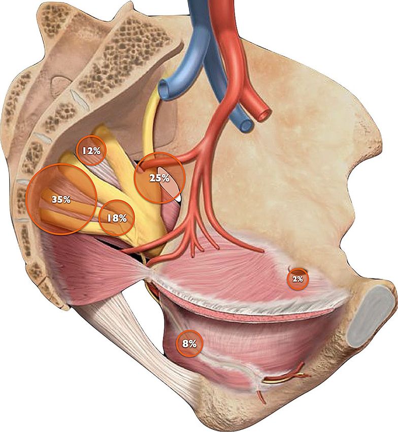
www.nature.com/scientificreports/
Methods
This is a retrospective-prospective analysis of 63 consecutive patients who underwent a LANN detrapment of
intrapelvic portions of the lumbosacral plexus from December 2009 to December 2019 at our Neuropelveology
center in São Paulo, Brazil.
Patients were identified from our surgical database. All the surgical reports within the study period were
reviewed and all the patients who underwent LANN detrapment surgery were included. Data was retrospectively
extracted from their electronic medical records until 2018, when this research protocol was approved by the
research ethics board; data has been prospectively collected since. All data was extracted into a Microsoft Excel
database file which began to be prospectively updated in 2018.
The diagnosis of INEs was based on Neuropelveological assessment, following the standards of the Interna-
tional School of N europelveology10. This comprehensive assessment included a full Neuropelveology-oriented
history, neurological examination, urodynamic study, pelvic MRI and a diagnostic nerve block.
Pain was assessed with a Numeric Rating Scale (NRS), with 0 representing no pain and 10 the worst imagina-
ble pain, at the pre-operative and all subsequent post-operative follow-up visits. Clinically relevant pain reduction
was defined as a ≥ 50% reduction in NRS. It is widely accepted in the pain literature that a minimum reduction
of 30% or 2-point reduction in a numeric pain scale (from 0–10) defines a clinically meaningful improvement
in pain11. However, it is common for pain relief to deteriorate over time and therefore a higher cut off (50%) is
often used for longer-term follow-up s tudies12.
The primary outcome of this study was clinically relevant pain reduction at 1 year follow up. Data from the
patient’s most recent follow up was also extracted and analyzed.
In addition, the following pre-operative and intra-operative information was collected from the patients’
records: the time of the onset of symptoms, duration and dose of preoperative opioid use, entrapment location
and etiology, operative time, intraoperative blood loss and perioperative complications. From this information,
the diagnostic lag was calculated and was defined as the time between the onset of symptoms and the time of the
LANN detrapment surgery. The operative time was determined from video recordings, defined as the moment
the laparoscopic camera was inserted into the abdomen to the moment all ports were removed.
Post-operative complications were also extracted from follow-up visits notes. Post-decompression neuralgia
(PDN) was defined as pain on the same dermatomes where pain was experienced pre-operatively, with different
quality and/or intensity. Any motor deficit within the lumbosacral plexus myotomes developed after surgery
was also identified and collected. In the subsequent follow up appointments, the duration of PDN and motor
deficits were recorded.
Statistical analysis. Extracted data was put into an MS-Excel database and analyzed in Prism (version 6.0,
GraphPad, San Diego, CA, USA).
A chi-squared test was used to assess the proportions of clinically relevant pain reduction rates at 1 year and
at final follow-up, to ensure maintenance of pain reduction.
To account for patients whose one-year follow up data was not available, three different sub-analyses were
performed: patients with missing data excluded, patients with missing data considered a failure (worst case
scenario analysis) and considering the last follow up data to be the 1-year follow up data (carry-over analysis).
A Komolgorov-Smirnov test was used to assess the normality distribution for NRS scores, which showed
that this data did not follow a Gaussian distribution. As such, non-parametric tests were used in our analyses. A
paired Wilcoxon’s test was used to compare preoperative and 1-year follow up NRS scores. For this paired test,
patients with no 1-year follow up data were excluded from the 1-year analysis. A paired Wilcoxon’s test was also
used to compare preoperative NRS score with the NRS score of the last follow up. Finally, to determine if results
were unchanged over time, a Wilcoxon’s test was used to compare 1-year NRS scores with the scores from the
last follow up.
A chi-squared test was used to determine if preoperative chronic opioid use—defined as daily use ≥ 10 mg
morphine equivalent for at least 4 months—or if a diagnostic lag of at least two years affected rates of clinically
relevant pain reduction. A Mann–Whitney test was used to compare pre-operative NRS homogeneity of both
subgroups. Descriptive statistics were used for the remainder of the data.
Ethical proceedings. This study was approved by the Research Ethics Board of the Federal University of
São Paulo (Protocol # 3.272.139). Consent for collection and storage of images and clinical data for quality
improvement, medical education, scientific studies and publications was obtained from all subjects, as a stand-
ard practice in surgical consenting process. This consent for authorization of usage of clinical data and images
was approved by the REB as part of the ethical analysis. All the proceedings and methods of this study are in
accordance with Brazilian research ethics regulations and the Declaration of Helsinki.
This study was also registered at the Brazilian Clinical Trials Registry (UTN code: U1111-1261-4910).
Results
A total of 63 patients (56 females and 7 males) underwent laparoscopic nerve root decompression during the
study inclusion period. The average age of the patients included in this study was 39.9 ± 10.6 years (Table 1).
Two patients were lost to follow-up. Additionally, there was one patient that had surgery within less than
one-year of publication, and as such had not yet had a 1-year follow-up appointment. Therefore, one-year follow
up data was available for 95.2% (60/63) of patients.
Clinically relevant pain reduction, defined as a ≥ 50% improvement in NRS for pain, was 78.3% (47/60) 1 year
after surgery (p < 0.001; Chi-squared test). This improvement in pain scores was maintained through the patient’s
final follow-up appointment (73.3% (44/60); p = 0.670, Chi-squared test, for one-year versus last follow up), at
Scientific Reports | (2021) 11:10820 | https://doi.org/10.1038/s41598-021-90319-y 2
Vol:.(1234567890)www.nature.com/scientificreports/
Mean Median Standard deviation
Age (years) 39.7 37.8 10.5
Follow-up (years) 3.0 2.5 1.8
Pre-op NRS 8.7 9.0 1.8
Post-op NRS at 1 year post-op 2.7* 2.0 2.9
Post-op NRS at final follow-up 2.9* 2.0 3.2
Operating time (min) 167.5 144 87.8
Previous surgeries 1.1 1.0 1.4
Diagnostic lag (years) 4.5 3.0 4.1
Table 1. Patient demographics and surgical outcomes. *Paired Wilcoxon’s test comparing pre-op NRS and
post-op NRS at 1 year and final follow-up (p < 0.001).
an average of 3.2 ± 1.8 years. The mean NRS dropped significantly from 8.7 ± 1.8 pre-operatively, to 2.7 ± 2.9 at
the 1-year follow-up (p < 0.001), and 3.0 ± 3.2 at final follow-up (p < 0.001). NRS did not significantly differ from
the 1 year follow-up to last follow up (p = 0.0502; paired Wilcoxon test) time points, suggesting outcomes are
maintained over time (Table 1).
When patients with missing data were excluded, clinically significant pain reduction was achieved on 47/60
(78.3%) of patients. No statistically significant difference in results were observed when patients with missing
data were considered failures (worst case scenario analysis, 74.6% versus 78.3%, p = 0.6749) or when the result
from the last follow up was carried over to one-year follow up (carry over analysis, 79.4% versus 78.3%, p = 0.888).
Preoperatively, 13/60 patients (21.7%) consumed ≥ 10 mg morphine equivalents daily, for an average of
18.6 ± 9.6 months. Pre-operative chronic opioid use adversely affected the primary outcome, as the clinically
relevant pain reduction was observed in only 30.8% (4/13) in this group of patients, compared to a 91.5%
(43/47) in patients not taking opioid on a regular basis (p < 0.01). Preoperative NRS scores were not significantly
different between the two subsets of patients—8.6 (± 1.8; 3–10) for non-opiod vs. 9.0 (± 1.7; 5–10) for opioid
users (p = 0.4458). The mean time interval between onset of symptoms and surgery was 4.9 ± 4.3 (0.3–15) years
for opioid users and 4.6 ± 4.0 (0–15) years for non-opioid users. This difference was not statistically significant
(p = 0.7902).
The average interval between symptom onset and surgery was 4.73 ± 4.1 years, with patients having undergone
an average of 1.1 ± 1.5 years surgical procedures for treatment of their symptoms prior to our LANN detrapment
(Table 1). Three out of 21 (14.3%) patients had unsuccessful outcomes when surgery was performed within 2 years
of symptom onset compared to 10/39 (25.6%) when surgery was performed after 2 years from symptom onset,
however this was not statistically significant (p = 0.5461). Preoperative NRS were also homogeneous between
the two groups (p = 0.594). The time interval between onset of symptoms and surgery was 4.6 ± 4.0 (0–15) years
for patients with clinically significant pain reduction and 4.8 ± 4.3 (0.5–15) years for patients in whom clinically
significant pain reduction was not achieved. This difference was not statistically significant (p = 0.8308).
Distribution of nerve entrapments locations are displayed in Fig. 1. The most common entrapment site was
proximal S2/S3/S4 nerve roots, in 21 (35%) cases, followed by lateral sciatic/lumbosacral trunk in 15 (25%),
proximal pudendal/medial sciatic in 11 (18%) and S1/S2 nerve roots in 7 (12%). Entrapments at the Alcock’s
canal level were present in only 5 (8%) patients and one patient had an obturator nerve entrapment, caused by
a transobturator sling.
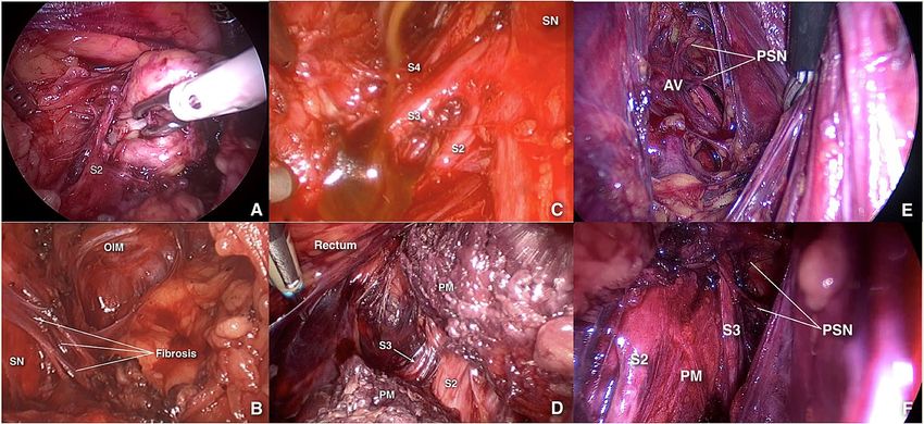
The five etiologies of nerve entrapments identified during surgery in these 60 patients were endometriosis,
fibrosis, piriformis entrapment by abnormal muscle fibres originating medial to the sacral foramina, neurovascu-
lar conflict, and neoplasm (Fig. 2). Neurovascular conflict and endometriosis were the most common etiologies
of entrapment in this group of patients (38.3% and 36.7%, respectively), followed by fibrosis (10%), abnormal
piriformis bundles originating medially to the sacral foramina (10%), and nerve sheath tumors (1.7%). Two
patients had both fibrosis and abnormal piriformis muscle bundles causing their INE (3.3%).
The average operating time was 168 ± 89.2 min (Table 1). Perioperative complications included 1 pudendal
nerve transection, 1 obturator nerve tear, 1 ureteral injury, 2 cases of genitofemoral neuropathy, 1 incisional her-
nia, 1 rectovaginal fistula, and 1 case of piriformis muscle adhesion to the sciatic nerve requiring a transgluteal
endoscopic procedure. One patient required a blood transfusion, 2 patients had transient urinary retention and
1 patient had impaired bladder sensation. Overall, 20% (12/60) of patients had a perioperative complication; of
these, the rectovaginal fistula and the ureteric injury were attributable to endometriosis treatment, not the nerve
detrapment per se, resulting in a LANN detrapment complication rate of 16.7%.
During the postoperative course, 56.7% (34/60) of patients experienced post-decompression neuralgia, lasting
on average 5.7 ± 3.4 (1–12) months. Fourteen (23.3%) patients experienced a post-decompression motor deficit,
lasting on average 3.2 ± 3.2 (0.5–12) months.
Discussion
It is well known that a large portion of the lumbosacral plexus is located intra-abdominally, in the retroperito-
neal space13. However, most literature descriptions of lesions on this plexus refer to its extra-abdominal parts,
in the spine or deep gluteal space, while reports of intrapelvic entrapments are traditionally limited to isolated
case reports.
Scientific Reports | (2021) 11:10820 | https://doi.org/10.1038/s41598-021-90319-y 3
Vol.:(0123456789)www.nature.com/scientificreports/
Figure 1. Distribution of nerve entrapments locations: 35% in proximal S2/S3/S4 nerve roots, 25% in sciatic/
lumbosacral, 18% in proximal pudendal/medial sciatic, 12% in S1/S2 nerve roots, 8% in Alcock’s canal level and
2% in obturator nerve entrapment.
Figure 2. The five etiologies of intrapelvic nerve entrapment: neoplastic (A), fibrotic (B), endometriotic (C),
abnormal piriformis muscle bundle originating medially to the sacral foramina (D) and neurovascular conflict
before (E) and after (F) decompression. SN sciatic nerve; PM pyriformis muscle; OIM obturator internus
muscle; PSN pelvic splanchnic nerves; AV abnormal vein.
Currently, intrapelvic entrapments are not well understood and rarely considered as a cause of sciatica and
pudendal neuralgia. One of the reasons for this is the deep retroperitoneal location of the plexus, in an area that
can only be adequately approached by laparoscopy, which provides better lighting, magnification and visualiza-
tion of this area. Therefore, since the specialists that usually deal with peripheral nerve surgery (neurosurgeons,
orthopedic and plastic surgeons) are not usually trained in laparoscopy and those specialists who routinely
perform advanced laparoscopic procedures (gynecologists, urologists and gastrointestinal surgeons) are not
familiarized with peripheral nerve surgery, a knowledge gap is c reated2. Therefore, none of the traditional and
widely available residency and fellowship programs offers training in the skills set required for the diagnosis
and management of INEs.
Scientific Reports | (2021) 11:10820 | https://doi.org/10.1038/s41598-021-90319-y 4
Vol:.(1234567890)www.nature.com/scientificreports/
In 2004, Possover et al. pioneered the Laparoscopic Neuronavigation (LANN) technique7, opening the doors
to accessing the intrapelvic portion of the lumbosacral plexus through a safe and minimally invasive way. This
led to the development of a new groundbreaking medical field—Neuropelveology8.
The LANN technique was initially developed to standardize nerve-sparing approaches in retroperitoneal
dissection during cervical cancer or deeply infiltrating endometriosis surgeries14. The LANN technique was
later proposed as a minimally invasive detrapment method to significantly reduce or completely resolve pain
secondary to INEs. This surgical intervention follows the same principles of surgeries that have been successfully
used in the treatment of other peripheral nerve entrapment s yndromes9,14,15.
In 2009, a series of 134 patients who underwent laparoscopic nerve decompression for refractory anogenital
pain showed improvement in VAS scores depending on etiology, with significant decrease in pain achieved in
83% of patients with Alcock’s canal syndrome, 62% of patients with endopelvic lesions, and 78% of patients with
endometriosis9. Additionally, a study involving 120 patients showed that 65.5% of patients experienced significant
improvement in somatic pelvic pain following laparoscopic nerve decompression6. Recently, the same group
reported similar outcomes in a series of 213 female patients with lumbosacral entrapment due to neurovascular
conflict and/or e ndometriosis1.
This is the first case series in the literature showing the reproducibility of the Neuropelveological approach
proposed by Possover et al.9 for INEs. It is also the first study with a systematic assessment, as previous reports
of INEs are generally narrative case series. In our study, we had a clear time point at one-year follow up for the
primary outcome. In addition, this is the first series to report the sustainability of such outcomes at mid to long-
term follow up of 3.2 years, on average.
This is also the first report to show the negative impact chronic opioid use has on long-term outcomes follow-
ing LANN detrapment of intrapelvic nerves. We show here that chronic opioid use reduces the odds of success of
this etiology-oriented treatment from 91.5% to 30.8%. This is of extreme importance as it supports the concept
that chronic exposure to opioids may predispose a patient to opioid induced hyperalgesia, thus resulting in a
lower pain threshold and thus poor analgesic o utcomes16,17. Prolonged preoperative opioid exposure was the only
predictor for poor surgical outcome identified in our cohort; preoperative pain scores and time between onset
of symptoms and surgery, two factors we considered would be associated with these results, were homogeneous
between chronic opioid users and opioid naïve patients. Such information should influence opioid prescription
habits and motivate the use of this medication class only as end-stage therapy. Alternatively, the use of multimodal
non-opioid pain management strategies should be promoted in patients with all other forms of chronic pain, as
opioids might jeopardize their chances of cure and make them prone to lifelong pain.
We have also described the different locations of entrapments, which is very important for understanding the
dermatomes and myotomes implicated in INEs. It is important to highlight the very small proportion of entrap-
ments at the level of Alcock’s canal (8%) observed in our series. This is very significant, given that “pudendal
neuralgia” is often used interchangeably in the literature with “Alcock’s Canal Syndrome”. Also, the most common
criteria for diagnosis of pudendal nerve e ntrapments16 was developed to diagnose entrapments at the level of
Alcock’s Canal and/or at the interligamentous plane. The fact that the most common site of pudendal entrapments
in our cohort are the pudendal nerve roots could possibly explain several cases of “atypical pudendal neuralgia”
or surgical failures seen in the literature. For the purpose of differential diagnosis, we should raise awareness for
the combined symptoms of pudendal pain and sciatica and/or gluteal pain, which could be explained by an INE
at the level of the sacral nerve roots.
Another important finding in our report is the long diagnostic lag experienced by our patients, who have suf-
fered for 4.7 years and undergone 1.1 previous failed surgeries in an attempt to treat their symptoms. This further
illustrates the knowledge gap and lack of awareness of INEs as an extraspinal cause of sciatica and pudendal pain.
This data is also consistent with the data reported on European cohorts, in whom an average diagnostic gap of
5.6 years was documented and an average of 4 surgical procedures were conducted before an INE was suspected6.
On top of the prolonged suffering, the long diagnostic gap can have other consequences. First, the success rates
of detrapment procedures would be adversely affected18. In our patients, the failure rate of LANN decompression
was 86% higher when surgery was performed more than 2 years from symptom onset. Although this difference
in failure rate did not reach statistical significance, likely due to our limited sample size, it is consistent with the
current literature, meaning this likely reflects a r eality4,15,19–21.
Secondly, lack of awareness of INEs increases the risk of patients undergoing ineffective surgeries which do
not address the underlying cause of pain and may cause added harm. Moreover, each surgical attempt further
prolongs the treatment delay, as patient and surgeon will need time to assess surgical outcomes. Patients who
have been waiting for a long time tend to c atastrophize22,23; under pressure, healthcare professionals may resort
to the use of o pioids24. This is of particular importance, as we have shown here that chronic opioid use decreases
the chances of a successful surgical outcome.
In this study we have also systematically assessed the postoperative course. Approximately half of our patients
experienced post-decompression neuralgia, which lasted on average about 6 months and almost a quarter of
patients experienced a post-decompression motor deficit, lasting approximately 3 months. It is important to
make patients aware that post-decompression neuralgia and motor deficits are part of the natural postoperative
course, since, if long lasting, they can be misinterpreted as permanent and cause significant distress.
In addition, the perioperative complication rate was 20% in our cohort, ranging from nerve injuries to tran-
sient bladder symptoms including retention and impaired sensation. For intricate surgery on peripheral nerves,
such as LANN detrapment, this complication rate is within the acceptable range, but discussing these risks with
patients is essential for appropriate preoperative counselling and consenting.
This current study is limited by the relatively small cohort, retrospective design and the use of NRS as the
singular outcome measure. However, the consistency with Possover et al.’s13 results, as well as the similarity of
Scientific Reports | (2021) 11:10820 | https://doi.org/10.1038/s41598-021-90319-y 5
Vol.:(0123456789)www.nature.com/scientificreports/
the outcomes with surgical treatment of other nerve entrapment syndromes suggests that our results can be
extrapolated to a larger population.
Future studies assessing the impact of laparoscopic nerve detrapment surgery on different variables, such as
sexual function, quality of pain, bowel/bladder dysfunction, and psychosocial components of chronic pain, will
help to create a more robust understanding of the outcomes of this approach. Additionally, following patients
for a longer post-operatively will result in further understanding of the maintenance of the effects of surgery and
the future need for additional pain management medications or procedures.
Nevertheless, our results validate the Neuropelveology approach to diagnose and treat INEs and highlight
the importance of raising awareness of the risk of non-specific pain management opioids, which significantly
reduces the likelihood of a successful surgical outcome.
Received: 17 January 2021; Accepted: 26 April 2021
References
1. Possover, M. The neuropelveology: From the laparoscopic exposure of the pelvic nerves to a new discipline in medicine?. Gynecol.
Surg. 8, 117–119 (2011).
2. Lemos, N. & Possover, M. Laparoscopic approach to intrapelvic nerve entrapments. J. Hip. Preserv. Surg. 2(2), 92–98 (2015).
3. Lemos, N. et al. Recognition and treatment of endometriosis involving the sacral nerve roots. Int. Urogynecol. J. 27(1), 147–150
(2016).
4. Lemos, N. et al. Sacral nerve infiltrative endometriosis presenting as perimenstrual right-sided sciatica and bladder atonia: Case
report and description of surgical technique. J. Minim. Invasive. Gynecol. 19(3), 396–400 (2012).
5. Possover, M. & Lemos, N. Risks, symptoms, and management of pelvic nerve damage secondary to surgery for pelvic organ pro-
lapse: A report of 95 cases. Int. Urogynecol. J. 22(12), 1485–1490 (2011).
6. Possover, M., Schneider, T. & Henle, K. P. Laparoscopic therapy for endometriosis and vascular entrapment of sacral plexus. Fertil.
Steril. 95(2), 756–758 (2011).
7. Possover, M., Rhiem, K. & Chiantera, V. The “Laparoscopic Neuro-Navigation”— LANN: From a functional cartography of the
pelvic autonomous neurosystem to a new field of laparoscopic surgery. Minim. Invasive Ther. Allied Technol. 13(5), 362–367 (2004).
8. Possover, M. et al. Neuropelveology: New groundbreaking discipline in medicine. J. Minim. Invasive Gynecol. 22(7), 1140–1141
(2015).
9. Possover, M. Laparoscopic management of endopelvic etiologies of pudendal pain in 134 consecutive patients. J. Urol. 181(4),
1732–1736 (2009).
10. Possover, M. International School of Neuropelveology: Clinical Neuropelveology. https://isnp.possover.com/catalog/info/id:126.
11. Farrar, J. T. et al. Clinical importance of changes in chronic pain intensity measured on an 11-point numerical pain rating scale.
Pain 94(2), 149–158 (2001).
12. Davis, T. et al. Prospective, multicenter, randomized, crossover clinical trial comparing the safety and effectiveness of cooled
radiofrequency ablation with corticosteroid injection in the management of knee pain from osteoarthritis. Reg. Anesth. Pain Med.
43(1), 84–91 (2018).
13. Gray, H. & Lewis, W. H. Anatomy of the Human Body 20th edn. (Lea & Febiger, Philadelphia and New York, 1918).
14. Possover, M., Quakernack, J. & Chiantera, V. The LANN technique to reduce postoperative functional morbidity in laparoscopic
radical pelvic surgery. J. Am. Coll. Surg. 201(6), 913–917 (2005).
15. Taner, U. et al. Robot-assisted laparoscopic management of a vascular entrapment of the sacral nerve roots causing pelvic pain. J.
Obstet. Gynaecol. Res. 45(8), 1603–1607 (2019).
16. Lee, M. et al. A comprehensive review of opioid-induced hyperalgesia. Pain Physician 14(2), 145–161 (2011).
17. Roeckel, L. A. et al. Opioid-induced hyperalgesia: Cellular and molecular mechanisms. Neuroscience 338, 160–182 (2016).
18. Russell, S. M. Examination of peripheral nerve injuries: An anatomical approach. Bone Joint J. 89B(6), 178 (2007).
19. Ramsden, C. E. et al. Pudendal nerve entrapment as source of intractable perineal pain. Am. J. Phys. Med. Rehabil. 82(6), 479–484
(2003).
20. Kale, A. et al. Vascular entrapment of both the sciatic and pudendal nerves causing persistent sciatica and pudendal neuralgia. J.
Minim. Invasive Gynecol. 26(2), 360–361 (2019).
21. Lemos, N. et al. Superior gluteal vein syndrome: An intrapelvic cause of sciatica. J. Hip. Preserv. Surg. 6(2), 104–108 (2019).
22. Choiniere, M. et al. The Canadian STOP-PAIN project—Part 1: Who are the patients on the waitlists of multidisciplinary pain
treatment facilities?. Can. J. Anaesth. 57(6), 539–548 (2010).
23. Guerriere, D. N. et al. The Canadian STOP-PAIN project—Part 2: What is the cost of pain for patients on waitlists of multidisci-
plinary pain treatment facilities?. Can. J. Anaesth. 57(6), 549–558 (2010).
24. Possover, M. & Forman, A. Pelvic Neuralgias By Neuro-Vascular entrapment: Anatomical findings in a series of 97 consecutive
patients treated by laparoscopic nerve decompression. Pain Physician 18(6), E1139–E1143 (2015).
Acknowledgements
Funding for this study was provided by Associação de Ensino, Pesquisa e Assistência à Saúde (AEPAS), a fully
registered charity in Brazil. Research coordination was provided by Cátia Mendes Pereira. Nucelio Lemos has
an eventual speaker contract with Medtronic Inc and has received honoraria for one event in 2019; this event
is not related to this study.
Author contributions
N.L.—study design, data collection, data analysis and manuscript preparation. C.S.—data analysis and manu-
script preparation. G.F.—study design and data collection. A.M.R.—study design and data collection. A.R.—
data analysis and manuscript preparation. Z.Y.Z.—data analysis and manuscript preparation. M.J.B.C.G.—data
analysis and manuscript preparation. P.P.—data analysis and manuscript preparation.
Competing interests
Nucelio Lemos has also received speaker honoraria for one event promoted by Promedon Inc. in March 2020.
Nucelio Lemos received research support from Medtronic Inc for a researcher initiated project, in the form of
Scientific Reports | (2021) 11:10820 | https://doi.org/10.1038/s41598-021-90319-y 6
Vol:.(1234567890)www.nature.com/scientificreports/
equipment donation for the study, which is not related to this publication. No honoraria was received. The other
authors declare no competing interests.
Additional information
Correspondence and requests for materials should be addressed to N.L.
Reprints and permissions information is available at www.nature.com/reprints.
Publisher’s note Springer Nature remains neutral with regard to jurisdictional claims in published maps and
institutional affiliations.
Open Access This article is licensed under a Creative Commons Attribution 4.0 International
License, which permits use, sharing, adaptation, distribution and reproduction in any medium or
format, as long as you give appropriate credit to the original author(s) and the source, provide a link to the
Creative Commons licence, and indicate if changes were made. The images or other third party material in this
article are included in the article’s Creative Commons licence, unless indicated otherwise in a credit line to the
material. If material is not included in the article’s Creative Commons licence and your intended use is not
permitted by statutory regulation or exceeds the permitted use, you will need to obtain permission directly from
the copyright holder. To view a copy of this licence, visit http://creativecommons.org/licenses/by/4.0/.
© The Author(s) 2021
Scientific Reports | (2021) 11:10820 | https://doi.org/10.1038/s41598-021-90319-y 7
Vol.:(0123456789)You can also read