Laser speckle contrast imaging of cerebral blood flow in freely moving animals
←
→
Page content transcription
If your browser does not render page correctly, please read the page content below

Laser speckle contrast imaging of cerebral
                 blood flow in freely moving animals

                 Peng Miao
                 Hongyang Lu
                 Qi Liu
                 Yao Li
                 Shanbao Tong

Downloaded From: https://www.spiedigitallibrary.org/journals/Journal-of-Biomedical-Optics on 29 Jul 2021
Terms of Use: https://www.spiedigitallibrary.org/terms-of-use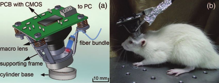
JBO Letters

                Laser speckle contrast                                                              that can monitor the real-time neurovascular changes in freely
                                                                                                    moving small animals, such as rats.
                imaging of cerebral blood                                                               The imager system consists of five major components. i.
                                                                                                    A cylinder base (height, 4.20 mm; radius, 5.50 mm; thickness,
                flow in freely moving                                                               0.5 mm) is fixed onto the scalp over the imaging area with dental
                                                                                                    cement. The body of the imager is attached to the cylinder base
                animals                                                                             through a screw connection during imaging. ii. A multimode
                                                                                                    optical fiber bundle (50 fibers; diameter, 0.25 mm; 0.45 NA;
                                                                                                    SMOIF, Shanghai, China) is used for laser light delivery. Coher-
                Peng Miao,a Hongyang Lu,b Qi Liu,b Yao Li,b                                         ent light from a laser diode (780 nm; 10 mW; L780P010, Thor-
                and Shanbao Tonga,b                                                                 labs, U.S.A.) powered by a driver module (LDC220C, Thorlabs,
                a Shanghai Jiao Tong University, School of Biomedical Engineering,                  U.S.A.) is coupled into the fiber bundle using a collimating lens
                800 Dongchuan Road, Shanghai, 200240, China                                         (f = 8 mm; C240TM, Thorlabs, U.S.A.). The end of the fiber
                b Shanghai Jiao Tong University, Med-X Research Institute, 1954
                Huashan Road, Shanghai, 200030, China
                                                                                                    bundle is enclosed in an aluminum tube (length, 7.5 mm; ra-
                                                                                                    dius, 1.54 mm) that is fixed onto the supporting frame as the
                Abstract. We designed a miniature laser speckle imager                              light source of LSCI. iii. The system also has a printed circuit
                that weighs ∼20 g and is 3.1-cm high for full-field high-                           board (PCB; 3.6 × 3.6 cm; Thorlabs, U.S.A.) with a CMOS
                resolution imaging of cerebral blood flow (CBF) in freely                           sensor (1024 × 1396 pixels; 8-bit precision; Thorlabs, U.S.A.),
                moving animals. Coherent laser light illuminates the cortex                         as well as image acquisition and transferring circuits, which are
                through a multimode optical fiber bundle fixed onto the                             connected to a computer via USB cable. iv. A macrolens sys-
                supporting frame of the imager. The reflected lights are then                       tem (radius, 3.44 mm; focal length, 5.05 mm; minimum focus
                collected by a miniature macrolens system and imaged by a                           distance, 20 mm; 0.011 NA; custom-made to our design by Gi-
                high-resolution CMOS camera at a high frame rate (50 fps).                          antTec, Shanghai, China) is located within a plastic tube fixed
                Using this miniature imager, we achieve high spatiotem-                             onto the PCB, whose parameters were carefully designed to be
                poral resolution laser speckle contrast imaging of CBF in                           adapted to the illumination tube (length, 7.5 mm), CMOS sensor
                freely moving animals in real time. C 2011 Society of Photo-Optical                (size, 2/3 in.) in order to obtain good imaging quality. v. A sup-
                Instrumentation Engineers (SPIE). [DOI: 10.1117/1.3625231]
                                                                                                    porting frame is used to fix the PCB, the macrolens system, and
                Keywords: laser speckle contrast imaging; cerebral blood flow; high                 fiber bundle. Four springs are added in the screw connections be-
                spatiotemporal resolution; freely moving animal.                                    tween the PCB and the supporting frame to reduce the influence
                Paper 11253LR received May 20, 2011; revised manuscript received                    of motion artifacts. The connection screws are also adjustable
                Jul. 16, 2011; accepted for publication Jul. 19, 2011; published online             for focusing the macrolens onto the imaging plane. The entire
                Sep. 1, 2011.
                                                                                                    imager weighs ∼20 g and is 3.1 cm high. Figure 1(a) illustrates
                                                                                                    the design of the miniature head-mounted imager, and Fig. 1(b)
                As a full-field high-resolution optical imaging technique,1, 2 laser                is a snapshot of a fully conscious rat wearing the imager during
                speckle contrast imaging (LSCI) has gained increasing atten-                        the experiment.
                tion in the neuroscience community.3 It is used for monitor-                            Before LSCI, the imager is fixed onto the cylinder base. Laser
                ing cerebral blood flow (CBF) under different physiological4, 5                     light (e.g., 780 nm in this study) reflected by the cortex is col-
                or pathological6–8 conditions. Until recently, the use of almost                    lected by the macrolens system and then imaged by the CMOS
                all CBF monitoring devices equipped with LSCI has been re-                          sensor. The imager can be configured by software through a
                stricted to anesthetized animals.9 Anesthesia always introduces                     USB interface (pixel clock of 35 MHz, frame rate of 50 fps,
                various types of physiological changes such as local blood flow                     exposure time of 5 ms, area of interest of 640×640 pixels).
                changes10 and suppression of CBF response,11 which may result                       A custom-developed software program reads the raw images
                in biased experimental results. LSCI has shown its considerable                     (25 frames/stack) through a USB cable for further CBF analysis
                potential in the study of the cerebral blood flow;3, 7, 8, 12 there-                on the computer. To eliminate motion artifacts, the speckle im-
                fore, the head-mounted imager for small animals facilitates the                     ages in each stack (i.e., 25 frames) are aligned to the first frame
                study of cerebral blood flow changes without anesthesia.                            by registered laser speckle contrast analysis (rLASCA).13 The
                    Two major challenges are encountered in designing a                             raw speckle images were preprocessed with a 3×3 convolution
                laser speckle imager for small fully conscious animals. First,                      kernel, and then registered by normalized correlation metric and
                the imaging system, which includes an optical fiber bundle,                         finally resampled with cubic B-spline interpolator.13 After the
                macrolens system, and imaging sensor and circuits, should be                        registration, raw speckle images were processed by the random
                sufficiently small and lightweight. Second, severe motion arti-                     process estimator (RPE) method, utilizing the Gaussian property
                facts must be corrected. These problems occur only to a minimal                     of estimation noise in integrated intensity random process, to
                extent in anesthetized animals, but are exacerbated under free                      obtain the contrast image with high signal-to-noise ratio.14 Ac-
                motion. To overcome the above-mentioned problems, we de-                            cording to theory of laser speckle contrast imaging,1, 9, 15 blood
                signed a miniature head-mounted laser speckle imager (20 g)                         flow speed v is related to contrast value K [Eq. (1)].9

                                                                                                                                τc   τ2
                Address all correspondence to: Shanbao Tong, Shanghai Jiao Tong                                    K2 = β          + c 2 (e−2T /τc − 1) .           (1)
                University, Med-X Research Institute, 1954 Huashan Road – Shanghai, Shang-                                      T   2T
                hai 200030 China; Tel: + 86-21-34205138; Fax: + 86-21-34204717; E-mail:
                shanbao.tong@gmail.com.                                                             1083-3668/2011/16(9)/090502/3/$25.00
                                                                                                                                         C 2011 SPIE

                Journal of Biomedical Optics                                               090502-1                                         September 2011 r Vol. 16(9)

Downloaded From: https://www.spiedigitallibrary.org/journals/Journal-of-Biomedical-Optics on 29 Jul 2021
Terms of Use: https://www.spiedigitallibrary.org/terms-of-use
JBO Letters
Fig. 1 (a) Design of the miniature head-mounted laser speckle im-
ager. The system includes a i. cylinder base, ii. multimode optical fiber
bundle, iii. CMOS sensor on a PCB, iv. macrolens system, and v. sup-
porting frame. (b) A snapshot of the imager mounted onto the head of
a conscious rat during the experiment.
where the correlation time τc is assumed to be inversely pro-
portional to the blood flow speed v. β = 1/N , where N is the
number of speckle in each pixel area.16 T is the exposure time.
The performance of the imager was tested using a standard
USAF 1951 resolution test chart, which resulted in a 16 lp/mm
resolution andJBO Letters
the major vessels under different physiological or pathological 2. R. Bandyopadhyay, A. S. Gittings, S. S. Suh, P. K. Dixon, and D. J.
conditions. Durian, “Speckle-visibility spectroscopy: A tool to study time-varying
dynamics,” Rev. Sci. Instrum. 76(9), 093110 (2005).
The laser speckle images of the i. anesthetized rat and ii. the
3. A. K. Dunn, H. Bolay, M. A. Moskowitz, and D. A. Boas, “Dynamic
conscious and freely moving rat were continuously compared imaging of cerebral blood flow using laser speckle,” J. Cereb. Blood
for 20 min. For free motion, the first 25 frames of the speckle Flow Metab. 21, 195–201 (2001).
images at each minute were used for rLASCA and RPE analy- 4. P. Li, S. Ni, L. Zhang, S. Zeng, and Q. Luo, “Imaging cerebral blood
sis. Two crossing vessels (Fig. 3) were purposely selected to test flow through the intact rat skull with temporal laser speckle imaging,”
Opt. Lett. 31, 1824–1826 (2006).
whether the coordinates of their crossing center, at which point
5. H. Cheng, Y. Yan, and T. Q. Duong, “Temporal statistical analysis of
the pixel’s contrast value is a local minimum [see embedded im- laser speckle images and its application to retinal blood-flow imaging,”
age in Fig. 3(c)], would change in a moving animal. Figure 3(c) Opt. Express 16(14), 10214–10219 (2008).
shows that rLASCA successfully eliminated the motion artifacts 6. B. Choi, N. M. Kang, and J. S. Nelson, “Laser speckle imaging for
and resulted in images with highly stable contrast. These images monitoring blood flow dynamics in the in vivo rodent dorsal skin fold
model,” Microvasc. Res. 68, 143–146 (2004).
were comparable to those of the anesthetized rat. 7. C. Ayata, H. K. Shin, S. Salomone, Y. Ozdemir-Gursoy, D. A. Boas,
Finally, to test the stability of the optical focusing of the A. K. Dunn, and M. A. Moskowitz, “Pronounced hypoperfusion during
imager, we analyzed the diameter of the superior sagittal si- spreading depression in mouse cortex,” J. Cereb. Blood Flow Metab.
nus along the white line in Fig. 2(b) based on the manual 24, 1172–1182 (2004).
segmentations.13 The vessel diameter from 10 volunteers’ seg- 8. J. S. Paul, A. R. Luft, E. Yew, and F. Sheu, “Imaging the development of
an ischemic core following photochemically induced cortical infarction
mentations showed very low variance (Mean ± Std: 56.6 ± in rats using Laser Speckle Contrast Analysis (LASCA),” Neuroimage
0.05 pixels), indicating high stability of the imager in optical fo- 29(1), 38–45 (2006).
cusing. This high stability is attributed to several special consid- 9. D. A. Boas and A. K. Dunn, “Laser speckle contrast imaging in biomed-
erations in the design: i. the fast frame rate of the CMOS sensor ical optics,” J. Biomed. Opt. 15(1), 011109 (2010).
10. T. Maekawa, C. Tommasino, H. M. Shapiro, J. Keifer-Goodman, and
(50 fps); ii. springs around the connection screws between the
R. W. Kohlenberger, “Local cerebral blood flow and glucose utilization
supporting frame and PCB; and iii. efficient registration of the during isoflurance anesthesia in the rat,” Anesthesiology 65(2), 144–151
rLASCA algorithm. (1986).
In summary, we designed a miniature head-mounted laser 11. K. Sicard, Q. Shen, M. E. Brevard, R. Sullivan, C. F. Ferris, J. A. King,
speckle imager for full-field high resolution imaging of CBF and T. Q. Duong, “Regional cerebral blood flow and BOLD responses in
conscious and anesthetized rats under basal and hypercapnic conditions:
in freely moving rats. The new laser speckle imager provides
implications for functional MRI studies,” J. Cereb. Blood Flow Metab.
the possibility of studying the structural and functional CBF of 23, 472–481 (2003).
small conscious and freely moving animals. 12. P. Zakharov, A. C. Völker, M. T. Wyss, F. Haiss, N. Calcinaghi, C.
Zunzunegui, A. Buck, F. Scheffold, and B. Weber, “Dynamic laser
Acknowledgments speckle imaging of cerebral blood flow,” Opt. Express 17(16), 13904–
13917 (2009).
This work is supported by National Science Foundation of China 13. P. Miao, A. Rege, N. Li, N. V. Thakor, and S. Tong, “High resolu-
(Grant No. 81071192). Peng Miao is also supported by Scholar- tion cerebral blood flow imaging by registered laser speckle contrast
ship Award for Excellent Doctoral Student (granted by Ministry analysis,” IEEE Trans. Biomed. Eng. 57(5), 1152–1157 (2010).
14. P. Miao, N. Li, N. V. Thakor, and S. Tong, “Random process estimator
of Education). The authors are also grateful to Dr. Guo-Yuan
for laser speckle imaging of cerebral blood flow,” Opt. Express 18(1),
Yang for advice in animal experiments. 218–236 (2010).
15. D. D. Duncan and S. J. Kirkpatrick, “Can laser speckle flowmetry
References be made a quantitative tool,” J. Opt. Soc. Am. A 25(8), 2088–2094
(2008).
1. J. D. Briers and S. Webster, “Laser speckle contrast analysis (LASCA): 16. P. A. Lemieux and D. J. Durian, “Investigating nonGaussian scattering
a nonscanning, full-field technique for monitoring capillary blood flow,” processes by using nth-order intensity correlation functions,” J. Opt.
J. Biomed. Opt. 1(2), 174–179 (1996). Soc. Am. A 16, 1651–1664 (1999).
Journal of Biomedical Optics 090502-3 September 2011 r Vol. 16(9)
Downloaded From: https://www.spiedigitallibrary.org/journals/Journal-of-Biomedical-Optics on 29 Jul 2021
Terms of Use: https://www.spiedigitallibrary.org/terms-of-useYou can also read