LES AIRES CORTICALES HUMAINES IMPLIQUEES DANS LE TRAITEMENT VISUEL : LES QUESTIONS D HOMOLOGIES - Guy A. Orban Cours 4
←
→
Page content transcription
If your browser does not render page correctly, please read the page content below
LES AIRES CORTICALES HUMAINES IMPLIQUEES
       DANS LE TRAITEMENT VISUEL :
       LES QUESTIONS D´HOMOLOGIES

                Guy A. Orban

             Chaire européenne 2006-2007

                    Cours 4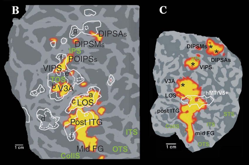
La triade des neurosciences cognitives

Enregistrements unitaires                                        IRMf
       singe vigile                  IRMf singe vigile          humaine
                    Base neuronale
                    du signal IRMf                 Homologies
                    Reconnaissance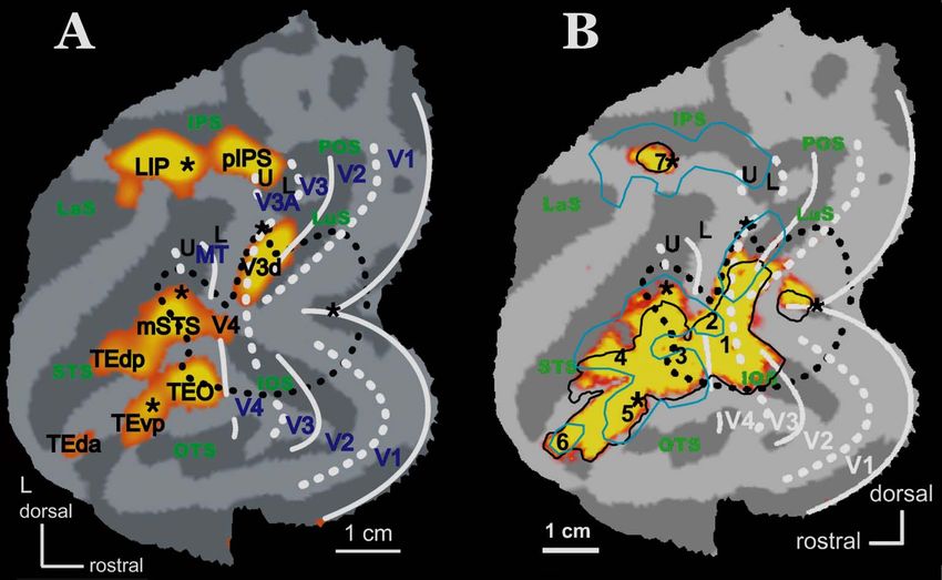
Homologie Cérébrale
• Des aires corticales sont homologues
  lorsqu’elles dérivent d’un ancêtre commun

• Cette propriété peut seulement être déduite de
  l’étude d’espèces existantes

• la certitude de cette déduction dépend du
  nombre d’espèces étudiées et du nombre de
  critères utilisés pour caractériser une aire
Homologies Cérébrales
             (certitude décroissante)

1. Aires homologues parmi les mammifères

2. Aires homologues parmi les primates

3. Aires homologues entre Homo et Maccaca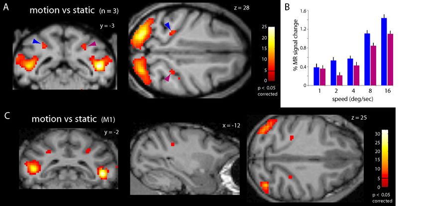
Homologies Cérébrales
             (certitude décroissante)

1. Aires homologues parmi les mammifères

2. Aires homologues parmi les primates

3. Aires homologues entre Homo et Maccaca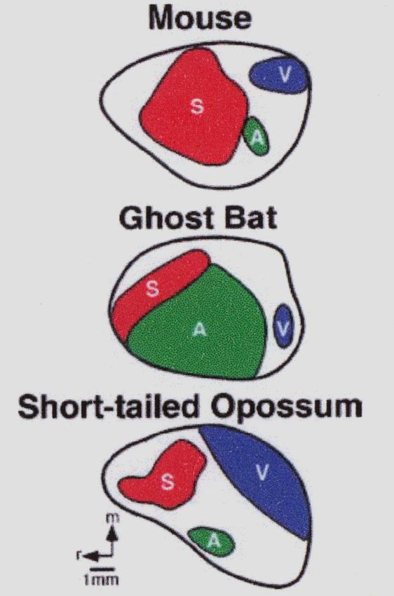
Aires homologues parmi les mammifères

                                        J.H. Kaas ´04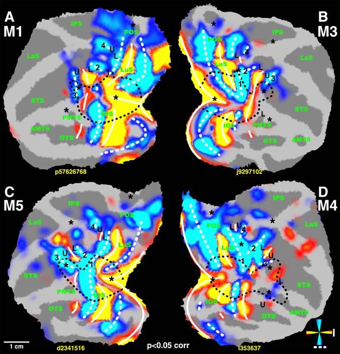
Aires homologues parmi les mammifères

Variations in cortical field organization of different mammals with vastly different lifestyles. In all mammals observed, there are cortical fields that are
common (e.g. SI, VI, AI, SII, PV, and M), and patterns of callosal and subcortical connections are fairly constant across different lineages, despite
differences in size, shape and geographic location of different fields. However, there are large shifts in the geographic location of homologous fields as well
as changes in their size and shape.
Rostral is to the left, medial is up.

                                                                                                                              L. Krubitzer and D.M. Kahn ´03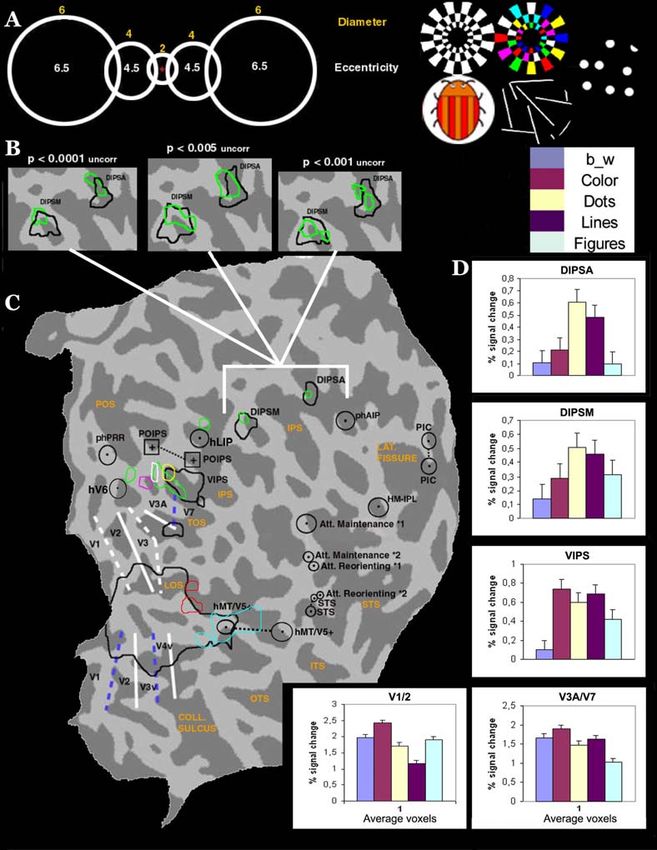
Aires homologues parmi les mammifères
                                        Primary cortical areas in three species of
                                        mammals that have approximately the same
                                        size cortical sheet, but different amounts of
                                        cortex allotted to different sensory systems,
                                        related to specialized sensory receptor
                                        arrays and use of particular sensory receptor
                                        arrays. For example, in the mouse, which
                                        relies heavily on tactile inputs from the
                                        whiskers for survival, the primary
                                        somatosensory cortex (red) and the rest of
                                        somatosensory cortex is enlarged, and the
                                        portion of cortex representing the whiskers is
                                        magnified, compared with the ghost bat and
                                        short-tailed opossum. Similarly, the primary
                                        auditory cortex and surrounding fields in the
                                        cortex of the echolocating ghost bat (green)
                                        is expanded, while the primary visual area
                                        (blue) and somatosensory area is relatively
                                        small. Finally, the cortex of the highly visual
                                        short-tailed opossum is dominated by V1
                                        (blue) and other visual areas. Although the
                                        size, shape, and the details of internal
                                        organization of particular cortical fields vary
                                        depending on use (activation from peripheral
                                        receptors), certain aspects of organization
                                        are conserved in these brains, even in the
                                        absence of apparent use. The similarity in
                                        relative location of cortical domains and
                                        fields therein suggests that the geographic
                                        organization and overall pattern of
                                        thalamocortical projections of the brain is
                                        constrained by developmental mechanisms.
                                        On the other hand, the differences in size,
                                        shape, and detailed organization of primary
                                        cortical fields indicate that input from the
                                        periphery is a crucial factor in guiding many
                                        of the details of organization of the
                                        neocortex. Medial is up and rostral is to the
                                        left, scale bar=1 mm.

                                                        L. Krubitzer and D.M. Kahn ´03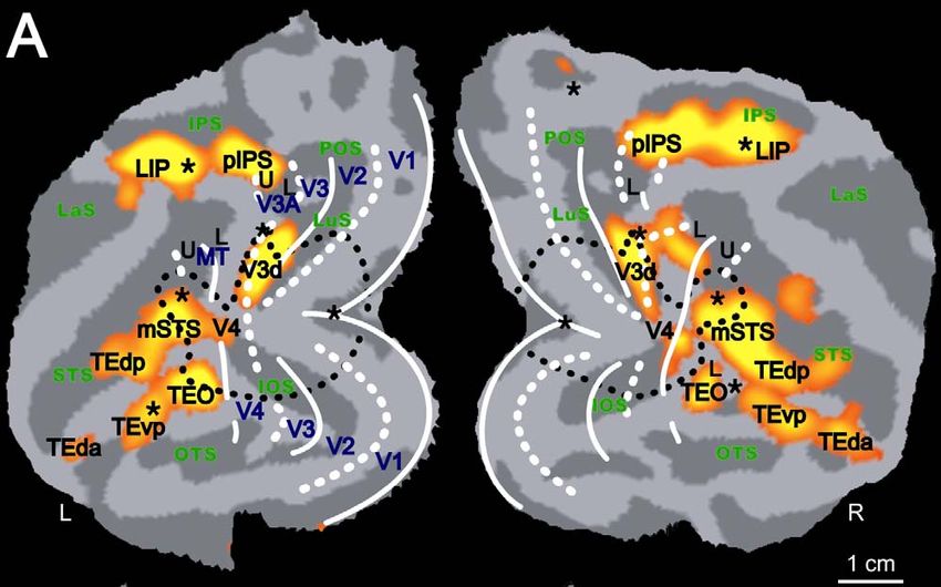
Aires homologues parmi les mammifères

                                        L. Krubitzer and K.J. Huffman ´00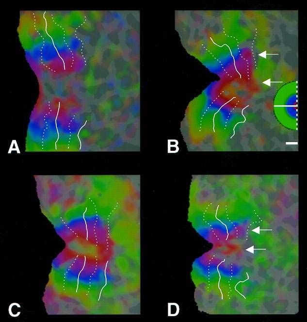
Aires homologues parmi les mammifères

                                        J.H. Kaas ´04
Aires homologues parmi les mammifères
Illustrations of how specific patterns of cell division in the ventricular zone (VZ) give rise to the patterns of clonally related neurons in the neocortex. In part
A, asymmetric divisions from a single progenitor cell (P) (black arrows) generate "sibling" cells that migrate sequentially to different layers of the cortical
plate (CP). This type of cell division determines cortical thickness. Symmetric divisions from a single progenitor cell (colored arrows) generate several
progenitor cells that in turn simultaneously generate "cousin" cells that then migrate, in parallel, to the same cortical layer. This type of division determines
cortical sheet size. Duration (B) and number (C) of cell cycle divisions differs dramatically in the mouse (pink) and the rhesus monkey (blue). In part C, black
bars represent the length of gestation in the mouse (19 days) and the monkey (165 days). In the mouse (pink rectangle) neurogenesis lasts 6 days, from
embryonic (E) day E11 to E17. In the monkey, neurogenesis lasts 60 days, from E40 to E100. The expanded duration and the increased number of cell
cycles could be one mechanism involved in expansion of the primate neocortex. IZ, intermediate zone (white matter), M, marginal zone (layer I), SP,
subplate zone (data used to construct this figure is taken from the work of [Kornack and Rakic, 1998] and [Kornack, 2000]).
L. Krubitzer and D.M. Kahn ´03Aires homologues parmi les mammifères
L. Krubitzer and D.M. Kahn ´03Homologies Cérébrales
(certitude décroissante)
1. Aires homologues parmi les mammifères
2. Aires homologues parmi les primates
3. Aires homologues entre Homo et MaccacaAires homologues parmi les primates
Collicule superieur: hemichamp controlateral
J.H. Kaas ´04Aires homologues parmi les primates
Quatre
Couches
Genouillees
2 Parvo
2 Magno
J.H. Kaas ´04Aires homologues parmi les primates
An evolutionary tree depicting the
phylogenetic relationship of major
orders of mammals and the cortical
organization of some of the sensory
fields that have been described in
particular species.
Electrophysiological, anatomical,
histochemical and molecular
analyses have revealed that certain
cortical regions, such as S1, S2, A1,
V1, and V2, are common to all
mammals and most likely are
homologous areas that arose from a
common ancestor. On the other
hand, some regions, such as MT
(pink), have been observed in only a
few orders, such as primates, and
likely evolved independently in these
lineages. A comparative analysis of
the neocortex, using the criteria
described above, allows one to infer
the organization of an unknown
mammal, such as the common
ancestor or human. If a number of
species are compared, one can be
fairly confident when assigning
features of cortical organization to
the unknown state, even in the
absence of direct data. S1: primary
somatosensory area (red), S2:
second somatosensory area
(orange), A1: auditory (green), V1:
primary visual area (dark blue), V2:
second visual area (light blue),
rostral is left, medial is up.
V1 V2 communes aux mammiferes
MT (et V3) commune aux primates
L Krubitzer and D M Kahn ´03Homologies Cérébrales
(certitude décroissante)
1. Aires homologues parmi les mammifères:
2. Aires homologues parmi les primates
3. Aires homologues entre Homo et MaccacaAires retinotopiques
R.B.H. Tootell et al, ´97Aires retinotopiques
R.B.H. Tootell et al, ´97Aires retinotopiques
R.B.H. Tootell et al, ´97Aires retinotopiques
R.B.H. Tootell et al, ´97Aires retinotopiques
IRMf primate non humain vigile
C
(Produit de
Contraste)
D
Mov - Sta
W. Vanduffel et al, Neuron, ´01Aires retinotopiques
V3A
D. Fize et al, ´03Aires retinotopiques
V1,V2,V3,V3a et V4v homologues; quid de V4d?
Macaque Humain
A B
G. Orban et al, TICs ´04Aires retinotopiques
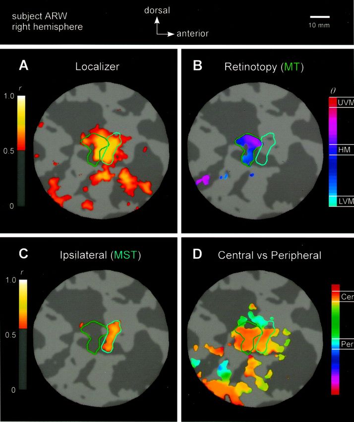
R.B.H. Tootell et al, ´97hMT/V5+
M.C. Morrone et al, Nature Neurosci, ´00hMT/V5+
A.C. Hulk et al, Nature Neurosci, ´02hMT/V5+
K. Nelissen et al, JoNS, ´06hMT/V5+
K. Nelissen et al, JoNS, ´06hMT/V5+
D
K. Nelissen et al, JoNS, ´06hMT/V5+
K. Nelissen et al, JoNS, ´06Aires pariétales
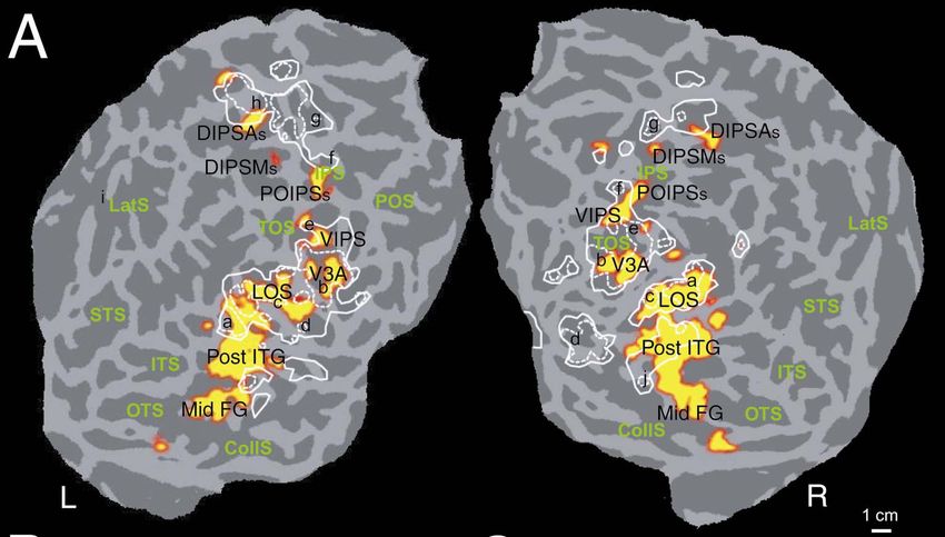
G. Orban et al, Neuropsychologia, ´06Aires pariétales
Passifs Actifs
A. Expertiment 3 A. Expertiment 4
Diameter Diameter
Eccentricity
Eccentricity
B.
G. Orban et al, Neuropsychologia, ´06Aires pariétales
Anatomie fonctionnelle de la sensibilité à la forme 2D
A C
B D
K. Denys et al, JoNS, ´04Aires pariétales
Anatomie fonctionnelle de la sensibilité à la forme 2D
A C
B D
K. Denys et al, JoNS, ´04Aires pariétales
G. Orban et al, Neuropsychologia, ´06Aires pariétales
G. Orban et al, Neuropsychologia, ´06Aires pariétales
A B
G. Orban et al, Neuropsychologia, ´06Aires pariétales
Anatomie fonctionnelle des mouvements oculaires
Macaque Humain
hFEF
M. Koyama et al, Neuron, ´04Aires pariétales
Correspondance fonctionnelle entre régions intrapariétales
des primates non humain et humain
Macaque Humain
Ant AIP hAIP
Post AIP DIPSA
Ant LIP DIPSM
LIPd hLIP
pIPS POIPS
? VIPS
V3A hV3A
Orban ´07Conclusions ● Homologies entre aires corticales humain-macaque sont difficiles à établir : Nécessité d’employer plusieurs critères (dont la position géographique) ● Homologies entre complexes d’aires plus faciles à établir qu’entre une aire individuelle ● Aires rétinotopiques : V1,V2,V3 et V3A homologues ( ~primates) Complexe MT/V5+ homologie du complexe pas encore des aires individuelles Aires pariétales : triade d’aires homologues centrées sur LIP (hypothèse)
You can also read