MAMMALS Common Diseases of Urban Wildlife - Australian Registry of Wildlife Health
←
→
Page content transcription
If your browser does not render page correctly, please read the page content below
Common Diseases of Urban Wildlife

                          MAMMALS

                          MISSION STATEMENT:
The Australian Registry of Wildlife Health is committed to contributing to the
preservation of Australia’s biodiversity through increased understanding of the
  interactions among animals, the environment, and disease causing agents.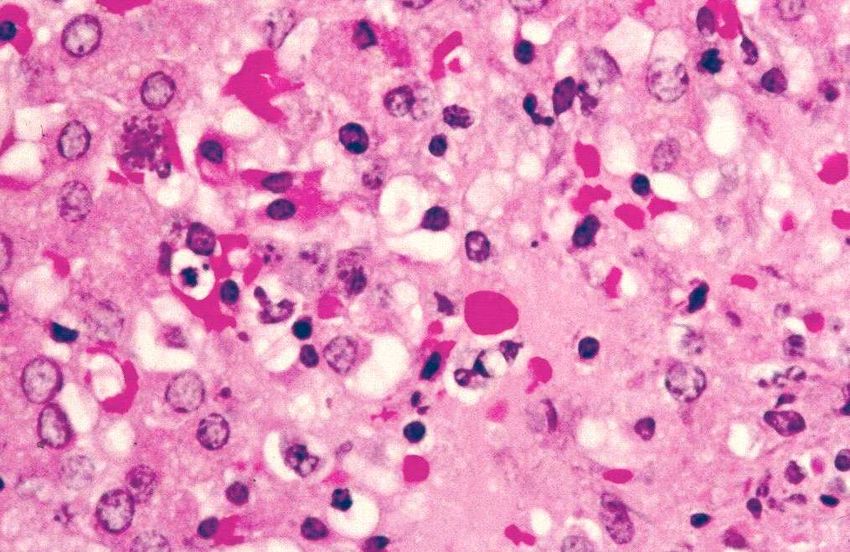
Common Diseases of Urban Wildlife: MAMMALS
K. Rose, June 2005

1 Common Diseases of the Common Wombat
1.1 Introduction
The common wombat (Vombatus ursinus) primarily lives within borrows located near
the margins of forest and scrubland.         Common wombats are fairly solitary,
herbivorous and nocturnal animals. The nocturnal habits of wombats place them at
risk of motor vehicle associated trauma. Wombat pouch young often survive the
traumatic injury and are hand raised by wildlife rehabilitators. These hand raised sub-
adult wombats are commonly presented to urban veterinarians for medical care.

1.2 Parasitic Disease

1.2.1 Sarcoptic Mange
Aetiology
Sarcoptes scabiei var wombati is a mite of uncertain host specificity. There are
reports of transmission of sarcoptic mange from wombats to humans and dogs (Booth,
1994, Skerratt and Beveridge, 1999). Persons handling infested animals should take
precautions.

Native animals reported to have suffered sarcoptic mange include the common
wombat, koalas, agile wallabies and the common ringtail possum.            Intermittent
outbreaks of infestation occur within the southern hairy-nosed wombat, but the
parasite is not reported to occur in the endangered northern-hairy nosed wombat
(Skerratt et al., 1998).

The common wombat is known to suffer severe debilitation as a result of S. scabiei
infestation. Sarcoptic mange occurs throughout the range of the common wombat,
including Tasmania and Flinder’s Island. Outbreaks of severely debilitating sarcoptic
mange occur only sporadically; nonetheless, these outbreaks may pose a risk to the
long-term survival of some of the smaller sub-populations of the common wombat
(Martin et al., 1998).

                                      -2-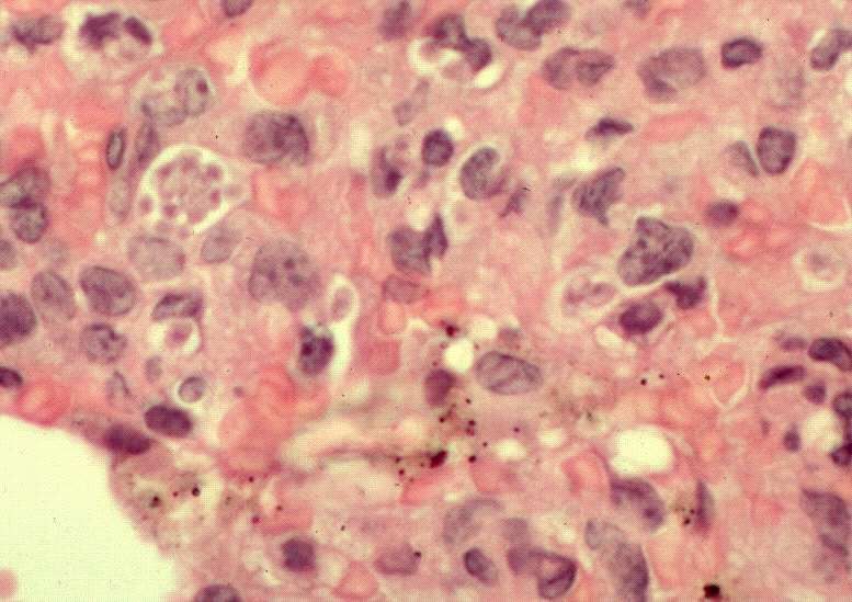
Common Diseases of Urban Wildlife: MAMMALS
K. Rose, June 2005

S. scabiei is transmitted during direct contact between animals and through sharing
contaminated rubbing sites and burrows. It is an obligate skin parasite and does not
live longer than a few weeks in the environment. It is uncertain whether the mites
responsible for sarcoptic mange in wombats were introduced to Australia with dingo,
or with the European red fox (Skerratt et. al, 1998).

Clinical signs
Sarcoptic mites burrow into the keratinising layers of the epidermis causing pruritis,
hyperkeratosis, epidermal thickening, and alopecia.                 Evidence of infestation may
begin as thickened skin on the head, and progress to thickened skin and hyperkeratotic
crust formation along the shoulders, flanks and limbs. The thick crust is composed of
keratin, bacterial colonies, cellular debris, mites and degenerating neutrophils
(Skerratt et al., 1998). Deep cracks may extend through the hyperkeratotic crust into
the dermis, resulting in myiasis or invasion of opportunistic bacteria.

The        severe           and
extensive nature of skin
lesions       in          some
wombats can lead to
impaired                 vision,
disturbance                  in
mastication, emaciation
and abnormal activity
                                   Sarcoptic mange, common wombat
throughout         the      day
(Booth, 1994). Death may result from secondary infection, dehydration or starvation.

Pathogenesis
It has been proposed that wombats exhibit marked skin lesions in response to low
numbers of Sarcoptic scabiei mites due to hypersensitivity reaction (Munday, 1988;
Skerratt, 2003). Systemic effects can also be severe. When greater than 30% of the
skin surface is covered by a hyperkeratotic crust, wombats generally have marked
reduction of muscle mass, and atrophy of fat. These severely infested wombats may

                                        -3-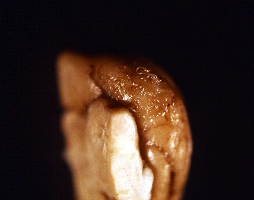
Common Diseases of Urban Wildlife: MAMMALS
K. Rose, June 2005

have anaemia, leucocytosis, lymphopaenia, and a reduced serum protein concentration
(Skerratt, 1989). The most commonly infested locations included the anterior lateral
aspect of the body, the posterior lateral surface of the body, and occasionally the
ventral and dorsal midline. Lymphadenitis is commonly encountered in the peripheral
lymph nodes that drain severely infested portions of the skin (Skerratt, 1998, Skerratt,
2003a and b). Sarcoptic mange in wombats may be associated with overcrowding,
habitat degradation, and a high density of foxes or feral dogs (Booth, 1998).

Diagnosis
Diagnosis of sarcoptic mange relies on identifying the mite within skin scrapings or
samples of the hyperkeratotic crust.

Treatment
Treatment should be initiated early in the progression of the disease whenever
possible. Prior to initiating therapy, the availability of suitable habitat to release the
animal should be considered.          Severely infested wombats commonly become re-
infected with sarcoptic mites and may transmit the mite to others in the wild
population. Euthanasia should be considered for seriously infested and debilitated
wombats.

Treatment of mildly to moderately affected wombats consists of application of either a
topical acaricide solution, or systemic therapy with anti-parasitic agents. Prior to the
first application of topical solutions, the animal may require a bath and keratolytic
agents to remove some of the hyperkeratotic crust (Booth, 1994).                Additional
information on treatment is described by Skerratt (2003b).

1.2.2 Coccidiosis

Aetiology
Eimeria arundeli is a frequently encountered enteric coccidian of the common
wombat. This parasite has been associated with enteritis in sub-adult wild and hand-
reared wombats. The life cycle of the coccidian parasites of wombats has not been
thoroughly investigated, but it is thought to be similar to other eimerian parasites.

                                      -4-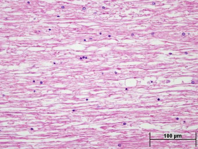
Common Diseases of Urban Wildlife: MAMMALS
K. Rose, June 2005

Clinical signs
Oocysts of E. arundeli are often found within the faeces of healthy adult common
wombats. Wild and captive sub-adult wombats appear to be the most likely animals
to exhibit clinical signs of enteritis associated with coccidial infection. Wombats with
a severe burden of coccidia may develop mucoid to liquid green diarrhoea,
progressively loose weight and become bloated.        The onset of enteritis is often
associated with the onset of grazing, which occurs at approximately 10 months in wild
wombats, but is often earlier in hand raised wombats.            Presumably, animals
immunologically naïve to coccidia are suddenly exposed to large numbers of infective
oocysts present on contaminated pasture.

Pathology
Gross post mortem examination often reveals a gastrointestinal tract diffusely
distended with fluid and gas. The character of the luminal fluid varies from yellow,
mucoid to green/grey and thick. Fibrin strands are often scattered throughout the
luminal fluid. The wall of the proximal small intestine is often segmentally thickened
and bears a reticular pattern created by small yellow foci raised above the more
normal mucosa. Intestinal villi may visibly hypertrophic (Hum, et al., 1991).

Histological examination of sections of the affected intestine typically reveals
congestion of the mucosa, and large masses of coccidial gametocytes within distended
host cells in the mucosa and lamina propria.       The abundance of these coccidial
gametocytes results in the hypertrophic villi seen upon gross examination (Hum, et al.,
1991). The presence of the coccidial organisms has been associated with oedema and
an inflammatory cell infiltrate throughout the lamina propria.

Wombats that die with intestinal coccidiosis often have concurrent salmonellosis
(ARWH).

Diagnoses

                                      -5-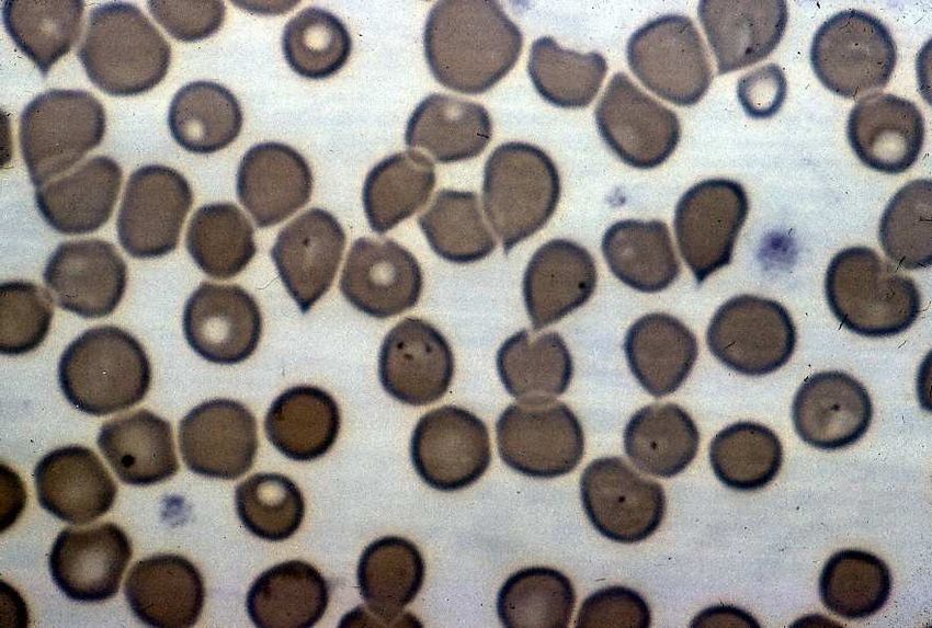
Common Diseases of Urban Wildlife: MAMMALS
K. Rose, June 2005

Diagnosis of coccidiosis is achieved through examination of faeces within a wet
preparation using light microscopy at a magnification of 400x.          Standard faecal
flotation techniques are also useful in the diagnosis of coccidiosis.

Treatment
Once clinical signs of enteritis have developed, treatment becomes very difficult.
Fluid therapy can be difficult to deliver to wombats, and response to anti-coccidial
therapies that are used in other species is often poor.

1.2.3 Other Parasites
Toxoplasmosis can cause mortality in hand-raised wombats. This disease is discussed
in detail in section 5.2.1

Sheep liver fluke infection (Fasciola hepatica) also occurs commonly in wombats.
Although one often does not see adult flukes or not large numbers of them, the
immunological response of the wombat to the fluke is such that the entire liver may
become completely fibrosed. (David Spratt, personal communication).

1.3 Fungal Disease
Spores of the fungus Emmonsia parvum and E. crescens are frequently encountered
during histologic examination of the pulmonary tissue. These fungi are most often
localised within the alveoli at the periphery of the lung, and are unaccompanied by
significant inflammation. This appears to be an incidental finding in a burrowing
species.

1.4 Nutritional Disease

Captive wombats offered energy or carbohydrate-rich diets, such as lucerne and fruit,
can become bloated. Bloated wombats have a distended abdomen, abdominal pain,
and flatulence. Diet modification and short-term use of agents such as Milanta® can
be effective remedies for these wombats.

                                      -6-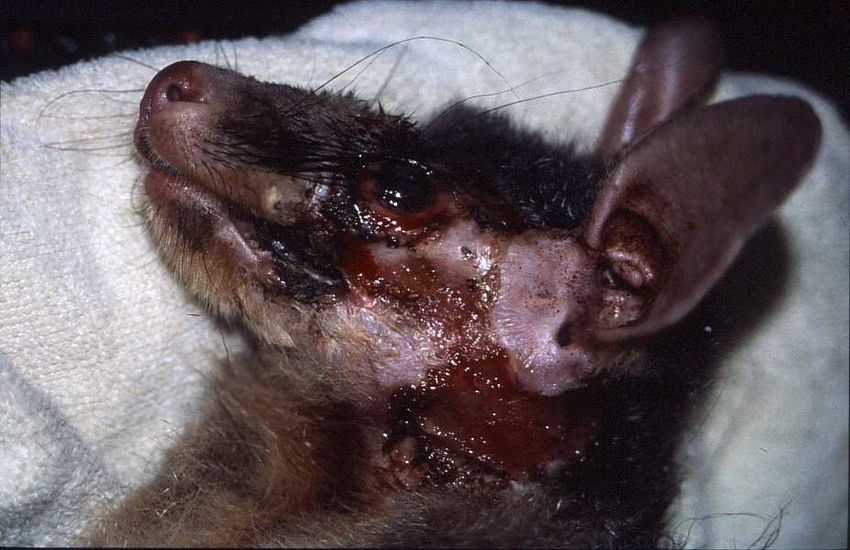
Common Diseases of Urban Wildlife: MAMMALS
K. Rose, June 2005

Wombats have continuously growing teeth. Teeth that are fractured during trauma
may be filed or clipped.         Malocclusion of incisors and molars may result from
irregular tooth wear, insufficient tooth wear or traumatic injury. Elongated teeth are
filed or clipped while the animal is under general anaesthesia. Provision of sufficient
browse should help to alleviate malocclusion in captive wombats; however, repeated
tooth clipping is often required.

A syndrome of multisystemic mineralisation has historically been reported in captive
common wombats.           Each wombat examined had significant arteriosclerosis and
medial mineralisation of the aorta, pulmonary artery, and sub-endocardial tissue of the
right atrium. Many of the animals examined also had marked calcification of the
footpads. Chronic interstitial nephritis with multifocal interstitial mineralisation was
also evident upon post mortem examination. Mineral deposits within the renal tubular
lumina or within the interstitium consisted of concentric rings of basophilic,
mineralised material (Griner, 1983). Several cases of multisystemic mineralisation in
wombats are present within the Australian Registry of Wildlife Health at Taronga Zoo
(ARWH). Affected animals had been maintained on a diet composed primarily of dog
food. Based on the gross and microscopic post mortem findings, in conjunction with
the known diet, vitamin D intoxication is the most likely cause of this syndrome.

1.5 Traumatic Injury

Wombats that are injured by motor vehicles tend to suffer spinal injuries, pelvic
fractures, concussion, or massive contusions.

2 Common Diseases of the Short-beaked Echidna
2.1 Introduction

The short-beaked echidna (Tachyglossus aculeatus) is an insectivorous monotreme
that inhabits virtually every habitat-type throughout Australia. Short-beaked echidna
have crepuscular habits in summer and are diurnal during winter. The normal body
temperature for the short-beaked echidna ranges between 27 and 32oC. Whenever
environment temperatures fall below 10oC and food availability declines, short-beaked

                                      -7-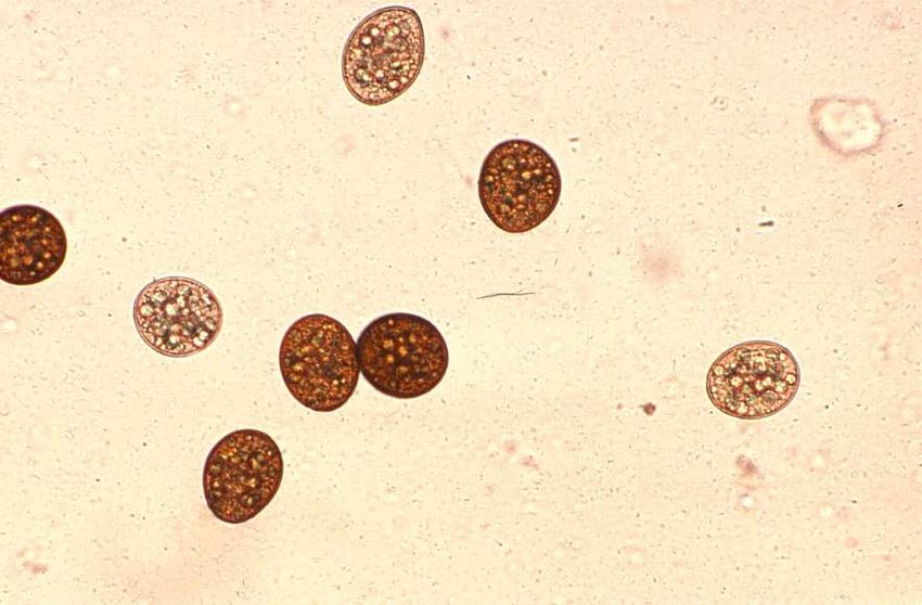
Common Diseases of Urban Wildlife: MAMMALS
K. Rose, June 2005

echidna will dig into the substrate and begin hibernation. During hibernation, the
animal’s body temperature may fall to as low as 5oC (Whittington, 1993).

2.2 Parasitic Disease

2.2.1 Coccidiosis

Aetiology
Eimeria echidna, E. tachyglossi, and Octosporella hystrix are enteric coccidia of the
short-beaked echidna (Barker et al., 1985). Coccidial oocysts are a frequent finding
within the faeces of healthy echidna; however, coccidia are occasionally associated
with marked enteritis or disseminated disease.

Clinical signs
Short-beaked echidna with coccidial enteritis or disseminated coccidiosis are most
often found dead without premonitory clinical signs.

Pathology
Enteric coccidiosis:
Short-beaked echidna with enteric coccidiosis may have haemorrhagic and necrotising
enteritis.    The intestinal mucosa is often thickened due to the presence of large
numbers of coccidial microgametocytes, macrogametocytes and oocysts.                                Villous
atrophy has been reported in some animals with enteric coccidiosis. An intense
mononuclear cell infiltration is usually evident throughout the intestinal lamina
propria of echidna with coccidial enteritis.

Haemorrhagic enteritis caused        by   disseminated   Intestinal coccidiosis, small intestine, short-beaked
coccidiosis, short-beaked echidna.                       echidna. Note formation of oocysts with eosinophilic

                                           -8-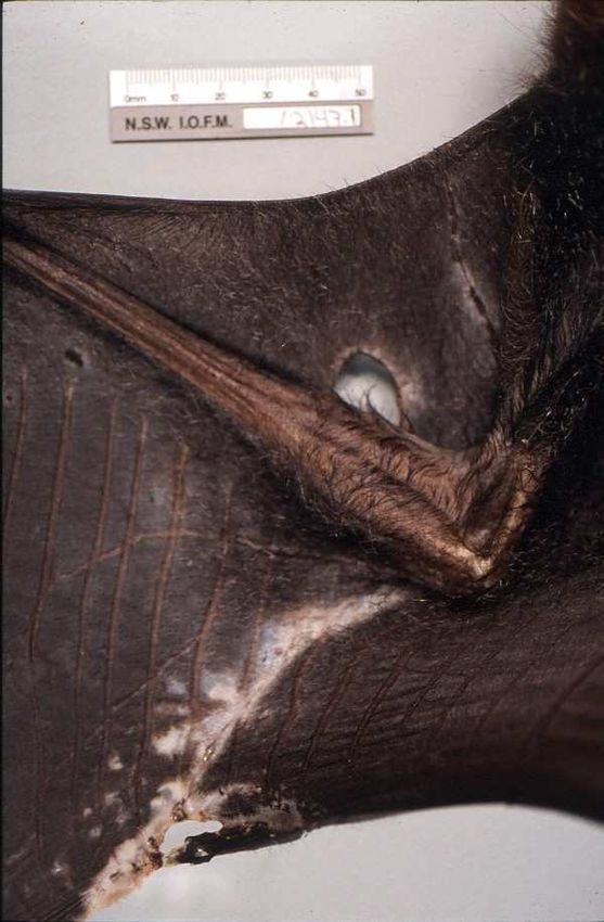
Common Diseases of Urban Wildlife: MAMMALS
K. Rose, June 2005

                                                        amylopectin bodies.

Disseminated coccidiosis:
Echidna                     with
disseminated
coccidiosis also have
microgametocytes,
macrogametocytes            and
oocysts,         within     host
cells of the intestinal
mucosa           and      lamina
propria. Desquamation
of         the     superficial     Hepatic necrosis, disseminated coccidiosis (see arrow).

enterocytes, hypertrophy of the villous epithelium, and a mononuclear cell infiltrate
throughout the lamina propria are also evident within affected segments of the
intestine (Dubey and Hartley, 1993). In addition, the disseminated form of coccidiosis
is characterised by asexual and sexual stages of the coccidian lifecycle scattered
throughout the endothelium and parenchyma of the lung, liver, heart, kidney, spleen
and gastrointestinal tract (Dubey and Hartley, 1993).

Coccidial schizogeny (arrow), lung, short-beaked       Coccidial schizogeny (arrow) in the endothelium of
echidna.                                               pulmonary blood vessel, short-beaked echidna.

                                          -9-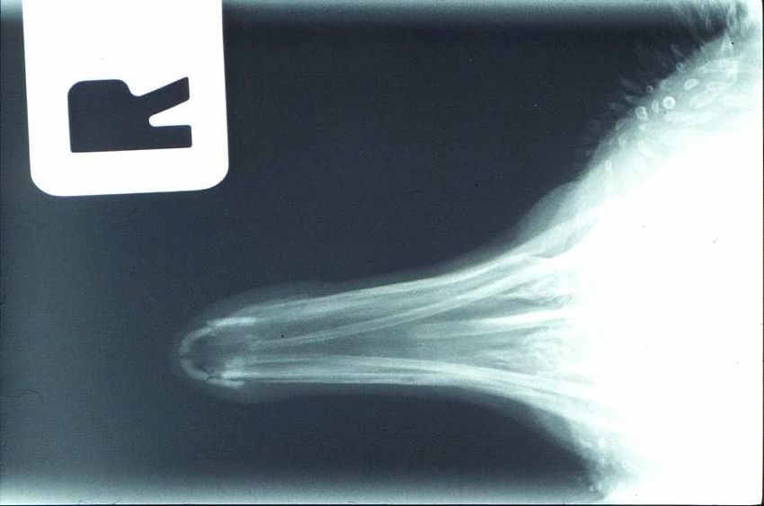
Common Diseases of Urban Wildlife: MAMMALS
K. Rose, June 2005
Monotremes have a
diffuse lymphoid system
composed of nodules,
which are suspended by
a pedicle, dispersed
throughout the
lymphatic vessels. The
lymphoid nodules
within the intestinal
mesentery are grossly
visible as small white,
Mesenteric lymph nodes, short-beaked echidna
raised foci near the
attachment of the
mesentery to the intestinal serosa. Echidna with disseminated coccidiosis had foci of
necrosis throughout the spleen and the disseminated lymph nodules. Presumably,
these foci of necrosis represent sites of schizogony, where cell damage is associated
with the release of merozoites from ruptured schizonts.
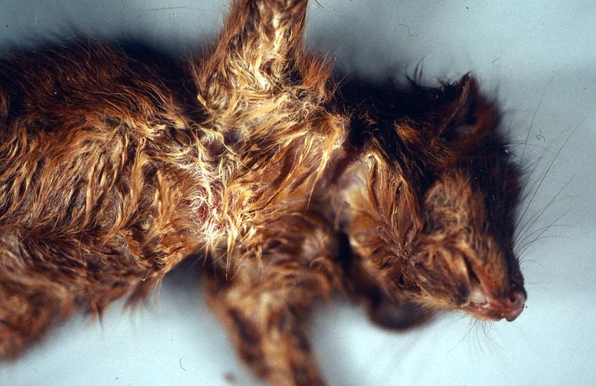
Several short-beaked echidna
with multisystemic coccidiosis
have had Atoxoplasma-like blood
parasites circulating within
monocytes. Examination of
blood films from short-beaked
echidna can be used to diagnose
Systemic coccidiosis, short-beaked echidna (blood smear).
clinical systemic coccidiosis. Note organisms within mononuclear cells
Many echidna have very small number of circulating organisms, but large numbers of
organisms evident in monocytes is associated with clinical disease (ARWH).
Echidna that die with systemic coccidiosis often also have acute Salmonella species
septicaemia (ARWH).
- 10 -Common Diseases of Urban Wildlife: MAMMALS
K. Rose, June 2005
Diagnosis
Diagnosis of coccidiosis is achieved
through examination of faeces within
a wet preparation using light
microscopy at a magnification of
400x. Standard faecal flotation
techniques are also useful in the
diagnosis of coccidiosis. Collection
Coccidial oocysts, faeces, short beaked echidna, 400x
of a blood sample and examination of
the blood film is encouraged to identify systemic coccidiosis. Buffy coat preparations
concentrate the leucocytes and can aid in the identification of intracellular parasites.
Treatment
Short-beaked echidna with coccidial enteritis or systemic coccidiosis are most
commonly presented moribund or dead. Treatment methods have not been thoroughly
investigated.
2.2.2 Other Parasites
Short-beaked echidna are commonly infested with ticks. Aponomma concolor is the
“echidna tick”, which infests both captive and wild short-beaked echidna. Large tick
burdens can result in anaemia and dermatitis. Ticks are also capable of transmitting a
number of haemoprotozoa and viruses.
Sparganosis due to the plerocercoid Spirometra eranacei has been reported to cause
large tumour-like masses within the subcutaneous tissues of the ventral and lateral
abdomen of short-beaked echidna. The masses can be as large as 12 cm diameter and
are composed of a central sparganum, surrounded by an intense non-suppurative
inflammatory infiltrate, and a thick layer of fibrous tissue. Several infected echidna
have had numerous plerocercoids replacing a large proportion of the pulmonary
parenchyma (Whittington et al., 1992, ARWH). Presumably, the echidna ingest
- 11 -Common Diseases of Urban Wildlife: MAMMALS
K. Rose, June 2005
infected copepods and act as incidental intermediate hosts. The definitive hosts of
Spirometra eranacei are introduced felids and canids.
A wide variety of nematodes parasitise the gastrointestinal tract, pulmonary tissues,
and subcutaneous tissues of the short-beaked echidna, including: Parastrongyloides
spp., Nicollina spp., Tachynema spp., Tasmanema spp, Ophidascaris sp., and
Dipetalonema sp. Clinical signs are rarely associated with nematode parasitism.
Two of the most common parasites of the echidna are the cestodes Echidnotaenia
tachyglossi which occurs in northern echidna and Linstowia echidnae which occurs in
southern echidna and in which upwards of 500 individuals can occur in a single host
(David Spratt, personal communication).
Theileria tachyglossi, and Babesia tachyglossi are common incidental haemoprotozoa
of the short-beaked echidna. An Anaplasma marginale-like organism has also been
identified within circulating red blood cells of normal echidna (Whittington, 1993).
Theileria tachyglossi in Howell-Jolly body Babesia tachyglossi, short-beaked echidna
2.3 Bacterial Disease
Many species of Salmonella have been isolated within healthy short-beaked echidna,
yet several species of Salmonella have been identified as the cause of haemorrhagic
enteritis and septicaemia.
Multisystemic mycobacteriosis is a fairly common post mortem finding in short-
beaked echidna. Granulomatous lesions occur in a variety of tissues, but are most
- 12 -Common Diseases of Urban Wildlife: MAMMALS
K. Rose, June 2005
common in the liver. Beaded, red bacilli are easily demonstrated within the
granulomas upon acid fast staining.
Staphyloccocus spp., Streptococcus spp, Aeromonas spp., and Proteus spp. have been
associated with septicaemia in the short-beaked echidna. Edwardsiella sp. has been
isolated from the pulmonary tissue of echidna suffering from suppurative
bronchopneumonia. Edwardsiella sp. may be a primary pathogen of the short-beaked
echidna (Whittington, 1993).
2.4 Viral Disease
Multisystemic herpes virus infection and inclusion body hepatitis associated with
herpes virus have been reported in short-beaked echidna (Whittington, 1992).
Poxvirus is occasionally found within proliferative dermal lesions in juvenile short-
beaked echidna. It is uncertain whether poxvirus infection is the cause of a fairly
common seborrheic condition of hand reared juvenile short-beaked echidna
(Whittington, 1993).
Cytomegalo-like adenovirus inclusion bodies have been identified within the renal
epithelium of short-beaked echidna, but were considered to be an incidental finding
(Whittington, 1993).
2.5 Fungal Disease
Oesophagitis and gastritis are frequently diagnosed in short-beaked echidna that have
been hand raised or have concurrent disease. Affected animals may experience a short
course of diarrhoea, but are often found dead. Microscopic examination of the
oesophagus and squamous portions of the stomach reveal foci of mucosal necrosis,
inflammation and erosion. Corynebacterium, Candida and Fusobacterium spp. have
been isolated from these ulcerative lesions (Whittington, 1993). Among short-beaked
echidna presented to Taronga Zoo for post mortem examination (ARWP), Candida
albicans, or one of its telomorphs, is most commonly identified in microbial culture,
and during histologic examination of these lesions.
- 13 -Common Diseases of Urban Wildlife: MAMMALS
K. Rose, June 2005
Microsporum gypseum has been isolated from short-beaked echidna with fractured
quills and Cryptococcus neoformans has been isolated within pulmonary granulomas
of the short-beaked echidna.
2.6 Traumatic Injury
The oronasal structure of the short-beaked echidna is a sensitive and fragile organ.
Unfortunately, the bones
of the beak often sustain
comminuted fractures, and soft
tissues are severely damaged during
motor vehicle induced injury.
Internal fixation of these bones is not
possible, and the animals will not
tolerate external fixation. In
addition, the senses necessary for Fractured beak, Tachyglossus aculeatus
effective foraging are often
permanently damaged. Cage rest may help minor injuries to the beak resolve, but
euthanasia is often indicated for severe damage.
External examination is not a sensitive tool for the detection of traumatic injuries in
short-beaked echidna. Radiography is warranted when any echidna presents with
evidence of traumatic injury, since the presence of cutaneous spines prevent thorough
palpation of the skeletal system.
3 Common Diseases of Possums
3.1 Introduction
The common ringtail possum (Pseudocheirus peregrinus) and the common brushtail
possum (Trichosurus vulpecula) are widespread throughout the east coast of Australia
and southwest Australia. These herbivorous marsupials have nocturnal habits and
nest in tree hollows, logs, and roof spaces. While brushtail possums are solitary and
territorial, ringtail possums often have overlapping home ranges.
- 14 -Common Diseases of Urban Wildlife: MAMMALS
K. Rose, June 2005
3.2 Parasitic Disease
3.2.1 Toxoplasmosis
Aetiology
Toxoplasma gondii is a coccidian parasite with worldwide distribution and a broad
host range. All vertebrates may be infected with the asexual stages of reproduction;
however, some taxonomic groups are more susceptible to suffering clinical illness.
Marsupials are remarkably susceptible to developing toxoplasmosis.
Asexual (merogony) and sexual (gametogony)
stages of the T. gondii lifecycle take place within
the intestinal mucosa of felids, which are the
definitive hosts. Unsporulated oocysts are shed in
the faeces and become infective when they
sporulate, 24 to 96 hours after leaving the host.
Felids may shed millions of oocysts in their faeces
during their first infection with T. gondii. Felids
Toxoplasma gondii. (see arrow)
are usually infected when they eat birds or rodents
infected with T. gondii tissue cysts.
Marsupials are primarily infected with T. gondii when they ingest vegetation
contaminated with felid faecal material containing sporulated oocysts; however,
transplacental transmission of the parasite has been reported. Sporozoites are
released from ingested oocysts and replicate in the tissues of the intestine and
associated lymphoid tissue. The organisms are referred to as tachyzoites as they
undergo several generations of rapid replication, before spreading through the
circulatory system to numerous tissues and forming cysts. Cysts, which contain
bradyzoites, are identified most commonly in the tissues of the brain, liver, muscles
and retina. Cysts may remain dormant for prolonged periods, but are capable of
releasing their bradyzoites, which become tachyzoites and re-initiate active infection.
The organism has a high affinity for the central nervous system, lung, pancreas,
- 15 -Common Diseases of Urban Wildlife: MAMMALS
K. Rose, June 2005
lymphoid system, myocardium, adrenal gland and ocular tissues. Necrosis and
inflammation accompany the replication of tachyzoites in these tissues.
Both ringtail and brushtail possums share their habitat with pet and feral cats, and as a
consequence toxoplasmosis is not uncommon in these species.
Clinical signs
Toxoplasma gondii infection is often inapparent. Clinical illness associate with T.
gondii occurs primarily in animals that are immunosuppressed or hand raised.
Clinical signs of toxoplasmosis are primarily associated with lesions in the central
nervous system, lungs, and liver. The severity of the illness is highly variable, ranging
from mild malaise to peracute mortality. Animals clinically affected by toxoplasmosis
may be depressed, weak, anorexic, pyrexic, dyspnoeic, ataxic, hemiplegic,
quadriplegic, comatose, convulsive, or may exhibit muscle stiffness, diarrhoea,
emesis, uveitis, retinitis or cataract formation. Slow growth rates have been reported
in some hand raised animals in association with Toxoplasma gondii infection.
Infection or expression of disease may occur at any time of the year.
Serum concentrations of AST, CK and alanine amino transferase (ALT) are often
elevated in animals suffering from toxoplasmosis but this depends upon the organs
affected.
Pathology
Non-suppurative inflammation and necrosis are the most prevalent morphologic
changes associated with active T. gondii infections. Although bradyzoites can remain
dormant with little or no host inflammatory response, tachyzoites of T. gondii are
highly immunogenic, inciting a strong humeral and cell mediated immune response in
the host. Lymphocyte, plasma cell and macrophage infiltration is commonly
associated with the presence of trophozoites. The cause of tissue necrosis associated
with T. gondii trophozoites is uncertain (Canfield et al., 1990, Dubey and Hartley,
1991).
- 16 -Common Diseases of Urban Wildlife: MAMMALS
K. Rose, June 2005
Toxoplasma – subclinical infection, brain, wombat. Toxoplasma encephalitis, wombat
Gross post mortem lesions may be inapparent, however, pulmonary congestion,
oedema, consolidation, hepatosplenomegaly, lymphadenopathy, or multisystemic pale
foci may be evident. Histologic examination of tissues from clinically affected
animals may reveal any of the following lesions: retinitis, lymphadenitis, hepatitis,
myositis, pneumonia, pancreatitis, myelitis and encephalitis.
Individual tissue cysts are commonly found within the muscle and nervous tissue of
animals during histologic examination. Unless these cysts are associated with tissue
necrosis or inflammation, they most likely represent subclinical infection.
Diagnosis
Ante mortem diagnosis of toxoplasmosis relies on serological testing to detect rising
IgG T. gondii concentrations. Serial four fold increases in T. gondii IgG titres
represent active infection. IgM antibodies are more indicative of active infection than
IgG antibodies, and a single high T. gondii IgM concentration in serum may reflect
active infection. Most commercial veterinary labs offer indirect haemagglutination
inhibition tests for the detection of T. gondii IgG. Indirect fluorescent antibody tests
may be used to determine serum IgM concentrations.
Post mortem diagnosis of toxoplasmosis usually occurs through histopathologic
examination of tissues, or cytological examination of impression smears of the spleen,
liver, or lung. Immunoperoxidase staining, tissue antigen ELISA, or PCR are used to
- 17 -Common Diseases of Urban Wildlife: MAMMALS
K. Rose, June 2005
detect small numbers of T. gondii organisms in section, and differentiate T. gondii
from other protozoa.
Treatment
Treatment of toxoplasmosis is based upon the use of drugs that arrest replication of
the parasite. A drug that will eliminate the organism from tissues has not yet been
discovered. Thus, a competent host immune system is required to affect recovery.
Response to therapy may vary depending on the degree of tissue damage already
sustained. If therapy is likely to be effective, clinical improvement should be noticed
within the first seven days.
Prevention
Toxoplasmosis prevention relies on decreasing the opportunity for immunologically
naïve or otherwise susceptible hosts to be exposed to large numbers of infective
oocysts. Effective cat control and proper storage of animal foodstuffs are the
cornerstones of toxoplasmosis prevention. Clinical trials using commercial T gondii
vaccines, developed for use in sheep, have resulted in the development of fatal
toxoplasmosis in Tammar wallabies (Lynch et al., 1993). An oral vaccine consisting
of Hammondia hammondi, has effectively prevented the development of clinical
disease in Tammar wallabies challenged with T. gondii (Reddacliff, 1993).
Hammondia hammondi is a non-pathogenic coccidian parasite of cats that is very
similar to T. gondii. Clinical trails of vaccines using temperature sensitive strains of
T. gondii are under investigation. Additionally, the management of stressors, or other
immunosuppressive factors, are important in reducing the expression of clinical
disease.
3.2.2 Angiostrongylus cantonensis
Aetiology
Angiostongylus cantonensis is a metastrongylid nematode that has for some time been
a cause of eosinophilic meningoencephalitis in mammals and birds in north-eastern
Australia. It is extending its range along the central to south eastern seaboard and
cases are becoming common in Sydney.
- 18 -Common Diseases of Urban Wildlife: MAMMALS
K. Rose, June 2005
Parasitic meningoencephalitis due to infection by the metastrongylid nematode,
Angiostrongylus cantonensis, has been reported in ringtail possums, brushtail
possums, primates, flying foxes, yellow tailed black cockatoos, and tawny frogmouths
in Queensland and New South Wales. An outbreak of disease was also reported in
captive rufous bettongs in Queensland (Higgins et al., 1997). A. cantonensis can
cause central nervous system disorder and death in wild and captive native fauna.
This parasite was originally thought to be geographically restricted to Queensland, but
recent reports of the infection in dogs, zoo and wild animals indicate that the parasite
is now widespread in New South Wales (Carlisle et al., 1998, Collins and Rothwell,
1992, Reddacliff et al., 1999). A. cantonensis is a zoonotic threat. Eosinophilic
encephalomyelitis has been reported to occur within children that ingest snails or slugs
(Paul Prociv, personal communication).
Rats are the definitive host of A. cantonensis. Adult parasites live within the
pulmonary arteries and release eggs that are trapped in the pulmonary
microvasculature. Larvae emerge from the eggs and migrate into the airways and then
to the intestinal tract. Rats excrete first-stage larvae in their faeces, and these larvae
either directly penetrate, or are ingested by terrestrial snails or slugs. Larvae develop
into the infective third-stage in the molluscs, which are then eaten by rats. When
ingested by rats, infective larvae undertake a prolonged migration through the central
nervous tissue before they mature to adulthood in the pulmonary artery. In non-
adapted hosts, the parasite often undergoes ongoing migration through the central
nervous tissues, and rarely matures and migrates to the pulmonary arteries (Carlisle et
al., 1998).
Clinical signs
Animals suffering from A. cantonensis meningoencephalitis may exhibit a variety of
clinical signs related to focal lesions within the central nervous system. The most
common clinical signs of infection include hind limb ataxia, paresis, intention
tremors, which progress to forelimb paresis, profound central depression, coma,
seizures, and often death.
- 19 -Common Diseases of Urban Wildlife: MAMMALS
K. Rose, June 2005
Pathology
Gross post mortem examination is often unrewarding in animals with eosinophilic
meningoencephalitis. Occasionally nematode parasites are visible within the
subarachnoid spaces.
A. cantonensis within the meninges
Nematode parasites are evident upon histologic examination of serial sections of the
brain and spinal cord. Parasites may be unaccompanied by an inflammatory response,
but are often associated with a mixed inflammatory cell infiltrate. This infiltrate is
composed of lymphocytes, macrophages, plasma cells and scattered eosinophils. Foci
of malacia may occur when parasite migration has resulted in ischaemic tissue
necrosis.
Cross-sections of A. cantonensis , spinal cord, brushtail Wallerian degeneration, spinal cord, brushtail possum.
possum. Note axonal swelling and digestion chambers(
phagocytic cells within axonal chambers - arrow)
Diagnosis
- 20 -Common Diseases of Urban Wildlife: MAMMALS
K. Rose, June 2005
Ante mortem diagnosis of A. cantonensis is often difficult. Eosinophilia within the
cerebrospinal fluid is highly suggestive of parasitic meningoencephalitis; however,
infected animals often only show non-suppurative inflammation, which does not assist
in the differentiation from viral or protozoal infections (ARWH). Infected animals
rarely have systemic eosinophilia. ELISA has been used to detect infection in humans
and dogs, and preliminary work has been carried out to develop an ELISA for use in
marsupials (Smaller, 2004).
Treatment
Corticosteroids are employed in the treatment of angiostrongyliasis to reduce
inflammation within the central nervous system. Some researchers suggest that the
use of antiparasitic drugs can result in increased inflammation throughout the central
nervous system as a result of massive release of antigen from degenerating parasites.
Fenbendazole, has been used to successfully treat A. cantonensis infection in a grey-
headed flying-fox (Reddacliff et al., 1999). The prognosis for infected animals,
however, is generally poor.
Prevention
Prevention of eosinophilic meningoencephalitis caused by A. cantonensis relies on
eradicating intermediate hosts from the enclosures of captive mammals.
3.2.3 Other Parasites
Ectoparasites
The paralysis tick, Ixodes holocyclus, is commonly found in the pelage of brushtail
possums. Anecdotal reports have described tick paralysis in brushtail possums.
Notoedres muris, the mange mite of rats, has been associated with thick crusty
dermatitis of the ears, tail, snout and periocular tissues of brushtail possums in
Victoria (Booth, 1994b).
Cestodes
- 21 -Common Diseases of Urban Wildlife: MAMMALS
K. Rose, June 2005
Bertiella trichosuri is an anoplocephalid cestode that parasitises the intestinal tract of
brushtail possums. Many possums are infected with this tapeworm, yet clinical signs
have not been attributed to infection. Possums living in suboptimal habitats appear to
be most susceptible to infection with this parasite (Booth, 1994b).
Trematodes
Fasciola hepatica is the liver fluke of sheep. This parasite enjoys a wide host range,
and has been known to infect brushtail possums grazing on wet pastures. Snails
function as the intermediate host for this parasite.
Nematodes
A variety of nematode parasites infect the intestinal tract of possums; however,
parasitism rarely results in clinical disease.
Marsupiostrongylus minesi is most often an incidental lungworm found in possums
and gliders. On occasion, this parasite may occur in the airways and causes
pulmonary consolidation or bronchopneumonia (Booth, 1994b).
Brushtail possums grazing pastures inhabited by sheep may develop diarrhoea,
dehydration and eventually die as a result of heavy intestinal burdens of
Trichostrongylus axei and T. colubriformis.
Protozoa
A variety of protozoa are observed
within the faeces or intestinal lumen of
brushtail possums and ringtail
possums. Enteritis associated with the
presence of these parasites has not
been described.
Hepatozoon spp are occasionally found
Hepatozoon spp, ringtail possum
in the erythrocytes of possums and
gliders. Their clinical significance is unknown.
- 22 -Common Diseases of Urban Wildlife: MAMMALS
K. Rose, June 2005
3.3 Bacterial Disease
3.3.1 Yersiniosis
Aetiology
Yersinia pseudotuberculosis is a ubiquitous gram-negative bacterium of the family
Enterobactericeae that is commonly identified within the faeces of wild birds and
mammals. Transmission of this bacterium occurs via the faecal-oral route.
Clinical signs
Although many animals will harbour Y. pseudotuberculosis within the intestinal tract
without effect, the organism is capable of causing multisystemic illness. Yersiniosis
results in either rapidly fatal enteritis and septicaemia, or subacute to chronic
multisystemic abscessation. Clinical signs in animals experiencing the rapid,
septicaemic form of the disease may include, profound depression, dehydration,
diarrhoea and melaena. Animals suffering from the multisystemic form will have
clinical signs associated with the organs infected. Outbreaks of yersiniosis are
thought to be associated with stress or immunosuppression.
Pathology
An outbreak of yersiniosis in captive brushtail possums occurred in Melbourne Zoo in
1987. The incidence of disease was between 13 and 15 percent in this population, and
seven deaths were recorded (Y. pseudotuberculosis was isolated in 6 of 7 animals).
Gross post mortem examination conducted on these animal revealed multifocal
abscesses throughout the liver, spleen, and kidneys (Booth, 1994b).
Enteritis associated with yersiniosis is evident as foci of mucosal necrosis.
Histological examination of affected portions of the small intestine confirms the
mucosal necrosis and reveals mats of bacterial colonies along the mucosal surface and
within the necrotic debris. Animals with the more chronic form of yersiniosis will
have pale foci scattered throughout many organs upon gross post mortem
- 23 -Common Diseases of Urban Wildlife: MAMMALS
K. Rose, June 2005
examination. These pale areas represent foci of necrosis, which are infiltrated with
neutrophils and macrophages.
Diagnosis
Diagnosis of yersiniosis is achieved by isolating the organism within lesions. Y.
pseudotuberculosis can be difficult to isolate in microbial culture, however, cooling
tissue samples briefly may increase the likelihood of isolating the organism.
Treatment
Successful treatment is best achieved based on microbial culture and antimicrobial
sensitivity testing; however, once clinical signs are apparent, animals may respond
poorly to therapy.
Prevention
High standards of husbandry and hygiene are used to prevent yersiniosis. It is
important to protect food and water supplies from contamination with the faeces of
wild birds. Minimising stress experienced by captive wildlife may assist in the
prevention of yersiniosis.
3.3.2 Salmonellosis
Many Salmonella species have been isolated from the tissues or faeces of wild and
captive possums. Rarely have Salmonella spp. been responsible for disease in wild
possums. Salmonellosis is primarily a disease of captive possums, and disease often
occurs in hand raised animals or those animals subject to stressful events. Outbreaks
of samonellosis have been recorded in young ringtail possums being reared in aviaries
(ARWH). Rapidly fatal, haemorrhagic enteritis and septicaemia are the
manifestations of salmonellosis in young possums. Foci of hepatic necrosis and
paratyphoid nodule formation are reported in possums (Booth, 1994b).
3.3.3 E. coli
Escherichia coli infection is not uncommon in captive ringtail possums and the
findings are as described above for salmonellosis. A single wild ringtail possum has
- 24 -Common Diseases of Urban Wildlife: MAMMALS
K. Rose, June 2005
had severe granulomatous lymphadenitis associated with E. coli infection
(coligranulomas - ARWH).
3.3.4 Leptospirosis
Brushtail possums throughout New South Wales, Victoria, and the North Island of
New Zealand are often infected with Leptospira interrogans balcanica. This
organism does not appear to be host specific, and can infect sheep and cattle.
Infection in these species, however, is most often inapparent. Transmission between
possums occurs primarily via urine; however, sexual transmission has also been
reported. This organism is most often responsible for mild, transient clinical signs of
malaise in possums.
3.3.5 Mycobacteriosis
Mycobacteriosis has not been identified in free-living possums in Australia. Since
1967, feral brushtail possums in New Zealand have been a reservoir for
Mycobacterium bovis in areas where the cattle and possums share habitat at the
forest/pasture margin.
3.3.6 Tyzzer’s Disease
Aetiology
Tyzzer’s disease is the clinical syndrome caused by infection with the bacterium
Bacillus piliformis. B. piliformis is a gram negative aerobic, spore forming rod shaped
bacterium. Tyzzer’s disease has been reported in koala, wombat and dasyurids, but it
most often occurs as a fatal necrotising hepatitis and myocarditis in young possums
(Canfield and Hartley, 1991). In laboratory animals, mortality associated with B.
piliformis usually occurs subsequent to stressful situations such as weaning, poor
sanitation, overcrowding, transport, or concurrent disease.
Clinical signs
- 25 -Common Diseases of Urban Wildlife: MAMMALS
K. Rose, June 2005
Most possums will die acutely or exhibit non-specific signs of illness 24 to 48 hours
before death (Canfield and Hartley, 1991). Clinical signs of illness in laboratory
animals may include diarrhoea, jaundice, and elevated CK, AST, and ALT.
Pathology
Pale foci throughout the hepatic parenchyma and myocardium are often visible upon
gross post mortem examination of animals suffering from Tyzzer’s disease.
Microscopic examination of the hepatic and myocardial lesions reveals foci of
coagulation necrosis. When lesions are more chronic, neutrophils may be scattered
throughout the necrotic tissue. Slender bacilli may be evident packeted within
hepatocyte cytoplasm at the margins of the necrotic foci. Silver stains render the
organisms more visible using light microscopy
Diagnosis
Presumptive diagnosis of Tyzzer’s disease is based upon visualising intracytoplasmic
bacilli within hepatocytes or cardiac myocytes adjacent to necrotic foci. Definitive
diagnosis, however, relies on isolation of B. piliformis from the lesions.
Prevention
The only means of preventing Tyzzer’s disease is to attempt to reduce the stressors
placed on young hand raised possums, and adherence to the highest standards of
hygiene.
3.4 Nutritional Disease
Occasionally hand raised possums are presented with concurrent enteritis and bilateral
cataracts. These lesions are highly suggestive that the animal has been fed cow’s milk
or some other milk substitute containing high concentrations of lactose and galactose.
The milk of many marsupials contains scant lactose, and the intestinal tract of young
marsupials contains very low lactase activity (Jenness et al., 1964). Diarrhoea occurs
as a result of the osmotic effect of undigested lactose within the small intestine.
Cataract formation in these animals presumably occurs due to osmotic effects caused
by the conversion of galactose to dulcitol. Dulcitol is a sugar alcohol that has the
- 26 -Common Diseases of Urban Wildlife: MAMMALS
K. Rose, June 2005
ability to draw fluids into the lens. Toxoplasmosis should be considered as a
differential diagnosis in any young possum with cataracts.
3.5 Fungal Disease
Ringtail possums being hand reared can be susceptible to overgrowth of Candida sp.
yeast within their gastrointestinal tracts. A small number of wild, young ringtail
possums have also been found with a moist dermatitis caused by Candida sp.
Candida spp are yeasts that are commensal within the upper gastrointestinal tract.
Disease associated with an overabundance of this organism occurs most commonly in
young, hand-raised possums, particularly under conditions of poor hygiene,
inappropriate hand rearing formulas, antibiotic therapy, or significant stress. Adult
brushtail and ringtail possums that are administered broad-spectrum antibiotics orally,
or those possums that have concurrent systemic illness, are also susceptible to
candidiasis.
Young, wild ringtail possum with moist dermatitis, Candida Budding yeasts and hyphae. Candida albicans
albicans
Overgrowth of Candida sp. within the oral cavity, oesophagus, and stomach results in
weight loss, depression, anorexia, regurgitation, and diarrhoea. Oral infections may
result in visible white plaques along the mucosa.
Diagnosis of candidiasis relies upon microscopic examination of smears made from
oral lesions, moist skin lesions, or wet preparations of faeces. Gram stains and Diff
- 27 -Common Diseases of Urban Wildlife: MAMMALS
K. Rose, June 2005
Quik ® stains are useful to illustrate the presence of yeast within smears. Candida
spp. are commensal within the gastrointestinal tract, and scattered yeast cells within
tissue smears or faeces are not unusual. The presence of large numbers of budding
yeast, and pseudohyphae reflect active infection with Candida sp. Candida sp can
also be identified through standard fungal culture techniques.
3.6 Traumatic Injury
Possums are subject to a variety of traumatic injuries. Vehicular trauma often results
in multisystemic injuries. Ringtail possums and young brushtail possums are also
highly susceptible to cat bite injuries.
Ringtail possums have prehensile tails and can incur tail pull injuries resulting in
spinal cord damage. Significant trauma to the tail renders most ringtail possums in a
condition unfit to be released into the wild. Ringtail possums also frequently suffer
traumatic unilateral luxation of the coxofemoral joint. Treatment of this condition has
not been successful.
Brushtail possums that sustain severe traumatic injury frequently have hypersecretion
of fluids into the stomach. Subacute painful injuries in this species can be
accompanied by gastric ulceration. Ringtail or brushtail possums that are trapped
within the confines of a building will often be found emaciated, with scant intestinal
content, and severe gastric ulceration (ARWH).
3.7 Diseases of Uncertain Aetiology
3.7.1 Swollen Paw Syndrome
Clinical syndrome
Ringtail possums within the Sydney region have been presented to wildlife
rehabilitation facilities with swollen and gangrenous paws since 1990. The apparently
high prevalence of possums exhibiting similar clinical signs quickly lead to the
description of a syndrome known as Swollen Paw Syndrome (SPS).
- 28 -Common Diseases of Urban Wildlife: MAMMALS
K. Rose, June 2005
Ringtail possums with SPS most often originate from the northern suburbs of Sydney
and they are discovered throughout the year. SPS affects sub-adult and adult possums
of both sexes. This is primarily a disease of wild ringtail possums, but SPS
occasionally develops in captive possums.
Animals with SPS are bright, alert and continue to eat and drink. Clinical signs of
SPS begin with oedema of the front paws. Animals with more advanced disease
exhibit many of the following lesions:
• swollen and ulcerated
paws,
• moist or ulcerative
dermatitis of the bridge of
the nose,
• alopecia, curling and
necrosis of the tips of the
Swollen paw syndrome
pinna, or moist dermatitis
of the tips of the pinna, and/or
• multifocal dry, encrusted, or ulcerated lesions on the tail tip.
Ulcerative lesions occur on either the ventral, dorsal or medial surfaces of the paws.
Suppurative tenosynovitis may occur in Ringtail possums within deep ulceration,
primarily in the paws of the hind limbs. Some animals with SPS will have complete
avascular necrosis of the paws. Ringtail possums are occasionally observed in the
wild missing one or more of the distal extremities. Other possums are found with
alopecia along the sites described above. It has been speculated that these animals
have suffered avascular necrosis associated with swollen paw syndrome.
Pathology
Ringtail possums affected by SPS usually have reduced fat stores, but have reasonable
muscle mass. Gross post mortem examination does not reveal significant findings in
addition to those seen on external examination.
- 29 -Common Diseases of Urban Wildlife: MAMMALS
K. Rose, June 2005
Microscopic examination of the tail and feet of possums with SPS reveal vast areas of
coagulation necrosis, with variable degrees of epidermal ulceration. The dermis is
usually markedly oedematous and contains scattered fibrin. Inflammatory infiltrates
consisting of a mixture of lymphoid cells and neutrophils are present within the
dermis underlying foci of ulceration. Cocci are evident within the superficial necrotic
material covering the ulcerated epidermis. Staphylococcus aureus is the most
common organism isolated from wound sites. Aeromonas sobria, E. coli, Klebsiella
pneumoniae, and Alcaligenes faecalis have also been isolated from swabs of these
ulcerative lesions.
The pathogenesis of this disease is poorly understood. Microcirculatory compromise
or photosensitization are the most plausible processes responsible for such acute,
severe necrosis of the extremities. Although the pattern of skin lesions and
pathological findings are reasonably consistent with photosensitization, it would be
highly unusual for this disease to occur in a nocturnal animal. Additional aetiologic
agents that have been considered during investigation of SPS include bacterial or viral
infection, thermal injury, electrocution, mycotoxicosis (ergot poisoning), and toxicosis
from plant derived alkyloids (Henry Collins, personal communication, ARWH).
Elucidation of the aetiologic agent responsible for SPS is likely to be a difficult task.
Based on the character of the lesions, exposure to the inciting factor occurs a
significant time prior to the animal’s presentation with clinical signs.
Treatment
An effective treatment regime has not yet been established.
3.7.2 Exudative Dermatitis
Clinical syndrome
Severe and extensive exudative dermatitis is a common finding in brushtail possums
- 30 -Common Diseases of Urban Wildlife: MAMMALS
K. Rose, June 2005
admitted to urban wildlife
care centres. Brushtail
possums with this syndrome
are found in an emaciated
state with full thickness
ulcerative lesions along the
skin of the lumbosacral
region, hips, flanks, tail base,
Exudative dermatitis, brushtail possum and lateral thighs. The skin
covering the head and forelimbs is less often affected with lesions. Lesions occurring
early in the progression of the disease are characterised by alopecia, matting of the
hair, and thickening of the skin. In more advanced lesions the skin is ulcerated and
exudative (Munday, 1988, Reddacliff, 1981).
The haemogram of possums
with exudative dermatitis
often demonstrates non-
regenerative anaemia,
leukocytosis, neutrophilia
and lymphopaenia (Hemsley
and Canfield, 1994).
Exudative dermatitis, brushtail possum.
Pathology and pathogenesis
The histologic appearance of these ulcerative lesions is highly variable. Presumably
this variability is a result of both the timing of sample collection with respect to the
stage of disease, and the role of secondary infection. Microscopic lesions may range
from proliferation and thickening of the epidermis and adnexal glands to marked
ulcerative dermatitis with dermal oedema and neutrophilic infiltrates.
The exact aetiologic agent responsible for exudative dermatitis is uncertain, and it
may be that initiation of this syndrome relies upon multiple factors. Proposed
aetiologies include: Trichosurolaelaps crassipes, the common mite of brushtail
- 31 -Common Diseases of Urban Wildlife: MAMMALS
K. Rose, June 2005
possums, hypersensitivity reactions, bacterial infection, fungal infection, fighting
wounds, and other traumatic injuries that become secondarily infected (Reddacliff,
1988). A wide variety of bacteria have been isolated within the exudative wounds of
brushtail possums: Coliforms, Staphylococcus spp, and Streptococcus spp. are among
those most commonly identified (Reddacliff, 1981). Exudative dermatitis has been
reported to occur most commonly in animals subject to social stress. High population
densities, heavy rains, and high relative humidity have been proposed as risk factors in
the development of exudative dermatitis. Disease may also be more common in sub-
adult males, who disperse in search of their own territories.
Treatment
Administration of broad-spectrum antibiotics and topical wound therapy will usually
result in clinical improvement of the skin lesions. Wound contracture may result in
limited mobility of limbs in severely affected animals. It is important to ensure that
suitable habitat is available to release the animal prior to undertaking both intensive
and long term wound therapy.
3.7.3 Chronic meningoencephalitis
Clinical syndrome
Chronic meningoencephalitis and optic neuritis occur in brushtail possums in eastern
Australia and Tasmania in a syndrome often referred to as Wobbly possum. A similar
syndrome occurs in brushtail possums in New Zealand (O’Keefe et al 1997;
Thompson et al 1999; Perrott et al 2000); however, it appears likely that different
aetiologic agents are involved.
In Australia, adult possums are most often affected; however, this disease has also
been reported in sub-adult animals and pouch young. Clinical signs of disease may
progress over a period of weeks to months. Depression, blindness, and ataxia are the
most consistent clinical findings. Many possums will also have ophthalmological
abnormalities consisting of foci of tapetal discoloration, a pale optic disc, or a disc
that lacks the normal vascular tuft. These animals are clinically blind, and have
dilated pupils (Hartley et al., in preparation).
- 32 -Common Diseases of Urban Wildlife: MAMMALS
K. Rose, June 2005
Between 1985 and 1993, 540 live brushtail possums were submitted to the wildlife
clinic at Taronga Zoo. Thirty of these animals had clinical signs of depression and
blindness. None of the animals recovered from their neurological defects to be
suitable for release back to the wild. Tissues from these animals and an additional 12
brushtail possums from New South Wales, Victoria and Tasmania were examined
histologically. Twenty three of the animals had chronic non-suppurative
meningoencephalitis (Hartley et al., in preparation).
Pathology
Gross post mortem examination of animals with chronic meningoencephalitis does
not reveal significant lesions.
Brushtail possums with chronic meningoencephalitis consistently have moderate to
marked non-suppurative inflammation within the meninges and surrounding the blood
vessels in the parenchyma of the brain. Wallerian degeneration and mild non-
suppurative inflammation is observed in the optic tract of many possums with this
syndrome. Possums with chronic meningoencephalitis may have atrophy of the
cerebellar folia or retinal atrophy (Hartley et al., in preparation).
A viral agent is considered to be the most likely aetiology of chronic
meningoencephalitis in brushtail possums in Australia, but as yet no aetiological agent
has been identified.
Treatment
A variety of therapeutic efforts have been unsuccessful.
4 Common Diseases of Megachiroptera
4.1 Introduction
The grey-headed flying-fox (Pteropus poliocephalus), little red flying-fox (Pteropus
scapulatus), and black flying-fox (Pteropus alecto) are occasionally presented to
- 33 -Common Diseases of Urban Wildlife: MAMMALS
K. Rose, June 2005
wildlife care centres on Australia’s east coast. These large frugivorous and
insectivorous bats roost in trees during the day and become active at dusk.
4.2 Parasitic Disease
4.2.1 Angiostrongylus cantonensis
Parasitic encephalitis has been documented in black flying-fox and little red flying-
foxes in Queensland since 1992 (Reddacliff et al, 1999; Barrett et al, 2002). A single
outbreak of parasitic encephalitis in captive grey-headed flying-foxes (GHFF) was
reported in Sydney in 1997 (Reddacliff et al., 1999). The parasite associated with the
neurological lesions was verified as Angiostrongylus cantonensis. The GHFF that
suffered from parasitic encephalitis were hand raised sub-adult animals. Ringtail
possums on the same property had previously been diagnosed with A. cantonensis
encephalitis. Four of five captive GHFF became paretic, depressed and anorexic over
a period of several weeks. Three of the GHFF died, and the fourth animal responded
to oral fenbendazole therapy (Reddacliff et al., 1999).
Megachiroptera with parasitic encephalitis often do not exhibit any morphologic
lesions on gross post mortem examination. Histologic lesions within the brain may
include: parasites in cross section in the nervous tissue or subdural spaces, and foci of
haemorrhage, necrosis, or polymorphonuclear cell infiltration.
For further information regarding Angiostrongylus infections please refer to section
5.2.2.
4.2.2 Toxocara pteropodis
Toxocara pteropodis is an ascarid of southeast Asian and Australian flying-foxes.
The adult form of this nematode inhabits the upper gastrointestinal tract of nursing
pups. The adult female flying-fox ingests eggs of T. pteropodis that are passed in the
pup’s faeces. Larvae develop in the dam’s intestinal tract, penetrate into the portal
circulation, and encyst in the liver. The parasite lifecycle then lies dormant, but can
be re-activated near the end of parturition. Re-activated larvae concentrate in the
- 34 -You can also read