Melanin formation in barley grain occurs within plastids of pericarp and husk cells - Nature
←
→
Page content transcription
If your browser does not render page correctly, please read the page content below
www.nature.com/scientificreports

            OPEN           Melanin formation in barley grain
                           occurs within plastids of pericarp
                           and husk cells
                           Olesya Yu. Shoeva 1*, Sergey R. Mursalimov1, Natalya V. Gracheva2,
                           Anastasiya Yu. Glagoleva1, Andreas Börner 3 & Elena K. Khlestkina1,4
                           Melanins are a class of darkly pigmented biopolymers which are widely distributed among living
                           organisms. The molecular and cellular mechanisms adopted by bacteria, fungi and animals to synthesize
                           melanin, have been well described, but less is known regarding their production in plants. Here, a pair
                           of barley near isogenic lines, bred to differ with respect to the pigmentation of the spike, was compared
                           in order to understand the tissue and cellular location of melanin deposition. The melanic nature of the
                           pigments purified from black spikes was confirmed by a series of solubility tests and Fourier transform
                           infrared spectroscopy. An analysis of grains harvested at various stages of their development revealed
                           that intracellular pigmented structures first appeared in the pericarp and the husk of black spike plants
                           at early dough stage. The co-localization of these structures with red autofluorescence suggested that
                           they form in chloroplast-derived plastids, here designated “melanoplasts”. Differences in dynamics
                           of plastid internal structure during grain ripening were detected between the lines by transmission
                           electron microscopy. Both lines accumulated plastoglobuli inside plastids, which persisted in black grain
                           pericarp tissue up to the hard dough stage, while neither plastoglobuli nor any plastids were observed in
                           grain of the control line at this stage. The role of plastoglobuli in melanin synthesis is discussed.

                           Melanin is a dark brown to black pigment present most notably in animal (including human) hair, skin and eyes,
                           but is also found in bacteria, fungi and plants1–3. Melanins are synthesized from phenolic precursors, which are
                           oxidized through the action of polyphenol oxidase (PPO) into quinone, which in turn is subsequently polym-
                           erized2,4. Based on the monomers represented and their mode of synthesis, three classes of melanin are rec-
                           ognized, namely the eumelanins, pheomelanins and allomelanins2. Eumelanin, the predominant form found
                           in animals and microorganisms, is produced by oxidative polymerization of tyrosine or phenylalanine into
                           L-3,4-dihydroxyphenylalanine, which is converted into dopachrome and then to melanin3,5. The pheomelanins,
                           found only in certain yellow, orange or reddish hair and feathers, are also formed from tyrosine, but contain
                           sulfur6. Plant and fungal melanins, classified as allomelanins2, are the least well understood, as well as the most
                           heterogeneous group: their precursors are particularly varied3,7. In plants, melanins protect against damage from
                           excessive light, but also give mechanical strength to the testa, thereby protecting the developing embryo8–11.
                               In the testa of some Asparagales species seed and in the fruits of certain Compositae species, melanin is depos-
                           ited as a layer between hypodermis and the fiber layer12,13. At present, the identity of the cellular structures pro-
                           ducing the melanin and the cellular processes involved in its secretion and polymerization are unclear. It is even
                           uncertain as to whether the melanin formed in Compositae species is chemically similar to what is formed during
                           PPO-induced tissue browning13,14. Melanins have been demonstrated as contributing to the dark pigmentation
                           developed by the testa of sunflower, watermelon1, tomato11, morning glory15, oat7 and garlic16.
                               The pigment responsible for the black spike formed by certain varieties of barley has long been suspected to
                           be a melanin17. The black spike trait is under monogenic control, with the gene responsible (Blp) mapping to
                           chromosome 1H18. This simple mode of inheritance has facilitated the breeding of lines which are near-isogenic
                           for Blp19. The objective of the present study was to exploit these near isogenic lines (NILs) to reveal the molecular
                           and cellular basis of melanin formation in barley.

                           1
                            Institute of Cytology and Genetics SB RAS, Novosibirsk, Russia. 2Volgograd State Technical University, Volgograd,
                           Russia. 3Leibniz Institute of Plant Genetics and Crop Plant Research, Gatersleben, Germany. 4N.I.Vavilov All-Russian
                           Research Institute of Plant Genetic Resources, Saint-Petersburg, Russia. *email: olesya_ter@bionet.nsc.ru

Scientific Reports |   (2020) 10:179 | https://doi.org/10.1038/s41598-019-56982-y                                                             1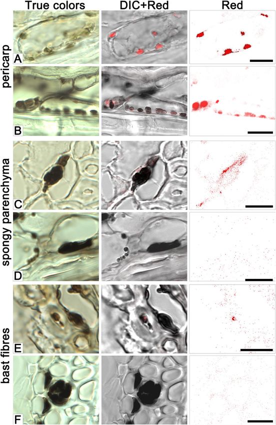
www.nature.com/scientificreports/ www.nature.com/scientificreports
Figure 1. Sample of the black pigment extracted from the husk and pericarp of the i:BwBlp NIL (A), FT-IR
spectrum of the pigment (B) and its expanded fingerprint region (C).
Results
Chemical analysis of the pigment in the i:BwBlp grain. The pigment material purified from the i:B-
wBlp NIL’s grain was a dark, glossy powder (Fig. 1A), which was insoluble in either water or any of the organic
solvents, except for hydroxymethylformamide. It was also partially soluble in 76% H2SO4, and fully soluble in
0.125 M NaOH (Supplementary Table S1). When exposed to either H2O2 or KMnO4, the pigment lost its color,
while exposure to FeCl3 resulted in the precipitation of a flocculent material which gradually redissolved when
the concentration of FeCl3 was raised. The behavior of the material was consistent with the presence of quinoid
and phenolic compounds. The FT-IR analysis revealed a profile characteristic of melanin (Fig. 1B,C): the broad
absorption band in the frequency range 3,200–3,600 cm−1 results from the stretching vibrations of the -OH and/
or -NH of amide, amine, carboxyl, phenolic or aromatic amino groups in the indole and pyrrole moieties; those in
the range 1,200–1,240 cm−1 are induced by vibrations of phenol C-O-H groups; those in the ranges 2,850–2,970
and 1,400–1,470 cm−1 are related to the stretching and deformation vibrations of aliphatic CH groups; those
in the range 1,640–1,650 cm−1 are attributed to C = O quinone vibrations, the one at ~1,509 cm−1 reflects the
stretching vibrations of an aromatic C-C bond; those in the range 1,000–1,075 cm−1 are diagnostic of either pri-
mary alcohol groups or the С-О-С-bonds present in aromatic ethers.
Development of the pigmentation during grain filling. By 42 days after sowing, the leading spike of
both NILs had emerged fully from the boot, and physiological maturity was reached by 67 days. The black pig-
mentation first appeared in the grain of the i:BwBlp NIL at the late milk or the early dough stage, beginning at the
tip of the spike, then spreading downwards to the base (Supplementary Fig. S1). The pigmentation developed in
an uneven manner (Supplementary Fig. S2). It first appeared both as a dark spot in the center of the dorsal side
of the grain and as stripes on the palea. By the early dough stage, the pigment began to form under the lemma, by
the later dough stage, it increasingly covered the lemmas and paleas at the tip of spike, and finally by the fully ripe
stage whole spike become black. Besides the grain, black pigmentation developed in awns first unevenly appeared
at the early dough stage and become clearly visible at the hard dough stage (Supplementary Fig. S1).
The microscopic structure of the developing grains. Grains were sampled for sectioning at various
developmental stages starting from the late milk stage (Fig. 2). Grains of cv. Bowman sampled at this stage exhib-
ited a high level of red autofluorescence in the spongy parenchyma of the husk, pericarp and aleurone (layer
nomenclature is presented in Supplementary Fig. S3), consistent with the presence of chloroplasts in these tissues
(Fig. 2A). In the i:BwBlp NIL at the early dough stage (the later one than studied in Bowman), the brown pigment
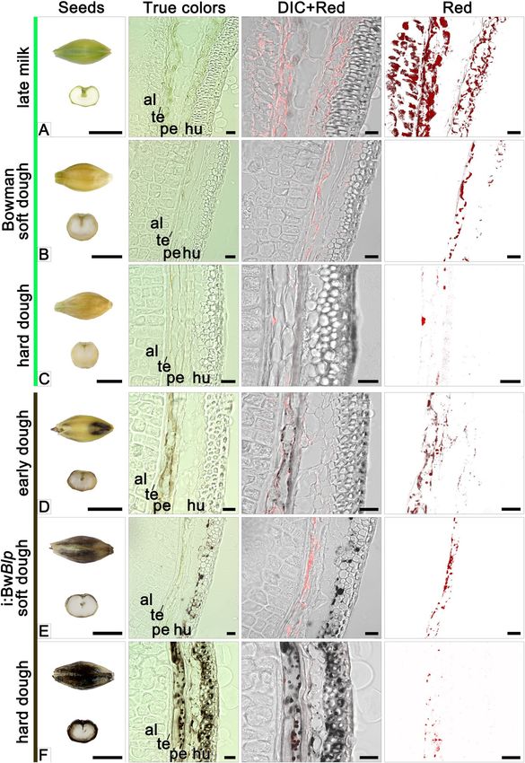
was seen exclusively in the pericarp, coinciding with the location of red autofluorescence (Figs. 2D and 3A).
Inspection of pericarp peeled from the grain of i:BwBlp revealed that the brown color was associated with plasids
which resembled the chloroplasts seen in the pericarp of cv. Bowman (Fig. 4A,C). Very little or no red autoflu-
orescence was emitted from the pigmented areas of the i:BwBlp grain. A chloroplast isolation kit was used to
prepare intact plastids from the pericarp tissue of both cv. Bowman and the i:BwBlp NIL (Fig. 4B,D): these were
colored brown in the latter. Brown cell inclusions were observed in spongy parenchyma and bast fibres, coincid-
ing, in a few examples, with the location of red autofluorescence (Fig. 3C,E).
Scientific Reports | (2020) 10:179 | https://doi.org/10.1038/s41598-019-56982-y 2
www.nature.com/scientificreports/                                                              www.nature.com/scientificreports

                           Figure 2. Cross-sections of grain set by cv. Bowman and the i:BwBlp NIL sampled at the late milk, early dough,
                           soft dough and hard dough stages. The images shown illustrate true colors, DIC+ red autofluorescence and red
                           autofluorescence. Scale bar for images of whole grains: 5 mm; and for micrographs: 20 um. Al: aleurone, hu:
                           husk, pe: pericarp; te: testa.

                               In grain sampled at the soft dough stage, the red autofluorescence emitted by pericarp tissue obtained from the
                           both NILs was less intense than in grains harvested at earlier development stages, and was barely detected in the
                           husk (Fig. 2B,E). The i:BwBlp NIL exhibited a marked irregularity in distribution of the brown pigment through
                           the pericarp and husk tissues. In some cases, the pigments were simultaneously detected in plastids of pericarp
                           cells and in spongy parenchyma cells, in a form of structurelles inclusions; in other grain sections, the structure-
                           less inclusions were observed in spongy parenchyma cells only (Fig. 2E); and there were grain sections with no
                           evidence of pigmented structures as well (not shown).
                               By the time of the hard dough stage, the decreased red autofluorescence was recorded in pericarp of the both
                           NILs (Fig. 2C,F). By this time, the grains set by cv. Bowman were almost colorless, with single plastids present in

Scientific Reports |   (2020) 10:179 | https://doi.org/10.1038/s41598-019-56982-y                                                            3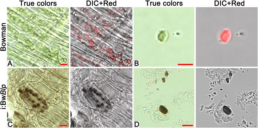
www.nature.com/scientificreports/                                                            www.nature.com/scientificreports

                           Figure 3. The accumulation of pigment in the pericarp (A, B), spongy parenchyma (C,D), and bast fibres (E,F)
                           cells of the iBwBlp NIL’s husk. Samples taken at the early (A,C,E) and hard (B,D,F) dough stages. The images
                           shown illustrate true colors, DIC + red autofluorescence and red autofluorescence. Scale bar: 10 um.

                           the pericarp; meanwhile the grains set by the i:BwBlp NIL had retained a population of the brown plastids in both
                           the pericarp and the spongy parenchyma (Figs. 2F and 3B), where the structureless inlcusions were observed too
                           (Fig. 3D). The brown pigment was present as structureless inclusions in bast fibres cells (Fig. 3F).

                           Ultrastructure of plastids. Ultrastructure of plastids in pericarp cells of grain was studied by transmission
                           electron microscopy (TEM) (Fig. 5). In grains of cv. Bowman sampled at the soft dough stage, part of the plas-
                           tids have normal chloroplast internal structure with thylakoid stacks without any visible signs of degradation

Scientific Reports |   (2020) 10:179 | https://doi.org/10.1038/s41598-019-56982-y                                                         4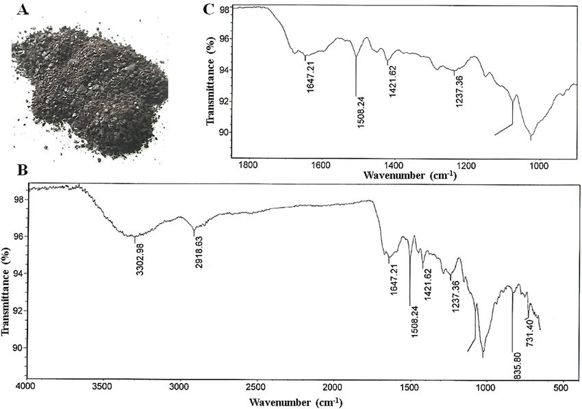
www.nature.com/scientificreports/                                                              www.nature.com/scientificreports

                           Figure 4. Microscopic analysis of pericarp peeled from cv. Bowman and the i:BwBlp NIL, scale bar: 10 um, and
                           isolated plastids (B,D), scale bar: 5 um. The images shown illustrate true colors, DIC + red autofluorescence.

                           Figure 5. Ultrastructure of plastids in pericarp cells of cv. Bowman (A–C) and the i:BwBlp NIL (D–F) at the
                           soft (A–D) and hard (E,F) dough development stages. Arrowheads show plastoglobuli. Scale bar: 5 um.

                           (Fig. 5A); meanwhile the majority of the plastids lose their internal structure and are assumed to be degraded
                           (Fig. 5B,C). By the hard dough stage, cells of the pericarp almost completely lost their internal structure and orga-
                           nelles, which could be identify as plastids were not observed.

Scientific Reports |   (2020) 10:179 | https://doi.org/10.1038/s41598-019-56982-y                                                             5
www.nature.com/scientificreports/ www.nature.com/scientificreports
In grains of the i:BwBlp NIL harvested at the soft dough stage, all observed plastids had normal chloroplast
internal structure without visible degradation signs (Fig. 5D) which become apparent later, at the hard dough
stage, when the internal membrane was organized in circle-shaped structure or was not detected at all (Fig. 5E,F).
The outer membrane of the plastids persisted up to the hard dough stage, the plastid borders were obvious, and
content of the plastids did not leave the organelles (Fig. 5F).
The osmiophillic particles plastoglobuli (PGs) were observed in plastids of the both NILs starting from the
soft dough stage (Fig. 5). In Bowman, PGs were distributed randomly within plastids, without any tendency to
aggregation, and had disappeared by the hard dough stage (Fig. 5A–C). In the i:BwBlp NIL, the PGs were also
distributed randomly at the soft dough stage, while they showed tendency to aggregation at the hard dough stage,
after internal membrane disruption (Fig. 5E,F).
Discussion
Melanins are synthesized by a wide range of organisms, which underlines their significance in determining evo-
lutionary success. Despite some heterogeneity in the identity of their precursors, the end products resemble one
another markedly with respect to not only their chemistry but also the pathways utilized for their synthesis.
Melanins are insoluble in most organic solvents, but dissolve readily in an alkaline medium and are discolored
by strong oxidizing agents; these hallmark properties have been used to devise a series of chemical tests for diag-
nostic use when encountering an unknown pigment1,20,21. These tests represented therefore the point of departure
for characterizing the dark pigment purified from the husk and pericarp of the barley NIL carrying the gene Blp
which is responsible for the formation of a black spike. The positive identification was supported by the outcome
of an FT-IR spectroscopy analysis, which revealed the presence of phenolic fragments, quinone and an aromatic
carbon backbone characteristic of melanin22–25.
When sectioned immature grains harvested from the i:BwBlp NIL were compared with those harvested from
cv. Bowman, it was determined that the melanin was synthesized in the pericarp within plastids exhibiting strong
red autofluorescence typical for chloroplasts26. During grain ripening the red autofluorescence observed by light
microscopy was drastically decreased in both lines. Decreasing of the red autofluorescence was coincided with
the degradation of the plastid internal structure, that was clearly observed by TEM in pericarp of Bowman and
the i:BwBlp NIL at the soft and hard dough stages, respectively. Besides pericarp tissue, melanin was observed in
the cells of the husk, where it was mostly present as structureless inclusion. The red autofluorescence in spongy
parenchyma and bast fibres cells noted at the early dough stage only may indicate that the melanin in these husk
tissues had initially formed in chlorophyll-containing plastids as was clearly the case in the pericarp. Then, proba-
bly after plastid degeneration during grain ripening the pigment released into cytoplasm and formed structureless
melanin fusion.
Supporting the notion that melanin synthesis in the black spike barley was initiated in chloroplasts is the
evidence that both the key melanogenesis enzyme PPO and the phenolic melanin substrates of PPO are present
in the chloroplast14,27,28. PPOs (also referred to variously as tyrosinases, polyphenolases, phenolases, catechol
oxidases, cresolases or catecholases) are ubiquitous in living matter. With the molluscan and arthropod oxygen
carrier proteins hemocyanins PPOs belong to the type-3 copper protein family which features a binuclear active
site composed of two copper atoms, each of which is coordinated by three conserved histidine residues29. They
are thought to have evolved in response to the photosynthesis-induced shift in the atmosphere from a reducing
to an oxidizing environment30. Although other classes of enzymes, notably laccases, peroxidases and polyketide
synthases are able to oxidize phenols and thus initiate the synthesis of melanin, the PPOs are typically responsible
for this process3. In plants, PPOs have been implicated in the browning response of damaged tissue, but their role
in intact tissue is uncertain27. Some evidence has been presented to suggest that they participate in the synthesis
of dark pigments in the rice husk31, but these particular pigments have not as yet been shown to be melanin.
Comparative TEM analysis of pericarp tissue of grains harvested at the early and hard dough stages showed
accumulation of PGs in the both NILs which in addition to observed chloroplasts breakdown, represent charac-
teristic feature of senescence gerontoplasts32. In the i:BwBlp NIL, aggregated PGs persisted in plastids up to the
hard dough stage, when in Bowman, neither PGs nor any plastids were detected. PGs represent lipoprotein par-
ticles surrounded by a membrane lipid monolayer with multiple functions in plastid metabolism, developmental
transition and environmental adaptation33. PGs are characterized by their own proteome and metabolome which
are different in distinct plastid types. They have striking dynamic nature and may vary substantially in form and
size in response to abiotic stress or development transition33. The observed differences in internal structure and
PGs dynamics between the NILs may be attributed to distinct metabolic processes inside these compartments
leading to melanin accumulation in the case of i:BwBlp, and to chloroplasts dismantling in the case of Bowman.
Comparative transcriptome analysis of the husk and pericarp tissues of the same NILs demonstrated differential
expression of more than a thousand genes with roles in the phenylpropanoid and fatty acid biosynthesis path-
ways were among the most represented and upregulated in the i:BwBlp NIL34 supported indirectly the metabolic
differences assumed between the NILs. For the new type of plastids which are assumed to accumulate melanin in
PGs and to be different from well-known senescent gerontoplasts the special term “melanoplasts” is suggested.
Although the relevant underling mechanism of melanogenesis in plant remains obscure the data obtained
here showed its intracellular accumulation within a membrane-delimited organelle, which is also the case in a
number of other organisms. In mammalian melanocytes, synthesis takes place within a melanosome3, while in
insects, melanin synthesis is associated with the formation of the cuticle35. Specialized hemocytes able to synthe-
size melanin in response to immune challenge have been reported in the larvae of both fruitfly36 and the mosquito
species Aedes aegypti37 and Armigeres subalbatus38. In fungi, melanin formation has been generally associated
with the cell wall, although the initial stages of its synthesis appear to be carried out in vesicles akin to mamma-
lian melanosomes before the product is transported to the cell wall39. In the Aspergillus species A. fumigatus and
A. nidulas, the enzymes involved in the initial stages of melanin synthesis are recruited by endosomes, whereas
Scientific Reports | (2020) 10:179 | https://doi.org/10.1038/s41598-019-56982-y 6www.nature.com/scientificreports/ www.nature.com/scientificreports
those involved in the later stages are active at the cell wall40. As the synthesis of melanin involves the formation
of cytotoxic intermediates (quinones), there is an evolutionary advantage in compartmentalizing the process. In
the barley plant, melanin appeared to be formed within senescing plastids in pericarp and husk tissues, so that
any harmful effects exerted by quinones on the plastids’ photosynthetic activity would have had only a minimal
impact on the plant’s overall photosynthesis. Further studies are required to clarify how melanogenesis in plants
is related to photosynthesis.
Methods
Plant material. Grain of the pair of NILs used for the analysis were obtained from the Nordic Gene Bank
(www.nordgen.org). The two lines were cv. Bowman (NGB22812) and the Blp carrier i:BwBlp (NGB20470).
Plants were raised in a greenhouse at Novosibirsk (Russia) providing a 12 h photoperiod and a temperature range
of 20–25 °C.
Extraction and characterization of spike pigment. For pigment extraction 300 g of the i:BwBlp NIL’s
grains were immersed for 2–4 h in cool water, after which the husk and pericarp were detached using a scalpel
and immersed for 48 h at room temperature in 1.5 L 0.5 M NaOH with constant stirring. The resulting pigmented
solution was filtered through cotton wadding and then through filter paper under vacuum. The pH of the filtrate
was reduced to 2.0 by the addition of concentrated HCl. The black precipitate which formed as a result was rinsed
in distilled water and centrifuged (3000 rpm for 20 min). The pellet was subsequently dissolved in a small volume
of 0.01 M NaOH and re-precipitated with HCl, a procedure which was repeated three times. Finally, the precip-
itate was rinsed with distilled water, dried at 20 °C and ground to a powder in a mortar. The presence of quinoid
and phenolic groups in the precipitated material was tested by its reaction with various oxidizing agents, namely
H2O241, KMnO442 and FeCl343. For the first of these reactions, an aliquot of 0.05% w/v of the powder dissolved in
0.1 M NaOH was combined with an equal volume of 10% H2O2 and left for 24 h; for the second, the 0.1 M NaOH
was replaced by 0.1 M KMnO4, while for the third, 0.5–1.0 mg/mL FeCl3 was added to 0.01% w/v of the powder
dissolved in 0.1 M NaOH. The solubility of the material was tested in a range of organic solvents (ethanol, isopro-
panol, hexane, petroleum ether, ethyl acetate, hydroxymethylformamide), in water, in concentrated (76%) H2SO4
and in 0.125 M NaOH. Fourier transform infrared (FT-IR) spectroscopy was carried out in KBr pellets using a
NicoletTM 6700 FT-IR device (Thermo Fisher Scientific, Waltham, MA, USA) set to the range 4,000–400 cm−1.
The resulting absorption peaks were interpreted as follows: 3,303 cm−1: -O(N)-H25; 2,921 cm−1 and 1,421 cm−1:
-CH-44,45; 1,648 cm−1: C = C conjugated with C = O22,46; 1,509 cm−1: Car = Car (conjugated carbons in the aro-
matic ring)23; 1,240 cm−1 and 1,020 cm−1: Car-O-R(H)24; 1,020 cm−1: C-OH or C-O-C16,45,47.
Cryosectioning and microscopy. Three grains were sampled at late milk (growth stage 77 based on the
BBCH-scale, BBCH-77), early dough (BBCH-83), soft dough (BBCH-85) and hard dough (BBCH-87) devel-
opmental stages from each of the two NILs, snap-frozen in liquid nitrogen and stored at −70 °C until required.
Prior to sectioning, the frozen grains were held at −20 °C for 30 min, mounted and embedded in Tissue-Tek
O.C.T.TM compound (Sakura Finetek Europe B.V., Alphen aan den Rijn, the Netherlands). Sectioning was car-
ried out at −20 °C using an HM 505 N cryostat microtome (Microm, Walldorf, Germany). Sections of thickness
15 um were mounted on a poly-L-lysine slide (Thermo Fisher Scientific) and fixed for 15 min in 8% formaldehyde
(Sigma-Aldrich, St. Louis, MO, USA) dissolved in phosphate buffered saline (pH 7.4). The slides were then rinsed
twice for 15 min in distilled water, mounted in glycerol and observed under microscope. Pericarp samples were
peeled from grains of both NILs at the soft dough stage, fixed in the same way as the sections, mounted on a glass
slide and observed under microscope. Chloroplasts were isolated from pericarp peels using a MinuteTM chloro-
plast isolation kit (Invent Biotechnologies, Inc., Plymouth, MN, USA). The suspended chloroplasts were mounted
on a glass slide and observed under microscope. Confocal laser scanning microscopy was achieved using an LSM
780 device (Zeiss, Oberkochen, Germany). Red autofluorescence was excited with a 633 nm laser. True colors
were captured from sections using an AxioCam HRc camera (Zeiss).
Ultrastructural analysis. Two grains per each NIL were sampled at the soft and hard dough stages.
Tissue fragments with pericarp were cut into pieces of 2–5 mm and fixed with 2.5% ice-cold glutaraldehyde
(Sigma-Aldrich, Germany) in phosphate buffer (pH 7.2) for 4 h. Then the material was washed three times for
15 min with phosphate buffer followed by postfixation with 1% osmium tetroxide (Azutite, Russia) for 4 h at a
room temperature, washed with phosphate buffer three times for 15 min, and dehydrated with ethanol solutions
of increasing concentrations. The samples were placed into acetone for 1 h and embedded into araldite epoxy
resin (Fluka, Switzerland). Ultrathin sections with a thickness of about 80 nm were made using an Ultracut UCT
(Leica, Switzerland) ultramicrotome and stained with lead citrate and uranyl acetate. The stained sections were
examined using a Jeol JEM-1400 (Japan) transmission electron microscope at an accelerating voltage of 80 kV.
Conclusion
The major finding from this investigation was that in the pigmented barley spike, melanin is synthesized in
chlorophyll-containing plastids in the grain pericarp; it represents, to the best of our knowledge, the first obser-
vation of intracellular melanin production in a plant. The discovery raises questions regarding the commonality
surrounding how and where melanin is synthesized, most particularly the identity of the key enzymes underlying
the process and the compartmentalization of the process, which would imply an ancient and perhaps monophyl-
etic origin of the cellular machinery involved.
Data availability
The extracted melanin samples are available from the corresponding author on reasonable request.
Scientific Reports | (2020) 10:179 | https://doi.org/10.1038/s41598-019-56982-y 7www.nature.com/scientificreports/ www.nature.com/scientificreports
Received: 6 August 2019; Accepted: 19 December 2019;
Published: xx xx xxxx
References
1. Nicolaus, R. A., Piattelli, M. & Fattorusso, E. The structure of melanins and melanogenesis-IV. On some nature of melanins.
Tetrahedron. 20, 1163–1172 (1964).
2. Britton, G. 1983. Biohimija prirodnyh pigmentov [The biochemistry of natural pigments, Cambridge university press, translated
from English] (russ. ed. Zaprometov, M.N.) 259-279 (Mir, 1986).
3. Solano, F. Melanins: skin pigments and much more – types, structural, models, biological functions, and formation routes. New J.
Sci. 2014, 498276 (2014).
4. Gerdemann, C., Eicken, C. & Krebs, B. The crystal structure of catechol oxidase: new insight into the function of type-3 copper
proteins. Acc. Chem. Res. 35, 183–191 (2002).
5. Langfelder, K., Streibel, M., Jahn, B., Haase, G. & Brakhage, A. A. Biosynthesis of fungal melanins and their importance for human
pathogenic fungi. Fungal Genet. Biol. 38, 143–158 (2003).
6. Prota, G. Progress in the chemistry of melanins and related metabolites. Med. Res. Rev. 8, 525–556 (1988).
7. Varga, M., Berkesi, O., Darula, Z., May, N. V. & Palágyi, A. Structural characterization of allomelanin from black oat. Phytochemistry.
130, 313–320 (2016).
8. Rogers, C. E. & Kreitner, G. L. Phytomelanin of sunflower achenes: a mechanism for pericarp resistance to abrasion by larvae of the
sunflower moth (Lepidoptera: Pyralidae). Environ. Entomol. 12, 277–285 (1983).
9. Duran, J. M. & Retamal, N. Coat structure and regulation of dormancy in Sinapis arvensis L. seeds. J. Plant Physiol. 135, 218–222
(1989).
10. Riley, P. Melanin. Int. J. Biochem. Cell Biol. 29, 1235–1239 (1997).
11. Downie, A. B. et al. Communication between the maternal testa and the embryo and/or endosperm affect testa attributes in tomato.
Plant Physiol. 133, 145–160 (2003).
12. Dahlgren, R. M. T. & Clifford, H. T. The monocotyledons: a comparative study (Academic Press, 1982).
13. Pandey, A. K., Stuessy, T. F. & Mathur, R. R. Phytomelanin and systematics of the Heliantheae Alliance (Compositae). Plant Div. Evol.
131/3, 145–165 (2014).
14. Nicolas, J. J., Richard-Forget, F. C., Goupy, P. M., Amiot, M. J. & Aubert, S. Y. Enzymatic browning reactions in apple and apple
products. Crit. Rev. Food Sci. Nutr. 34, 109–157 (1994).
15. Park, K. I. A bHLH protein partially controls proanthocyanidin and phytomelanin pigmentation in the seed coats of morning glory
Ipomoea tricolor Hort. Environ. Biotechnol. 53, 304–309 (2012).
16. Wang, L.-F. & Rhim, J.-W. Isolation and characterization of melanin from black garlic and sepia ink. LWT - Food Sci. Technol. 99,
17–23 (2019).
17. Harlan, H. V. Some distinctions in our cultivated barleys with reference to their use in plant breeding. US Dept. Agriculture. 137, 38
(1914).
18. Costa, J. M. et al. Molecular mapping of the Oregon Wolfe Barleys: a phenotypically polymorphic doubled-haploid population.
Theor. Appl. Genet. 103(2-3), 415–424 (2001).
19. Druka, A. et al. Genetic dissection of barley morphology and development. Plant Physiol. 155, 617–627 (2011).
20. Makordei, F. V., Venger, L. A., Slyusarenko, L. I. & Barba, I. N. Allomelanins. Isolation methods, physicochemical properties, and
possibilities of practical use. Izvestiya Vysshikh Uchebnykh Zavedenii. Khimiya i Khimicheskaya Tekhnologiya 37, 4–6 (1994).
21. Sava, V. M., Yang, S.-M., Hong, M.-Y., Yang, P.-C. & Huang, G. S. Isolation and characterization of melanic pigments derived from
tea and tea pholyphenols. Food Chem. 73, 177–184 (2001).
22. Booner, T. G. & Duncan, A. Infra-red spectra of some melanins. Nature. 194, 1078–1079 (1962).
23. Kazitsyna, L. A. & Kupletskaya, N. B. Primenenie UF, IK- i YaMR-spektposkopii v organicheskoy khimii [Application of UV, IR and
NMR spectroscopy in organic chemistry] 23–60, 235–257 In Russian (Vyssh. shk., 1971).
24. Bilinska, B. Progress of infrared investigations of melanin structures. Spectrochim. Acta Part A. 52, 1157–1162 (1996).
25. Magarelli, M., Passamonti, P. & Renieri, C. Purification, characterization and analysis of sepia melanin from commercial sepia ink
(Sepia officinalis). CES Medicina Veterinaria y. Zootecnia. 5, 18–28 (2010).
26. Krause, G. H. & Weis, E. Chlorophyll fluorescence and photosynthesis: the basics. Annu. Rev. Plant Physiol. Plant Mol. Biol. 42,
313–349 (1991).
27. Boeckx, T., Winters, A. L., Webb, K. J. & Kingston-Smith, A. H. Polyphenol oxidase in leaves: is there any significance to the
chloroplastic localization? J. Exp. Bot. 66, 3571–3579 (2015).
28. Boeckx, T., Winters, A., Webb, K. J. & Kingston-Smith, A. H. Detection of potential chloroplastic substrates for polyphenol oxidase
suggests a role in undamaged leaves. Front. Plant Sci. 8, 237 (2017).
29. Decker, H. et al. Similar enzyme activation and catalysis in hemocyanins and tyrosinases. Gene. 398, 183–191 (2007).
30. Decker, H. & Terwilliger, N. Cops and robbers: putative evolution of copper oxygen-binding proteins. J. Exp. Biol. 203, 1777–1782
(2000).
31. Fukuda, A. et al. Complementary genes that cause black ripening hulls in F1 plants of crosses between indica and japonica rice
cultivars. Plant Prod. Sci. 15, 270–273 (2012).
32. Wise, R. R. The diversity of plastid form and function in The structure and function of plastids (eds. Wise, R. R. & Hoober, J. K.) 3–26
(Springer, 2006).
33. van Wijk, K. J. & Kessler, F. Plastoglobuli: plastid microcompartments with integrated functions in metabolism, plastid
developmental transitions, and environmental adaptation. Annu. Rev. Plant Biol. 68, 253–289 (2017).
34. Glagoleva, A. et al. Metabolic pathways and genes identified by RNA-seq analysis of barley near-isogenic lines differing by allelic
state of the Black lemma and pericarp (Blp) gene. BMC Plant Biol. 17, 182 (2017).
35. Nappi, A. J. & Christensen, B. M. Melanogenesis and associated cytotoxic reactions: Applications to insect innate immunity. Insect
Biochem. Mol. Biol. 35, 443–459 (2005).
36. Rizki, T. M., Rizki, R. M. & Grell, E. H. A mutant affecting the crystal cells in Drosophila melanogaster. Wilehm Roux Arch. Dev. Biol.
188, 91–99 (1980).
37. Hillyer, J. F. & Christensen, B. M. Characterization of hemocytes from the yellow fever mosquito, Aedes aegypti. Histochem. Cell Biol.
117, 431–440 (2002).
38. Hillyer, J. F., Schmidt, S. L. & Christensen, B. M. Hemocyte-mediated phagocytosis and melanization in the mosquito Armigeres
subalbatus following immune challenge by bacteria. Cell Tissue Res. 313, 117–127 (2003).
39. Eisenman, H. C. & Casadevall, A. Synthesis and assembly of fungal melanin. Appl. Microbiol. Biotechnol. 93, 931–940 (2012).
40. Upadhyay, S. et al. Subcellular compartmentalization and trafficking of the biosynthetic machinery for fungal melanin. Cell Rep. 14,
2511–2518 (2016).
41. Lyakh, S. P. Microbial melanogenesis and its functions (Nauka, 1981).
42. Harki, E., Talou, T. & Dargent, R. Purification, characterization and analysis of melanin extracted from Tuber melanosporum Vitt.
Food Chem. 58, 69–73 (1997).
Scientific Reports | (2020) 10:179 | https://doi.org/10.1038/s41598-019-56982-y 8www.nature.com/scientificreports/ www.nature.com/scientificreports
43. Ruolin, H. et al. Characterization of the physicochemical properties and extraction optimization of natural melanin from Inonotus
hispidus mushroom. Food Chem. 277, 533–542 (2019).
44. Tarangini, K. & Mishra, S. Production, characterization and analysis of melanin from isolated marine Pseudomonas sp. using
vegetable waste. Res. J. Engineering Sci. 2, 40–46 (2013).
45. Mbonyiryivuze, A., Mwakikunga, B., Dhlamini, S. M. & Maaza, M. Fourier transform infrared spectroscopy for sepia melanin.
Physics Mater. Chem. 2, 25–29 (2015).
46. Bridelli, M. G., Tampellini, D. & Zecca, L. The structure of neuromelanin and its iron binding site studied by infrared spectroscopy.
FEBS Lett. 457, 18–22 (1999).
47. Paim, S., Linhares, L. F., Mangrich, A. S. & Martim, J. P. Characterization of fungal melanins and soil humic acids by chemical
analysis and infrared spectroscopy. Biol. Fertil Soils. 10, 72–76 (1990).
Acknowledgements
We thank Mrs. Kukoeva Tatjana for taking care about barley plants in greenhouse, the head of the Joint Access
Center for Microscopy of Biological Objects with the Siberian Branch of the Russian Academy of Sciences, Dr.
Sergey Bayborodin with the help in microscopy analysis, and Dr. Robert Koebner (www.smartenglish.co.uk) for
linguistic advice and valuable comments during the preparation of this manuscript. The study was supported by
the Russian Science Foundation № 16-14-00086. Growing of barley plants in ICG Plant Growth Core Facility was
supported by ICG project № 0324-2019-0039.
Author contributions
O.Y.S. designed and coordinated the study, participated in interpretation of the data, drafted the manuscript;
S.R.M. performed microscopy analysis, interpreted the data, participated in drafting the manuscript; N.V.G.
extracted and tested the pigments, analyzed FT-IR- absorption spectra, interpreted the data, participated in
drafting the manuscript; A.Y.G. observed the pigmentation formation, took photos of the developmental stages of
barley grain, peeled out the pericarp tissue for microscopy analysis and chloroplast isolation, isolated chloroplasts;
A.B. provided the near isogenic lines; E.K.K. initiated the study, revised the manuscript critically. All authors read
and approved the final manuscript.
Competing interests
The authors declare no competing interests.
Additional information
Supplementary information is available for this paper at https://doi.org/10.1038/s41598-019-56982-y.
Correspondence and requests for materials should be addressed to O.Y.S.
Reprints and permissions information is available at www.nature.com/reprints.
Publisher’s note Springer Nature remains neutral with regard to jurisdictional claims in published maps and
institutional affiliations.
Open Access This article is licensed under a Creative Commons Attribution 4.0 International
License, which permits use, sharing, adaptation, distribution and reproduction in any medium or
format, as long as you give appropriate credit to the original author(s) and the source, provide a link to the Cre-
ative Commons license, and indicate if changes were made. The images or other third party material in this
article are included in the article’s Creative Commons license, unless indicated otherwise in a credit line to the
material. If material is not included in the article’s Creative Commons license and your intended use is not per-
mitted by statutory regulation or exceeds the permitted use, you will need to obtain permission directly from the
copyright holder. To view a copy of this license, visit http://creativecommons.org/licenses/by/4.0/.
© The Author(s) 2020
Scientific Reports | (2020) 10:179 | https://doi.org/10.1038/s41598-019-56982-y 9You can also read