Melanoma Brain Metastasis Presentation, Treatment, and Outcomes in the Age of Targeted and Immunotherapies
←
→
Page content transcription
If your browser does not render page correctly, please read the page content below
Original Article
Melanoma Brain Metastasis Presentation, Treatment, and
Outcomes in the Age of Targeted and Immunotherapies
1,2
Evan D. Bander, MD ; Melissa Yuan, AB1; Joseph A. Carnevale, MD1,2; Anne S. Reiner, MPH 3
;
3 4,5 1 1
Katherine S. Panageas, PhD ; Michael A. Postow, MD ; Viviane Tabar, MD ; and Nelson S. Moss, MD
BACKGROUND: Historically, the prognosis for patients who have melanoma brain metastasis (MBM) has been dismal. However, break-
throughs in targeted and immunotherapies have improved long-term survival in those with advanced melanoma. Therefore, MBM pres-
entation, prognosis, and the use of multimodality central nervous system (CNS)-directed treatment were reassessed. METHODS: In
this retrospective study, the authors evaluated patients with MBM who received treatment at Memorial Sloan Kettering Cancer Center
between 2010 and 2019. Kaplan-Meier methodology was used to describe overall survival (OS). Recursive partitioning analysis and time-
dependent multivariable Cox modeling were used to assess prognostic variables and to associate CNS-directed treatments with OS.
RESULTS: Four hundred twenty-five patients with 2488 brain metastases were included. The median OS after an MBM diagnosis was
8.9 months (95% CI, 7.9-11.3 months). Patients who were diagnosed with MBM between 2015 and 2019 experienced longer OS compared
to those who were diagnosed between 2010 and 2014 (OS, 13.0 months [95% CI, 10.47-17.06 months] vs 7.0 months [95% CI, 6.1-8.3
months]; P = .0003). Prognostic multivariable modeling significantly associated shortened OS independently with leptomeningeal dis-
semination (P < .0001), increasing numbers of brain metastases at diagnosis (P < .0001), earlier MBM diagnosis year (P = .0008), higher
serum levels of lactate dehydrogenase (P < .0001), receipt of immunotherapy before MBM diagnosis (P = .003), and the presence of
extracranial disease (P = .02). The use of different CNS-directed treatment modalities was associated with presenting symptoms, diag-
nosis year, number and size of brain metastases, and the presence of extracranial disease. Multivariable analysis demonstrated improved
survival for patients who underwent craniotomy (P = .01). CONCLUSIONS: The prognosis for patients with MBM has improved within
the last 5 years, coinciding with the approval of PD-1 immune checkpoint blockade and combined BRAF/MEK targeting. Improving
survival reflects and may influence the willingness to use aggressive multimodality treatment for MBM. Cancer 2021;127:2062-2073.
© 2021 American Cancer Society.
LAY SUMMARY:
• Historically, melanoma brain metastases (MBM) have carried a poor survival prognosis of 4 to 6 months; however, the introduction of
immunotherapy and targeted precision medicines has altered the survival curve for advanced melanoma.
• In this large, single-institution, contemporary cohort, the authors demonstrate a significant increase in survival of patients with MBM to
13 months within the last 5 years of the study.
• A worse prognosis for patients with MBM was significantly associated with the number of metastases at diagnosis, previous exposure
to immunotherapy, spread of disease to the leptomeningeal compartment, serum lactate dehydrogenase elevation, and the presence of
extracranial disease.
• The current age of systemic treatments has also been accompanied by shifts in the use of central nervous system-directed therapies.
KEYWORDS: brain metastases, immunotherapy, melanoma, survival, targeted therapy.
INTRODUCTION
Melanoma is one of the primary causes of malignant metastases to the central nervous system (CNS), accounting
for 6% to 12% of all metastatic brain tumors.1-3 Survival after a diagnosis of melanoma brain metastasis (MBM)
has historically been dismal, with an overall survival (OS) of 4 to 6 months.4-8 However, over the recent decade,
numerous advances have been made in targeted therapy for melanoma, such as BRAF and MEK inhibition, and in
immunotherapy with approval of the checkpoint inhibitors ipilimumab, nivolumab, and pembrolizumab.9 These ad-
vances have resulted in significant improvements in the OS of patients with metastatic melanoma.10-18 Furthermore,
it has been demonstrated that patients with MBM also respond to these therapies.19-24 In the COMBI-MB trial
(ClinicalTrials.gov identifier NCT02039947), 58% of patients who had BRAF V600E-positive MBM responded
Corresponding Author: Nelson S. Moss, MD, Department of Neurosurgery and Brain Metastasis Center, Memorial Sloan Kettering Cancer Center, 1275 York Avenue, New
York, NY 10065 (mossn@mskcc.org).
1
Department of Neurosurgery and Brain Metastasis Center, Memorial Sloan Kettering Cancer Center, New York, New York; 2 Department of Neurosurgery, New York
Presbyterian Hospital/Weill Cornell Medical College, New York, New York; 3 Department of Epidemiology and Biostatistics, Memorial Sloan Kettering Cancer Center, New
York, New York; 4 Department of Medicine, Memorial Sloan Kettering Cancer Center, New York, New York; 5 Department of Medicine, Weill Cornell Medical College, New
York, New York
Additional supporting information may be found in the online version of this article.
DOI: 10.1002/cncr.33459, Received: May 29, 2020; Revised: December 17, 2020; Accepted: December 22, 2020, Published online March 2, 2021 in Wiley Online Library
(wileyonlinelibrary.com)
2062 Cancer June 15, 2021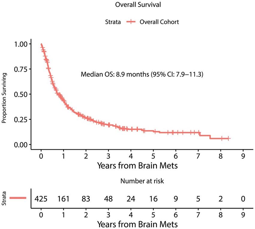
Contemporary Melanoma Brain Metastases/Bander et al
to combination dabrafenib and trametinib,23 and the immunotherapy, targeted BRAF/MEK therapy, stereo-
combination of nivolumab and ipilimumab produced tactic and/or whole brain radiation, and surgery, includ-
intracranial responses in 46% to 56% of patients with ing craniotomy and/or cerebrospinal fluid diversion);
MBM.22,24 Although clinical trials have begun to and the presence of progressive systemic and/or CNS
include more patients with MBM, little data exist to disease at the time of death (if known). Radiographic
assess how current treatments have changed the overall findings were based on radiologist interpretations of
prognosis of MBM diagnosis, affected central nervous magnetic resonance imaging and computed tomogra-
system (CNS)-directed local treatment algorithms with phy studies; these were further reviewed when quanti-
surgery and radiation, or enumerated factors that may tative or qualitative features of interest (size, number,
inform the survival of patients with MBM. Notably, location, hydrocephalus) were not commented upon.
the most recent iteration of the Graded Prognostic The presence of hemorrhage in metastases was based
Assessment tool called the Melanoma-molGPA demon- on radiology reports. Dominant metastasis was defined
strated the prognostic value of clinical features, includ- as the largest metastasis present on imaging, and size
ing age, Karnofsky performance status, the number of was determined based on greatest axial/coronal/sagittal
CNS metastases, the presence of extracranial metastases, dimension.
and BRAF status, in a cohort identified through 2015;
however, leptomeningeal disease (LMD) and the contri- Statistical Analysis
bution of immunotherapy were not investigated.25 This Descriptive statistics, such as frequencies, means, and
large, retrospective, single-institution study d escribes SDs, were used to characterize the cohort under study.
the presentation, treatments, and survival of patients OS was defined as the time from MBM diagnosis until
with MBM in the contemporary immunotherapy and the date of death or the date of last follow-up for patients
precision medicine era. who were censored. Recursive partitioning analysis was
used for exploration and visualization of empirically
identified cutoffs for associations of the number of
MATERIALS AND METHODS BMs, the MBM diagnosis year, and the size of largest
This study was approved by the Memorial Sloan MBM with OS. Univariable and multivariable Cox
Kettering Cancer Center (MSK) Institutional Review modeling was used to associate variables of interest with
Board. Patients (n = 440) were identified by an insti- OS. Variables that were significant in the univariable
tutional database search for all patients, regardless of models were brought forward for evaluation in the
treatments, with a diagnosis of cutaneous melanoma, multivariable analysis. LMD and all treatments after
no other systemic malignancy, and brain metastases a diagnosis of MBM were treated as time-dependent
(BMs) diagnosed from January 2010 through January variables in the Cox models. The time-dependent Cox
2019. Patients were excluded from the analysis if they models associating LMD with OS were stratified by
had primary LMD without parenchymal metastases at variables of interest, and heterogeneity was tested with
time of MBM diagnosis (n = 4) or if medical records nested models using the likelihood ratio test. Kaplan-
had incomplete clinical documentation for the param- Meier methodology was used to display survival curves.
eters enumerated below, represented a single encounter The cumulative incidence of LMD after a diagnosis of
without any follow-up, or were without baseline or fol- BM was calculated using competing risks methodology,
low up imaging (n = 11). A retrospective chart review and the Gray test was used to compare cumulative
was conducted to identify demographics, including age incidence curves stratified by pre-BM immunotherapy.
at diagnosis of MBM; the number, size, and location The Kruskal- Wallis test was used to investigate the
of BMs at diagnosis; CNS symptoms at diagnosis; the association between presenting MBM symptoms and
presence of metastasis- associated hemorrhage; serum the dominant size and number of BMs. The Fisher
lactate dehydrogenase (LDH) level at MBM diagno- test was used to explore the association of presenting
sis; the presence or absence of extracranial disease on MBM symptoms with dominant MBM location. The
the computed tomography scan of the chest, abdomen, Wilcoxon 2- sample test was used to investigate the
and/or pelvis most contemporaneous to the time of BM association between pre-BM diagnosis immunotherapy
diagnosis; diagnosis of LMD and/or hydrocephalus dur- and the number of BMs at diagnosis. A cause-specific,
ing treatment; OS; systemic and CNS-directed treat- time-dependent Cox model was used to model the
ments before and after BM diagnosis (chemotherapy, association of variables of interest with post- MBM
Cancer June 15, 2021 2063Original Article
treatments. All P values were 2-sided, with a level of TABLE 1. Melanoma Brain Metastasis Cohort
statistical significance 50.0
Institute Inc) and R version 3.6.0 (R Foundation for Dominant BM size—continuous, cm 425 (100)
Statistical Computing). Mean 2.1
Median 1.8
Range 0.2-8.8
RESULTS Serum LDH value (U/L)—continuous 233 (55)
Mean 389.1
Demographics, Survival, and BM Presentation Median 221
Range 110.0-4970.0
Four hundred twenty-five patients were diagnosed with Sex
a total of 2488 MBMs at MSK between 2010 and 2019 Women 121 (28)
(Table 1). The mean patient age was 59.3 years, and there Men 304 (72)
BRAF status
was a male predominance (men, 72%; women, 28%). Wild type 184 (43)
There were 324 deaths over the study duration. The Mutated 206 (49)
Unknown 35 (8)
median OS from the diagnosis of BMs was 8.9 months Systemic burden
(95% CI, 7.9-11.3 months) (Fig. 1). The median fol- No extracranial disease 42 (10)
Extracranial disease present 372 (88)
low-up was 22.5 months for survivors. The 3-year OS Unknown 11 (3)
rate for the cohort was 19.4% (95% CI, 15.5%-24.1%), Presenting symptoms
Asymptomatic 166 (39)
and the 5-year OS rate was 13.6% (95% CI, 10.0%- Headache 72 (17)
18.6%). Forty-nine percent of patients (n = 206) had Motor/sensory 83 (20)
Seizure 34 (8)
a BRAF mutation identified by immunohistochemistry, Mental status change 56 (13)
mass spectrometry, and/or targeted sequencing, whereas Other 14 (3)
43% had wild-type BRAF, and 8% had unknown BRAF Serum LDH (U/L)
Within normal limits 134 (32)
status. Eighty-eight percent of patients had extracranial Above normal limits 99 (23)
disease present at MBM diagnosis, 10% had BMs only Unknown 192 (45)
Hemorrhage present in BM at diagnosis
without evidence of extracranial disease, and 2% had No 177 (42)
unknown BM status. The median number of paren- Yes 248 (58)
Hemorrhage present in BM at last follow-up
chymal metastases at BM diagnosis was 3 (interquar- No 119 (28)
tile range, 1-6 parenchymal metastases; range, from 1 Yes 306 (72)
Dominant BM location
to >50 parenchymal metastases). In 90% of patients, Frontal 162 (38)
the dominant/largest BM was located in the supraten- Temporal 62 (15)
Parietal 80 (19)
torial compartment compared with the infratentorial
Occipital 43 (10)
compartment in 10%. The median size of the dominant Cerebellar/pontine 44 (10)
BM was 1.8 cm (interquartile range, 0.9-2.9 cm; range, Subcortical 34 (8)
Dominant BM supratentorial/infratentorial
0.2-8.8 cm). Fifty-eight percent of patients had radio- Supratentorial 381 (90)
graphic hemorrhage present at BM diagnosis, and 72% Infratentorial 44 (10)
Hydrocephalus
had hemorrhage present by the last follow-up. Serum No 382 (90)
LDH levels at the time of MBM diagnosis were above Yes 43 (10)
Cumulative incidence of LMD [95% CI], %
normal limits in 23%, within normal limits for 32%, At 1 y 12.3 [9.1-15.5]
and unknown for 45% of patients. Supporting Figure 1 At 3 y 15.33
[11.7-18.9]
illustrates the cumulative incidence of LMD diagnosis
2064 Cancer June 15, 2021Contemporary Melanoma Brain Metastases/Bander et al
TABLE 1. Continued (95% CI, 11.7%-18.9%) for patients with MBM at 3
No. of
years.
Variable Patients (%) Most patients were asymptomatic at the time of
Pre-BM immunotherapy BM diagnosis (39%), whereas 20% had focal motor or
No 226 (53) sensory complaints. Seizure was the initial presenting
Yes 199 (47)
Pre-BM BRAF-targeted therapy symptom in 8% of patients, and 33% presented with
No 366 (86) headache, mental status change, or other neurologic
Yes 59 (14)
Post-BM immunotherapy
complaint without focal deficit or seizure. Presenting
No 97 symptoms differed significantly in relation to dominant
Yes 326 (77)
Unknown 2 (0)
metastasis size (see Supporting Table 1). Asymptomatic
Post-BM BRAF-targeted therapy patients had a median dominant BM size of 1.0 cm ver-
No 317 (75) sus 3.1 cm for patients who presented with headache, 2.2
Yes 108 (25)
Surgery cm for those who presented with motor/sensory deficit,
None 260 (61) and 2.6 cm for those who presented with seizure (P <
Shunt 7 (2)
Craniotomy 147 (35) .0001). The dominant BM location was also significantly
Both 9 (2) associated with presenting symptoms (P = .01) (see
Radiation
None 92 (22)
Supporting Table 2). Headache was the most common
SRS 182 (43) presentation for patients with cerebellar/pontine BMs,
WBRT 103 (24)
Both 48 (11)
accounting for 34% of those cases. Seizures and motor/
sensory deficits occurred more frequently in patients who
Abbreviations: BM, brain metastasis; LDH, lactate dehydrogenase; LMD,
leptomeningeal disease; Max, maximum; Min, minimum; SRS, stereotactic
had frontal and parietal BMs compared with those who
radiosurgery; WBRT, whole-brain radiation therapy. had BMs in other locations. The number of BMs present
at diagnosis was not significantly associated with present-
ing symptom.
Prognostic Factors
Recursive partitioning analysis was used to explore the
cutoff point associated most with OS for each of the
following variables individually: number of MBM at di-
agnosis, year of MBM diagnosis, and size of the largest
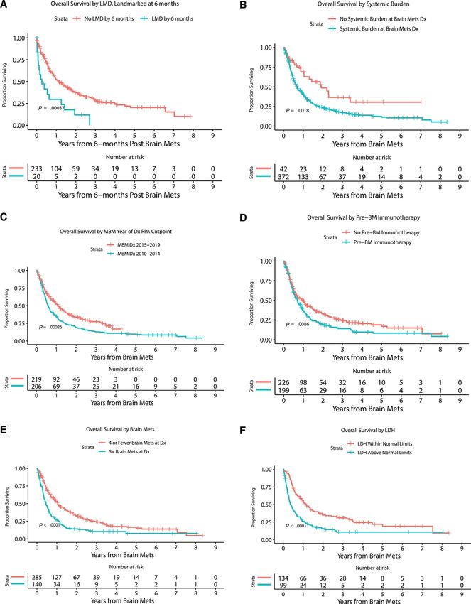
MBM (Fig. 2). The analysis demonstrated thatOriginal Article Figure 2. Kaplan-Meier estimates of overall survival are illustrated for patients (A) with or without leptomeningeal disease (LMD) 6 months after a melanoma brain metastases (MBM) diagnosis (Dx), (B) with or without extracranial systemic burden, (C) who had an MBM Dx between 2010 and 2014 or between 2015 and 2019, (D) who did or did not receive immunotherapy before MBM diagnosis, (E) with
Contemporary Melanoma Brain Metastases/Bander et al
TABLE 2. Factors Associated With Overall Survival in Patients With Metastatic Brain Metastasis
Unadjusted Adjusteda
Variable No. of Patients (%) HR [95% CI] P HR [95% CI] P
Age at MBM Dx—continuous 425 (100) 1.007 [0.999-1.014] .08
Dominant BM size—continuous, 425 (100) 0.99 [0.91-1.08] .84
cm
Year of MBM Dx—continuous 425 (100) 0.92 [0.87-0.96] .0004 0.92 [0.87-0.97] .0008
Serum LDH
Within normal limits 134 (32) Ref Ref
Above normal limits 99 (23) 2.14 [1.59-2.87]Original Article
BM diagnosis (HR, 0.98; 95% CI, 0.96-0.997; P = .02) P = .002). For each centimeter increase in dominant BM
retained a significant association in multivariable analy- size, patients were 72% more likely to receive a shunt and
sis; patients who received whole-brain radiation therapy 38% more likely to undergo craniotomy. Dominant BM
(WBRT) before they were diagnosed with LMD were size was not associated with the likelihood of ultimately
more likely to have an LMD diagnosis (HR, 3.02; 95% receiving SRS or WBRT. The presence of hemorrhage at
CI, 1.71-5.33; P = .0001). We note that undergoing cra- BM diagnosis was also significantly associated with an
niotomy (HR, 0.95; 95% CI, 0.54-1.68; P = .86) and the increased likelihood of undergoing craniotomy as first
number of BMs (HR, 1.01; 95% CI, 0.98-1.04; P = .58) treatment on multivariable analysis (HR, 1.68; 95% CI,
were not significantly associated with an LMD diagnosis. 1.09-2.58; P = .02). The presence of extracranial disease
was associated with a decreased likelihood of craniotomy
Treatments as first treatment (HR, 0.43; 95% CI, 0.28-0.68; P =
Before MBM diagnosis, 199 patients (47%) had received .001). Receipt of immunotherapy before BM diagnosis
immunotherapy, and 59 patients (14% of the total co- was associated with an increased likelihood of SRS as first
hort; 29% of patients with BRAF mutations) had received treatment (HR, 1.74; 95% CI, 1.22-2.46; P = .002). Age
BRAF-targeted therapy. By the time of the last follow-up, at BM diagnosis was not significantly associated with any
326 patients (77%) had ever received immunotherapy, of the CNS-directed treatment modalities. Year of BM
and 108 (25% of the total cohort; 52% of patients with diagnosis demonstrated a significant association with
BRAF mutations) had received BRAF-directed therapy. WBRT (HR, 0.79; 95% CI, 0.73-0.86; P < .0001): with
These treatments were not evaluated in a time-dependent each subsequent year, the likelihood of receiving WBRT
manner and thus do not fully reflect the at-risk popula- decreased by 21%.
tion. After a diagnosis of MBM, 39% of patients under- Table 4 provides details of the associations of local
went surgery (craniotomy, ventriculoperitoneal shunt, or treatment modalities, performed at any time during the
both), and 78% underwent either stereotactic radiosur- disease course, with OS. In multivariable analysis, all
gery (SRS), WBRT, or both for the treatment of MBM factors that were identified as significant in univariable
(Table 1; see Supporting Fig. 2). Each type of local ther- analysis retained significance, except for SRS. Patients
apy was examined as a first local/CNS treatment in rela- who underwent craniotomy experienced improved
tion to age, sex, year of BM diagnosis, extracranial disease, survival compared with those who did not (HR, 0.72;
pre-BM immunotherapy, the number of BMs, dominant 95% CI, 0.56-0.93; P = .01). Patients who underwent
BM size, the presence of hemorrhage at BM diagnosis, a shunt procedure (HR, 4.24; 95% CI, 2.48-7.24; P <
and presenting symptoms (Table 3). .0001) and WBRT (HR, 2.65; 95% CI, 2.08-3.38; P
In multivariable analysis, patients who presented < .0001) experienced shorter survival than those who
with headache (HR, 3.69; 95% CI, 2.05- 6.64; P < did not undergo one of these treatments. Although SRS
.0001), motor/sensory deficits (HR, 1.93; 95% CI, was associated with improved survival in univariable
1.03-3.60; P = .04), seizure (HR, 3.23; 95% CI, 1.52- analysis (HR, 0.59; 95% CI, 0.47-0.74; P < .0001),
6.83; P = .002), or mental status change (HR, 3.65; it did not maintain that association when adjusted in
95% CI, 1.94-6.85; P < .0001) were significantly more multivariable analysis (HR, 0.87; 95% CI, 0.68-1.13;
likely to undergo craniotomy as their first treatment P = .30).
compared with those who presented asymptomatically.
Symptomatic presentation was not significantly asso-
ciated with any other treatment modality. Fewer BMs, DISCUSSION
with quantity evaluated as a continuous variable, were In this large, retrospective evaluation of contemporary
significantly associated with craniotomy (HR, 0.87; 95% multimodality management of patients with MBM at a
CI, 0.81-0.93; P ≤ .0001) and SRS (HR, 0.97; 95% CI, large referral cancer center who were diagnosed between
0.94-1.00; P < .04), whereas higher BM quantity was 2010 and 2019, we identified a progressive improvement
associated the with receipt of WBRT (HR, 1.04; 95% in OS compared with historic cohorts, including controls
CI, 1.03-1.06; P < .0001). The number of BMs was not from our own institution and even within the latter one-
significantly associated with receiving a shunt. Dominant half of the cohort studied. The median survival was 8.9
BM size was significantly associated with a first treat- months (95% CI, 7.6-11.2 months), and the median OS
ment of craniotomy (HR, 1.38; 95% CI, 1.26-1.52; was 13.0 months among patients who were diagnosed
P < .0001) or shunt (HR, 1.72; 95% CI, 1.22-2.42; with MBM between 2015 and 2019. The 1-year rate
2068 Cancer June 15, 2021TABLE 3. The Association of Variables of Interest With Specific Central Nervous System-Directed Melanoma Brain Metastasis Treatments as First
Treatment
Cancer
First Treatment of Shunt First Treatment of Craniotomy First Treatment of SRS First Treatment of WBRT
a a a
Univariable Multivariable Univariable Multivariable Univariable Multivariable Univariable Multivariablea
No of No. of Cause- Cause- No of No. of Cause- Cause- No. of No. of Cause- Cause- No. of No. of Cause- Cause-
June 15, 2021
Variable of Patients Events Specific 95% Specific 95% Patients Events Specific 95% Specific 95% Patients Events Specific 95% Specific 95% Patients Events Specific 95% Specific 95%
Interest (%) (%) HR CI P HR CI P (%) (%) HR CI P HR CI P (%) (%) HR CI P HR CI P (%) (%) HR CI P HR CI P
No. of BM 425 (100) 10 (100) 0.73 0.48- .13 425 (100) 137 0.87 0.81-Original Article
TABLE 4. Association of Local and Central Nervous System Treatment Modalities With Overall Survival
Unadjusted Adjusted
Treatmenta No. of Patients (%) HR [95% CI] P HR [95% CI] P
Shunt
No 409 (96) Ref Ref
Yes, time-dependent variable 16 (4) 4.14 [2.45-6.99]Contemporary Melanoma Brain Metastases/Bander et al
undergoing SRS, and 11% undergoing both; and, indeed, have increasingly been treated with and responding to im-
the year of MBM diagnosis predicted CNS radiation mo- munotherapy.25 Given the success of immunotherapy and
dality. It is possible that the increasing use of SRS in com- BRAF/MEK inhibition in controlling systemic disease, and
bination with immunotherapy, as observed in our cohort, the concept of CNS privilege in particular for macromole-
could have played a role in improved survival through the cules, one might have expected an increase in the number
hypothesized abscopal effect.35 Coupling targeted thera- of patients presenting with MBM and no evidence of extra-
pies with SRS has also been shown to improve survival cranial disease at the time of BM diagnosis. However, the
in a retrospective analysis.36,37 However, the precise roles 10% rate of CNS-only disease is lower than our institu-
for SRS and immunotherapy remain controversial given tion’s prior report (16%) and may also reflect the reported
the CNS and extra-CNS efficacy of the latter and the risk CNS efficacy of the targeted agents, as discussed above.5
of symptomatic edema requiring corticosteroid use with Furthermore, treatment with immunotherapy before BM
the former.38 It is possible that the increasing use of SRS diagnosis did not significantly alter the number of BMs
contributed to the observed survival benefit after 2014; present at diagnosis, nor did it significantly alter the time-
however, our institution was an early adopter of SRS for line of development of LMD once diagnosed with BM.
oligometastatic disease, and patients were treated with However, it did portend a worse prognosis after a diagnosis
this modality before 2014. Surgery has remained a signif- of MBM, which is not surprising given that this scenario
icant component in the treatment of MBM, with 37% of is akin to treatment failure of immunotherapy, which has
patients undergoing craniotomy, which is comparable to more limited available salvage options.43
35.5% in the cohort reported by Raizer et al. Craniotomy LMD remained a dismal prognostic factor in our co-
was used for patients who had fewer, larger, and symp- hort, despite the treatment advances for extracranial and
tomatic BMs, and its association with improved survival CNS parenchymal control. Previous reports describe an OS
on multivariable analysis can be attributed both to its ef- of 1.2 to 4.0 months after a diagnosis of LMD, and the
ficacy, in line with the established literature demonstrat- current cohort falls within this range, with an OS of 2.3
ing survival and functional benefits for metastasectomy months after LMD diagnosis.5,6,44 Our cohort excluded
in both the palliative and local-control settings; and to 4 patients who had a diagnosis of primary LMD, defined
its reservation for selected patients who are motivated to as LMD without parenchymal BM at MBM diagnosis.
receive therapy.39 Shunting and WBRT both were associ- Primary LMD may represent a separate clinical entity with
ated with a worse prognosis and OS likely because of their a particularly poor prognosis that requires separate atten-
use as palliative, end-of-life treatments. tion and study. However, many patients are diagnosed with
The factors associated with a poorer prognosis in our LMD over the course of CNS disease. Although most LMD
cohort included pre- BM immunotherapy, the number diagnoses were made within the first 2 years after MBM
of metastases at MBM diagnosis, serum LDH level, the diagnosis, a plateau around 3 years was observed, with ap-
presence of extracranial disease burden, and LMD. These proximately 15.3% of patients with MBM diagnosed with
factors are consistent with prior reports.4-7,40,41 The HR of LMD at 3 years. A time-dependent analysis indicated that
1.67 (95% CI, 1.07-2.59) for patients with extracranial LMD diagnosis is a strong negative prognostic factor at
disease in the current cohort is similar to the HR of 2.13 in any time during the course of disease. The effects of small-
our institution’s previous report.5 LMD had been identified molecule serine- threonine kinase inhibitor therapy and
as a poor prognostic factor in prior cohorts; however, in the immunotherapy on LMD remain poorly understood given
current cohort, it was the strongest factor that remained the broad exclusion of patients who have LMD from the
statistically significant in our multivariable analysis.5,6 The larger clinical trials in general. Intrathecal administration
presence of ≥5 parenchymal metastases was associated with of immunotherapy has been proposed and, in the case of
significantly worse survival in this study. This is compa- intrathecal IL-2, has been suggested to improve survival.45
rable, although higher, than the previously reported 3 to However, it has not been demonstrated that systemic ad-
4 BM cutoff.5,6 This increase may be related to increased ministration of immunotherapies after an LMD diagnosis
evidence for and evident use of early SRS before WBRT significantly benefits patients who have LMD, except in case
for oligometastatic disease in the last decade.34,42 Although reports.46 Only 4 patients with LMD were treated in the
other analyses have reported an association between BRAF combination nivolumab plus ipilimumab immunotherapy
mutation and improved survival, our cohort did not iden- trial, with none achieving a complete response.24 Clearly,
tify a similar relation. This likely can be attributed to the further investigation is necessary to identify treatments that
improved survival of patients with wild-type BRAF, who can reduce LMD development or contribute to its control.
Cancer June 15, 2021 2071Original Article
In the current study, we assessed prognostic factors Genetics, Eisai, and Aduro, all outside the submitted work. Viviane Tabar
reports personal fees from BlueRock Therapeutics Inc, outside the submit-
among patients who were diagnosed with MBM in the ted work. Nelson S. Moss reports personal fees from AstraZeneca, outside
recent decade after the introduction of precision-targeted the submitted work. The remaining authors made no disclosures.
therapies and immunotherapies. Although all patients
diagnosed with MBM were included, this study was not AUTHOR CONTRIBUTIONS
designed to assess whether immunotherapy/precision Evan D. Bander: Conceptualization, data curation, formal analysis and
therapies decreased the rate or shifted the timeline of de- methodology, writing–original draft, writing–review and editing, and ap-
proved the final version. Melissa Yuan: Data curation and approved the final
velopment of BM in patients with advanced melanoma. version. Joseph A. Carnevale: Data curation and approved the final ver-
Whereas ipilimumab and vemurafenib were first approved sion. Anne S. Reiner: Conceptualization, formal analysis and methodology,
writing–original draft, writing–review and editing, and approved the final
by the US Food and Drug Administration in 2011, we version. Katherine S. Panageas: Conceptualization, formal analysis and
included patients from 2010 and later because of our in- methodology, writing–review and editing, and approved the final version.
stitution’s early involvement in the clinical trials for these Michael A. Postow: Formal analysis and methodology, writing–review and
editing, and approved the final version. Viviane Tabar: Conceptualization,
therapies. Given the heterogeneity of the cohort at diag- formal analysis and methodology, writing–review and editing, supervision,
nosis and its retrospective nature, this study also was not and approved the final version. Nelson S. Moss: Conceptualization, formal
analysis and methodology, writing–original draft, writing–review and edit-
designed to compare the effectiveness of treatment para- ing, supervision, and approved the final version.
digms. In particular, we did not specifically assess the ef-
fects of BRAF/MEK inhibition because of its application
REFERENCES
to only a smaller subset of patients. Furthermore, the focus
1. Schouten LJ, Rutten J, Huveneers HAM, Twijnstra A. Incidence of
of this study was to describe the treatment and prognosis brain metastases in a cohort of patients with carcinoma of the breast,
of all patients with MBM. The study may be biased by its colon, kidney, and lung and melanoma. Cancer. 2002;94:2698-2705.
doi:10.1002/cncr.10541
limitation to a single institution; however, is aided by the 2. Smedby KE, Brandt L, Backlund ML, Blomqvist P. Brain metas-
institution’s early involvement in immunotherapy and tar- tases admissions in Sweden between 1987 and 2006. Br J Cancer.
2009;101:1919-1924.doi:10.1038/sj.bjc.6605373
geted therapy trials for the metastatic melanoma popula- 3. Barnholtz- Sloan JS, Sloan AE, Davis FG, Vigneau FD, Lai P,
tion and a large patient population. This single-institution Sawaya RE. Incidence proportions of brain metastases in patients
study also provided a unique opportunity to compare out- diagnosed (1973 to 2001) in the Metropolitan Detroit Cancer
Surveillance System. J Clin Oncol. 2004;22:2865-2872.doi:10.1200/
comes with a similarly sized cohort at the same institution JCO.2004.12.149
from the preceding decade.5 Going forward, however, it 4. Sampson JH, Carter JH, Friedman AH, Seigler HF. Demographics,
prognosis, and therapy in 702 patients with brain metastases from
will be valuable to assess these prognostic factors in a vali- malignant melanoma. J Neurosurg. 1998;88:11-20.doi:10.3171/
dation cohort from other institutions. jns.1998.88.1.0011
5. Raizer JJ, Hwu WJ, Panageas KS, et al. Brain and leptomeningeal
metastases from cutaneous melanoma: survival outcomes based on
Conclusions clinical features. Neuro Oncol. 2008;10:199-207.doi:10.1215/15228
This study demonstrates that the prognosis for patients 517-2007-058
6. Davies MA, Liu P, McIntyre S, et al. Prognostic factors for survival
who have MBM has improved compared with historic co- in melanoma patients with brain metastases. Cancer. 2011;117:1687-
horts, and even within the later time period studied herein. 1696.doi:10.1002/cncr.25634
The number of BMs at diagnosis, the systemic disease bur- 7. Fife KM, Colman MH, Stevens GN, et al. Determinants of out-
come in melanoma patients with cerebral metastases. J Clin Oncol.
den, and the presence of LMD are important prognostic 2004;22:1293-1300.doi:10.1200/JCO.2004.08.140
indicators and can guide patient counseling. As treatment 8. Frinton E, Tong D, Tan J, et al. Metastatic melanoma: prognostic
factors and survival in patients with brain metastases. J Neurooncol.
paradigms continue to evolve, both CNS-directed and sys- 2017;135:507-512.doi:10.1007/s11060-017-2591-9
temic trials should be open to and accruing patients with 9. Oliva IG, Tawbi H, Davies MA. Melanoma brain metastases: current
areas of investigation and future directions. Cancer J. 2017;23:68-74.
MBM to understand treatment efficacy in this morbid, doi:10.1097/PPO.0000000000000237
difficult-to-treat, and increasingly prevalent disease stage 10. Pasquali S, Hadjinicolaou AV, Chiarion Sileni V, Rossi CR, Mocellin
and to continue improving their prognosis. S. Systemic treatments for metastatic cutaneous melanoma. Cochrane
Database Syst Rev. 2018;2:CD011123. doi:10.1002/14651858.CD011
123.pub2
11. Schadendorf D, Hodi FS, Robert C, et al. Pooled analysis of long-term
FUNDING SUPPORT survival data from phase II and phase III trials of ipilimumab in unre-
This research was funded in part by the National Institutes of Health/ sectable or metastatic melanoma. J Clin Oncol. 2015;33:1889-1894.
National Cancer Institute (cancer center support grant P30 CA008748). doi:10.1200/JCO.2014.56.2736
12. Larkin J, Chiarion-Sileni V, Gonzalez R, et al. Five-year survival with
combined nivolumab and ipilimumab in advanced melanoma. N Engl
CONFLICT OF INTEREST DISCLOSURES J Med. 2019;381:1535-1546.doi:10.1056/NEJMoa1910836
Michael A. Postow reports grants from Infinity, RGenix, and AstraZeneca; 13. Hodi FS, Chiarion-Sileni V, Gonzalez R, et al. Nivolumab plus ipilim-
grants and personal fees from Bristol- Myers Squibb, Merck, Array umab or nivolumab alone versus ipilimumab alone in advanced mela-
BioPharma, and Novartis; and personal fees from Incyte, New Link noma (CheckMate 067): 4-year outcomes of a multicentre, randomised,
2072 Cancer June 15, 2021Contemporary Melanoma Brain Metastases/Bander et al
phase 3 trial. Lancet Oncol. 2018;19:1480-1492.doi:10.1016/S1470 31. Chang EL, Wefel JS, Hess KR, et al. Neurocognition in patients
-2045(18)30700-9 with brain metastases treated with radiosurgery or radiosurgery plus
14. Wolchok JD, Chiarion-Sileni V, Gonzalez R, et al. Overall survival whole-brain irradiation: a randomised controlled trial. Lancet Oncol.
with combined nivolumab and ipilimumab in advanced melanoma. N 2009;10:1037-1044.doi:10.1016/S1470-2045(09)70263-3
Engl J Med. 2017;377:1345-1356.doi:10.1056/NEJMoa1709684 32. Brown PD, Jaeckle K, Ballman KV, et al. Effect of radiosurgery alone vs
15. Robert C, Grob JJ, Stroyakovskiy D, et al. Five-year outcomes with radiosurgery with whole brain radiation therapy on cognitive function
dabrafenib plus trametinib in metastatic melanoma. N Engl J Med. in patients with 1 to 3 brain metastases: a randomized clinical trial.
2019;381:626-636.doi:10.1056/NEJMoa1904059 JAMA. 2016;316:401-409.doi:10.1001/jama.2016.9839
16. Robert C, Karaszewska B, Schachter J, et al. Improved overall survival 33. Kocher M, Soffietti R, Abacioglu U, et al. Adjuvant whole-brain radio-
in melanoma with combined dabrafenib and trametinib. N Engl J Med. therapy versus observation after radiosurgery or surgical resection of one
2015;372:30-39.doi:10.1056/NEJMoa1412690 to three cerebral metastases: results of the EORTC 22952-26001 study.
17. Wolchok JD, Neyns B, Linette G, et al. Ipilimumab monother- J Clin Oncol. 2011;29:134-141.doi:10.1200/JCO.2010.30.1655
apy in patients with pretreated advanced melanoma: a randomised, 34. Yamamoto M, Serizawa T, Shuto T, et al. Stereotactic radiosur-
double-blind, multicentre, phase 2, dose-ranging study. Lancet Oncol. gery for patients with multiple brain metastases (JLGK0901): a
2010;11:155-164.doi:10.1016/S1470-2045(09)70334-1 multi-institutional prospective observational study. Lancet Oncol.
18. Wolchok JD, Kluger H, Callahan MK, et al. Nivolumab plus ipili- 2014;15:387-395.doi:10.1016/S1470-2045(14)70061-0
mumab in advanced melanoma. N Engl J Med. 2013;369:122-133. 35. Ngwa W, Irabor OC, Schoenfeld JD, Hesser J, Demaria S, Formenti
doi:10.1056/NEJMoa1302369 SC. Using immunotherapy to boost the abscopal effect. Nat Rev
19. Chapman PB. Changing the standard of care for treating melanoma Cancer. 2018;18:313-322.doi:10.1038/nrc.2018.6
brain metastases. Lancet Oncol. 2018;19:589-591.doi:10.1016/S1470 36. Knisely JPS, Yu JB, Flanigan J, Sznol M, Kluger HM, Chiang VLS.
-2045(18)30187-6 Radiosurgery for melanoma brain metastases in the ipilimumab era
20. Margolin K, Ernstoff MS, Hamid O, et al. Ipilimumab in patients with and the possibility of longer survival. J Neurosurg. 2012;117:227-233.
melanoma and brain metastases: an open-label, phase 2 trial. Lancet doi:10.3171/2012.5.JNS111929
Oncol. 2012;13:459-465.doi:10.1016/S1470-2045(12)70090-6 37. Ahmed KA, Abuodeh YA, Echevarria MI, et al. Clinical outcomes
21. Goldberg SB, Gettinger SN, Mahajan A, et al. Pembrolizumab for of melanoma brain metastases treated with stereotactic radiosurgery
patients with melanoma or non-small-cell lung cancer and untreated and anti- PD- 1 therapy, anti-CTLA- 4 therapy, BRAF/MEK inhib-
brain metastases: early analysis of a non- randomised, open- label, itors, BRAF inhibitor, or conventional chemotherapy. Ann Oncol.
phase 2 trial. Lancet Oncol. 2016;17:976-983.doi:10.1016/S1470 2016;27:2288-2294.doi:10.1093/annonc/mdw417
-2045(16)30053-5 38. Martin AM, Cagney DN, Catalano PJ, et al. Immunotherapy and
22. Tawbi HA, Forsyth PA, Algazi A, et al. Combined nivolumab and symptomatic radiation necrosis in patients with brain metastases
ipilimumab in melanoma metastatic to the brain. N Engl J Med. treated with stereotactic radiation. JAMA Oncol. 2018;4:1123-1124.
2018;379:722-730.doi:10.1056/NEJMoa1805453 doi:10.1001/jamaoncol.2017.3993
23. Davies MA, Saiag P, Robert C, et al. Dabrafenib plus trametinib 39. Patchell RA, Tibbs PA, Walsh JW, et al. A randomized trial of sur-
in patients with BRAFV600- mutant melanoma brain metastases gery in the treatment of single metastases to the brain. N Engl J Med.
(COMBI-MB): a multicentre, multicohort, open-label, phase 2 trial. 1990;322:494-500.doi:10.1056/NEJM199002223220802
Lancet Oncol. 2017;18:863-873.doi:10.1016/S1470-2045(17)30429-1 40. Petrelli F, Ardito R, Merelli B, et al. Prognostic and predictive role
24. Long GV, Atkinson V, Lo S, et al. Combination nivolumab and ip- of elevated lactate dehydrogenase in patients with melanoma treated
ilimumab or nivolumab alone in melanoma brain metastases: a mul- with immunotherapy and BRAF inhibitors: a systematic review and
ticentre randomised phase 2 study. Lancet Oncol. 2018;19:672-681. meta-analysis. Melanoma Res. 2019;29:1-12.doi:10.1097/CMR.00000
doi:10.1016/S1470-2045(18)30139-6 00000000520
25. Sperduto PW, Jiang W, Brown PD, et al. Estimating survival in 41. Long GV, Grob JJ, Nathan P, et al. Factors predictive of response,
melanoma patients with brain metastases: an update of the graded disease progression, and overall survival after dabrafenib and trame-
prognostic assessment for melanoma using molecular markers tinib combination treatment: a pooled analysis of individual patient
(Melanoma-molGPA). Int J Radiat Oncol Biol Phys. 2017;99:812-816. data from randomised trials. Lancet Oncol. 2016;17:1743-1754.
doi:10.1016/j.ijrobp.2017.06.2454 doi:10.1016/S1470-2045(16)30578-2
26. Iorgulescu JB, Harary M, Zogg CK, et al. Improved risk-adjusted sur- 42. Mazzola R, Corradini S, Gregucci F, Figlia V, Fiorentino A, Alongi F.
vival for melanoma brain metastases in the era of checkpoint blockade Role of radiosurgery/stereotactic radiotherapy in oligometastatic dis-
immunotherapies: results from a national cohort. Cancer Immunol Res. ease: brain oligometastases. Front Oncol. 2019;9:206. doi:10.3389/
2018;6:1039-1045.doi:10.1158/2326-6066.CIR-18-0067 fonc.2019.00206
27. Korn EL, Liu PY, Lee SJ, et al. Meta-analysis of phase II cooperative 43. Schvartsman G, Ma J, Bassett RL, et al. Incidence, patterns of pro-
group trials in metastatic stage IV melanoma to determine progression- gression, and outcomes of preexisting and newly discovered brain
free and overall survival benchmarks for future phase II trials. J Clin metastases during treatment with anti-PD-1 in patients with met-
Oncol. 2008;26:527-534.doi:10.1200/JCO.2007.12.7837 astatic melanoma. Cancer. 2019;125:4193-4202.doi:10.1002/
28. Peters S, Camidge DR, Shaw AT, et al. Alectinib versus crizotinib in cncr.32454
untreated ALK-positive non–small-cell lung cancer. N Engl J Med. 44. Ferguson SD, Bindal S, Bassett RL, et al. Predictors of survival in
2017;377:829-838.doi:10.1056/NEJMoa1704795 metastatic melanoma patients with leptomeningeal disease (LMD). J
29. Ballard P, Yates JW, Yang Z, et al. Preclinical comparison of osimerti- Neurooncol. 2019;142:499-509.doi:10.1007/s11060-019-03121-2
nib with other EGFR-TKIs in EGFR-mutant NSCLC brain metasta- 45. Glitza IC, Rohlfs M, Guha-Thakurta N, et al. Retrospective review of
ses models, and early evidence of clinical brain metastases activity. Clin metastatic melanoma patients with leptomeningeal disease treated with
Cancer Res. 2016;22:5130-5140.doi:10.1158/1078-0432.CCR-16-0399 intrathecal interleukin-2. ESMO Open. 2018;3:e000283. doi:10.1136/
30. Brown PD, Ballman KV, Cerhan JH, et al. Postoperative stereotactic esmoopen-2017-000283
radiosurgery compared with whole brain radiotherapy for resected 46. Smalley KSM, Fedorenko IV, Kenchappa R, Sahebjam S, Forsyth PA.
metastatic brain disease (NCCTG N107C/CEC·3): a multicentre, ran- Managing leptomeningeal melanoma metastases in the era of immune
domised, controlled, phase 3 trial. Lancet Oncol. 2017;18:1049-1060. and targeted therapy. Int J Cancer. 2016;139:1195-1201.doi:10.1002/
doi:10.1016/S1470-2045(17)30441-2 ijc.30147
Cancer June 15, 2021 2073You can also read