A Rational Evaluation of the Syncope Patient: Optimizing the Emergency Department Visit - MDPI
←
→
Page content transcription
If your browser does not render page correctly, please read the page content below
medicina
Review
A Rational Evaluation of the Syncope Patient: Optimizing the
Emergency Department Visit
Tarek Hatoum and Robert S. Sheldon *

                                          Libin Cardiovascular Institute of Alberta, University of Calgary, Calgary, AB T2N 4Z6, Canada;
                                          tarek.hatoum1@ucalgary.ca
                                          * Correspondence: sheldon@ucalgary.ca

                                          Abstract: Syncope accounts for up to 2% of emergency department visits and results in the hospital-
                                          ization of 12–86% of patients. There is often a low diagnostic yield, with up to 50% of hospitalized
                                          patients being discharged with no clear diagnosis. We will outline a structured approach to the
                                          syncope patient in the emergency department, highlighting the evidence supporting the role of
                                          clinical judgement and the initial electrocardiogram (ECG) in making the preliminary diagnosis and
                                          in safely identifying the patients at low risk of short- and long-term adverse events or admitting the
                                          patient if likely to benefit from urgent intervention. Clinical decision tools and additional testing
                                          may aid in further stratifying patients and may guide disposition. While hospital admission does not
                                          seem to offer additional mortality benefit, the efficient utilization of outpatient testing may provide
                                          similar diagnostic yield, preventing unnecessary hospitalizations.

                                          Keywords: syncope; emergency; risk stratification


Citation: Hatoum, T.; Sheldon, R.S.       1. Introduction
A Rational Evaluation of the Syncope            Syncope is a transient loss of consciousness due to cerebral hypoperfusion. It is brief,
Patient: Optimizing the Emergency         reversible—with full recovery of neurologic baseline function—and does not require spe-
Department Visit. Medicina 2021, 57,
                                          cific resuscitative measures. The incidence and prevalence of syncope vary among studies
514. https://doi.org/10.3390/
                                          due to heterogeneity in the definitions and populations studied. A lifetime incidence of syn-
medicina57060514
                                          cope is estimated to be at least 32–35% [1–3]. Women universally have a higher incidence
                                          of syncope than men, and the elderly fare worse compared to younger patients [4].
Academic Editor: Franca Dipaola
                                                Syncope accounts for 0.6–1.7% of emergency department (ED) visits, and subsequent
                                          admission rates range from 12–86%, varying among countries and healthcare systems [5].
Received: 29 April 2021
Accepted: 19 May 2021
                                          The economic impact of syncope care varies, as much of the cost is incurred for inpatient
Published: 21 May 2021
                                          care. In countries with lower hospitalization rates, as in the case of Canada, the cost of
                                          syncope care is significantly less than countries with high hospitalization rates [6,7]. The
Publisher’s Note: MDPI stays neutral
                                          most common cause of syncope is benign, with vasovagal syncope (VVS) and orthostatic
with regard to jurisdictional claims in
                                          hypotension (OH) accounting for up to two-thirds of the cases seeking ED care. Although
published maps and institutional affil-   estimates vary, syncope is ascribed to cardiac causes in about 7–10% of cases. Cardiac syn-
iations.                                  cope is more common in older populations, and a significant portion remains unexplained
                                          following the ED visit [5]. In a multicenter prospective study following 5000 patients for
                                          one month after ED discharge [8], discharge diagnosis was presumed to be VVS in 53.3%
                                          of patients and cardiac syncope was diagnosed in 5.4%.
Copyright: © 2021 by the authors.
                                                The mortality rate in patients presenting with syncope is estimated to be
Medicina 2021, 57, 514                                                                                           2 of 11

                         discharged from the ED [10]. Of note, almost half of admitted patients in this study were
                         older than 65 years and had significant cardiac comorbidities. In a propensity score analysis
                         of 8864 patients admitted from the ED within 48 h, Kaul et al. found that admitted patients
                         were significantly older, lived in rural areas, were mostly males, and had lower income [11].
                         Neither short- nor long-term mortality was reduced in hospitalized patients compared
                         to a matched cohort. Whether this is because sicker patients are hospitalized or whether
                         hospitalization itself increases mortality is not well established.
                              Vasovagal syncope is the most common cause of syncope at all ages and is most
                         common in the young. Other common and benign causes include initial orthostatic hy-
                         potension and classic orthostatic hypotension. Cardiac syncope, usually due to treatable
                         arrhythmias or aortic stenosis, is more common in the elderly. While a fainting spell might
                         appear to be a benign event, it must be scrutinized by the physician for potential treatable
                         cardiac causes. As well, syncope can lead to body injuries of variable severities. About 15%
                         of syncopal events lead to injuries, and 30% of syncope patients have been injured during
                         a faint at some time [12,13]. Despite warning signs and prodromes, 33% of patients with
                         VVS are injured during syncope at some point [14].
                              Syncope symptoms may be deemed severe enough to impair a patient’s lifestyle,
                         career prospects, or psychological wellbeing [15]. However, the mortality risk varies
                         considerably with underlying causes and co-morbidities and is not increased in VVS [16].
                         Classic OH carries a 1.5-fold mortality risk due to later major adverse cardiovascular
                         events compared to patients with no OH, and the risk is more pronounced in patients
                         under 65 years old [17]. Patients with syncope due to OH warrant referral for expert
                         evaluation, mostly to address comorbidities. Syncope, regardless of symptoms, in the
                         presence of structural or electrical heart disease warrants further assessment and possible
                         urgent intervention. The ED physician carries the burden of accurately identifying those at
                         higher risk of cardiac syncope who might benefit from urgent in-hospital evaluation and
                         intervention while safely discharging the vast majority of the patients who are at low risk.

                         2. Value of History and Clinical Exam
                               The first step in syncope evaluation in the ED is confirming or excluding the occur-
                         rence of transient loss of consciousness and triaging the patient into one of four broad
                         categories: traumatic or nontraumatic head injury (ischemic or hemorrhagic stroke), epilep-
                         tic seizure disorders, psychogenic collapses, and true syncope [18]. Syncope is due to global
                         cerebral hypoperfusion and is characterized by loss of muscle tone; it ends in collapse, brief
                         unresponsiveness, transient amnesia, absence of focal neurologic signs, and subsequent
                         full recovery.
                               Brief myoclonic convulsions are common and can lead to an initial assessment for
                         seizure disorders. With up to 70% of syncope patients having convulsive activity, distin-
                         guishing convulsive syncope from seizures remains an important task, as misdiagnosing
                         syncope for epilepsy is not uncommon [19]. A history of drug-refractory seizure disorder,
                         the absence of a postictal state, convulsions lasting less than a minute, and myoclonic
                         activity favor syncope rather than epilepsy. A point score developed by our group had 94%
                         sensitivity and specificity to distinguish syncope from seizures [20].
                               A thorough history, including witness and first responder accounts when available,
                         identification of risk factors for adverse outcomes, and a focused physical exam remain the
                         key elements in identifying the high-risk patient [4,21,22]. Establishing a preliminary diag-
                         nosis after a standardized history, physical exam, and an electrocardiogram (ECG) can be
                         achieved in almost two-thirds of patients with an 88% accuracy, thus avoiding unnecessary
                         testing and hospitalization [23]. A diagnosis of VVS carries an excellent prognosis, while
                         having an ED diagnosis of cardiac syncope predicts an unfavorable prognosis [8]. Having
                         a history of heart disease, male gender, an age of more than 40 years, a lack of prodromes,
                         and no more than two spells are major predictors of cardiac syncope [24], with heart disease
                         being an independent risk factor for cardiac syncope [25]. Furthermore, in patients with
                         known structural heart disease, a structured, evidence-based history can identify patients
Medicina 2021, 57, 514 3 of 11
with ventricular tachycardia with 99% sensitivity, 68% specificity, and 96% negative predic-
tive value [26]. The presence of nausea, diaphoresis, warmth, and dizziness before or after
the faint are predictive of VVS, as is post-syncopal fatigue, prolonged prodrome, syncope
while standing or sitting, and headaches [26,27]. In a meta-analysis of 11 syncope studies, a
history of ischemic heart disease or heart failure, palpitations preceding syncope, syncope
during exertion, and evidence of bleeding were strong predictors of adverse outcomes [28].
Table 1 summarizes high-risk features obtainable from patient history.
Table 1. High-risk historical features for syncope [24,28].
Major High-Risk Clinical Features
Male sex
Brief or no prodromes *
Age > 40 years
Palpitations preceding syncope *
Age in 10-year increments
Syncope during effort *
Structural heart disease *
Syncope while supine *
Clinical evidence of bleeding
No more than 2 spells *
* Evidence from index presentation or past history.
3. Value of the ECG
The ECG plays a crucial role, along with the history and physical examination, in
excluding serious cardiac risk factors. Several studies and risk scores have identified
“abnormal ECG” or “non-sinus rhythm” as predictors of adverse outcomes [29–33]. Non-
sinus rhythm and any left bundle branch conduction abnormality carried threefold odds
ratios of significant cardiac outcomes in patients with syncope or near syncope who were
more than 35 years old [34]. The heterogeneity and inclusive non-specificity of abnormal
ECG definitions in almost all early studies prompted Ottawa investigators to identify
more specific ECG risk factors. The Ottawa Electrographic Criteria identified the presence
on ECG of high-grade atrioventricular block, any bifascicular block, non-sinus rhythm,
new ischemic changes, left axis deviation, or ED monitoring abnormalities as strongly
predictive of 30-day serious cardiac outcomes in adults [35]. In a prospective study, this
group identified non-sinus rhythm and prolonged QTc as independent predictors of 30-day
arrhythmia or death [36]. Although a large proportion (30–65%) of adult patients with
syncope have an abnormal ECG, only the presence of atrial fibrillation, intraventricular
conduction delay, left ventricular hypertrophy, and pacemaker rhythm were independently
associated with one-year mortality [37]. A recent prospective multicenter study found non-
sinus rhythm, multiple premature ventricular contractions, short PR interval, first-degree
atrioventricular block, complete left bundle branch block, and ischemic Q/ST/T-segment
abnormalities to be associated with a two- to threefold increase in 30-day serious cardiac
arrhythmias in syncope patients older than 60 years, with similar sensitivity to other
findings of abnormal ECG but with slightly better specificity [38]. Table 2 highlights the
high-risk ECG features in young and older adults. Prolonged QTc in an older population
(>60 years old) in sinus rhythm and no conduction abnormalities was not an independent
risk marker for 30-day serious adverse outcomes [39]. However, in smaller retrospective
studies with more than 30 months mean follow-up, QTc > 450 ms in men and >460 ms in
women carried a 2.2-fold hazard ratio for long-term mortality [40], and QTc > 500 ms in
patients older than 65 years was associated with a 3.5-fold hazard ratio for mortality [41].
Early repolarization patterns did not seem to increase risk in this population or in patients
referred for tilt testing and followed up for two years [42].Medicina 2021, 57, 514 4 of 11
Table 2. High-risk ECG features predictive of 30-day adverse outcomes [38,43].
Patients Older Than 16 Years Patients Older Than 60 Years
QRS duration > 130 ms Non sinus rhythm
QTc interval > 480 ms Multiple PVCs
QRS axis < −30◦ or >100◦ Short PR interval
Acute or chronic ischemic changes
Left bundle branch block
First degree atrioventricular block
In a recent prospective multicenter international study, the BASEL-IX investigators
sought to integrate several ECG criteria to develop an ECG-based risk tool to identify
cardiac syncope in patients older than 40 years presenting to the ED [44]. The model
identified seven independent predictors of cardiac syncope that were used to build the
“Basel ECG Risk Calculator for Cardiac Syncope” (ALERT-CS); these predictors are shown
in Table 3. Based on a predefined 99% sensitivity and 95% specificity, a predicted probability
of 5.5% was set as a rapid rule-out and a derived probability of 37.5% for rule-in cardiac
syncope. The model showed a high diagnostic accuracy, with AUC of 0.8—better than
the EGSYS and SFSR. Using the ECG model alone identified 11% of the high-risk and 8%
of the low-risk patients. The tool also increased the accuracy of clinical judgement and
biomarkers significantly. Based on this prediction tool, 30-day major adverse cardiac events
were 37.5 times higher in the rule-in cohort compared to the rule-out group. The results of
the BASEL-IX were prospectively validated with the Syncope Risk Stratification in Older
Adults cohort, showing similar accuracy to the derivation cohort [38,39].
Table 3. The ECG variable predictors of cardiac syncope in patients older than 40 years in the
ALERT-CS † calculator [44].
ALERT-CS ECG Variables
Heart rate *
Non-sinus rhythm
Corrected QT interval *
Ventricular ectopy
ST segment depression
Bundle branch block
Atrio-ventricular block
* Continuous variables. † BAseL ECG Risk CalculaTor for Cardiac Syncope (ALERT-CS)
4. Risk Stratification
Several risk scores using ECG and history parameters have been developed to refine
patient stratification, estimate prognosis, and guide decisions for admission or urgent
specialist referral, including the Martin-Kapoor score [33], the San Francesco Syncope Rule
(SFSR) [29,45], the OESIL score (Osservatorio Epidemiologico sulla Sincopenel Lazio) [31],
and the EGSYS score (Evaluation of Guidelines in Syncope Study) [32]. However, the SFRS
performed poorly in validation studies due to high miss rates [46–48]. The Basel ECG
calculator showed better accuracy compared to EGSYS and SFSR [44]. When compared
to the simple and commonly used CHADS2 score (congestive heart failure, hypertension,
age > 75 years, diabetes (all 1 point each); previous stroke (2 points)), the CHADS2 score
had equal or better accuracy in predicting one-year mortality and major adverse cardiac
events (MACE) [49]. On the other hand, early clinical judgement performed better than
those syncope risk rules for diagnosing cardiac syncope. In another study, clinical judge-
ment had similar sensitivity but better specificity for recognizing patients at high risk for
short term adverse events [50], although the scores predicted fatal outcomes to a better
extent. A meta-analysis utilizing individual patient data failed to find additive value of the
OESIL, EGSYS, and SFSR beyond clinical judgement to predict serious adverse outcomes
in the ED or at 10 and 30 days [51]. Solbiati et al. used attribute matching—a tool thatMedicina 2021, 57, 514 5 of 11
allows for personalized risk prediction by computer generated modeling—in an effort to
refine 10-day risk prediction of serious adverse events, as compared to clinical judgment
and a regression model [52]. The matching cohort included 3388 patients from five previ-
ous prospective trials. Attribute matching was found inferior to regression models and
required a much larger cohort to match to all the variables. Due to inconsistent validity
of these rules, both American and European guidelines give prediction tools a class IIb
recommendation [21,22].
The Canadian Syncope Risk Score (CSRS) was developed to overcome limitations
observed in the earlier clinical decision tools [43]. The study prospectively evaluated
4030 adult syncope patients presenting to six Canadian emergency departments. The
point score model ranges from −3 to 11, and includes clinical data, investigations, and
presumptive ED diagnosis, as shown in Table 4. The model had a 99.2% sensitivity for
predicting 30-day serious events for a score of −2 or higher. Figure 1 shows the estimated
30-day risk of serious adverse events (SAE) according to CSRS value and risk category.
Following the publication of recent syncope guidelines, the CSRS was validated in nine
Canadian centers [53]. The observed risk of 30-day serious events was 0.3% in the very-
low-risk cohort compared to 51% in the very-high-risk group. The model had a 97.8%
sensitivity and 44.3% specificity at a score of −1 or higher. An Australian single center
validation study in 283 patients, however, demonstrated a lower sensitivity (71.4%) of a
CSRS −1 [54]; similar sensitivity was maintained at a CSRS threshold of 1, with improved
specificity (72.8%), and maintained a negative predictive value of 99%.
In a recent study comparing cardiac biomarkers to available risk scores, the CSRS per-
formed better than not only cardiac biomarkers at predicting death and adverse outcomes,
but also cardiac biomarkers combined with older risk scores [55].
Table 4. The Canadian Syncope Risk Score [43].
Risk Factors Points
Clinical Evaluation Predisposition to VVS −1
CVD 1
SBP < 90 or >180 mmHg 2
Investigations Elevated troponin 2
QRS axis < −30◦ or >100◦ 1
QRS duration > 130 ms 1
QTc interval > 480 ms 2
Clinical Diagnosis ED diagnosis of VVS −2
ED diagnosis of cardiac
2
syncope
Note: The Canadian Syncope risk score is used to identify patients with syncope at risk of SAE within 30 days
after disposition from the emergency department. The score is obtained by adding the points of each risk
factor. BP: systolic blood pressure, VVS: vasovagal syncope, ED: emergency department, CVD: history of
cardiovascular disease.Medicina 2021, 57, 514 6 of 11
Medicina 2021, 57, x FOR PEER REVIEW 6 of 11
CSRS value
−3 to −2 −1 to 0 1 to 3 4 to 5 6 to 11
100
28.9–83.6
30-days risk of SAE (Log10) 12.9–19.7
10
3.1–8.1
1.2–1.9 Very high risk
High risk
1 Intermediate
0.4–0.7
risk
Low risk
Very low risk
0.1
Figure 1.
Figure 1. Risk
Riskprediction
predictionat at
30 30
days from
days emergency
from evaluation
emergency based based
evaluation on CSRS
onvalue
CSRSand risk and risk
value
category [43]. CSRS: Canadian Syncope Risk Score, SAE: serious adverse events.
category [43]. CSRS: Canadian Syncope Risk Score, SAE: serious adverse events.
In aDiagnostic
5. Other recent studyTests
comparing
in The cardiac biomarkers
Emergency to available risk scores, the CSRS
Department
performed better than not only cardiac biomarkers at predicting death and adverse out-
Continuous cardiac rhythm monitoring remains an important complementary tool to
comes, but also cardiac biomarkers combined with older risk scores [55].
12-lead resting ECG while the patient is observed in the ED. The duration of ED monitoring
is5. controversial. In low-risk patients identified by the CSRS, a 2 h monitoring period
Other Diagnostic Tests in The Emergency Department
appears extremely safe in excluding serious arrhythmic events [56]. However, for medium-
Continuous cardiac rhythm monitoring remains an important complementary tool
and high-risk patients, despite identifying almost half the arrhythmic events within 6 h,
to 12-lead resting ECG while the patient is observed in the ED. The duration of ED moni-
a 5–18% residual risk remained, with 92% of arrhythmic events being identified within
toring is controversial. In low-risk patients identified by the CSRS, a 2 h monitoring period
Medicina 2021, 57, x FOR PEER REVIEWdays. Figure 2 summarizes the event-free estimates during the ED monitoring
15 period
7 of 11
appears extremely safe in excluding serious arrhythmic events [56]. However, for me-
and at 30 days.
dium- and high-risk patients, despite identifying almost half the arrhythmic events within
6 h, a 5–18% residual risk remained, with 92% of arrhythmic events being identified within
15100days. Figure 2 summarizes the event-free estimates during the ED monitoring period
and at 30 days.
95
90
85
80
75
70
65
60
55
50
Event-free estimates (95% CI) during Event-free estimates (95% CI) at 30 days
ED visit*
Low risk Intermediate risk High risk
Figure2.
Figure 2. Serious
Serious event-
event- and
and arrhythmia-free
arrhythmia-free estimates
estimatesbased
basedon
onCanadian
CanadianSyncope
SyncopeRisk
RiskScore
Score (CSRS)
(CSRS) classification from the time of emergency department (ED) presentation. * Within 2 h for
classification from the time of emergency department (ED) presentation. * Within 2 h for low-risk
low-risk and 6 h for intermediate- and high-risk patients and within 30 days [56].
and 6 h for intermediate- and high-risk patients and within 30 days [56].
Cardiac biomarkers were thought to improve diagnostic yield in identifying cardiac
causes of syncope, but biomarker testing should be driven by relevant history and ECG
findings. A patient-level meta-analysis of 4246 adult patients from 11 studies was per-
formed and found only modest ability of brain natriuretic peptide (BNP) and high-sensi-
tivity troponin (Hs-cTn) assays to identify the cardiac causes of syncope. It failed to pre-Medicina 2021, 57, 514 7 of 11
Cardiac biomarkers were thought to improve diagnostic yield in identifying cardiac
causes of syncope, but biomarker testing should be driven by relevant history and ECG find-
ings. A patient-level meta-analysis of 4246 adult patients from 11 studies was performed
and found only modest ability of brain natriuretic peptide (BNP) and high-sensitivity tro-
ponin (Hs-cTn) assays to identify the cardiac causes of syncope. It failed to predict patients
at risk of major cardiac events [57]. A recent prospective trial compared the utility of BNP,
pro-BNP, and Hs-Troponins in patients older than 45 years to the EGSYS risk score [55].
Biomarkers showed superior diagnostic accuracy for cardiac syncope, with an AUC of
0.77–0.78, and ability to rule in or out almost 30% of patients. However, although prognos-
tic value was superior to the Risk Stratification of Syncope in the Emergency Department
(ROSE), OESIL, and SFSR scores, its prognostic accuracy was inferior to the CSRS. NT-Pro
BNP was significantly elevated in syncope patients older than 16 years and predicted
serious adverse events occurring within 30 days of syncope presentation. However, it had
no incremental prognostic value above the CSRS [58].
Avoiding unnecessary testing in the ED preserves healthcare resources and reduces
ED visit times. In a study aimed at predicting significant echocardiographic findings, the
ROMEO (Risk of Major Echocardiographic findings in Older syncope patients) criteria
were developed [59]. The investigators enrolled 915 patients older than 60 years who
presented with syncope and had an echocardiography performed. Regression analysis
identified five variables (history of congestive heart failure or coronary artery disease,
abnormal ECG, elevated Hs-cTn or NT-pro BNP), with 99.5% sensitivity to exclude serious
echocardiographic findings; however, this tool has yet to be validated.
6. The Role of Hospitalization and Outpatient Referral
Discharging low-risk patients from the ED and admitting high-risk patients for further
management is a reasonable strategy, but hospital admission remains controversial for
patients with syncope of unclear etiology or moderate risk. The US guidelines recommend
hospital admission based on the critical nature of ED diagnosis [22], while the European
guidelines recommend admission on the basis of high-risk features upon evaluation [21].
Probst et al. found no significant difference after 30 days in serious events in syncope
patients more than 60 years old with no serious ED diagnosis in American patients who are
admitted to hospital when compared to the discharged group (propensity analysis) [60]. In
a province-wide analysis from Alberta, Canada, hospitalized syncope patients had higher
30-day (3.6% vs. 0.3%) and one-year (14.3% vs. 3.0%) mortality rates when compared to
discharged patients [11]. Upon discharge, 43% of the patients were discharged with a
primary diagnosis of syncope, while the rest were discharged with an alternate diagnosis.
The mortality seemed to be related to underlying comorbidities rather than the index
syncope, and hospital admission did not appear to reduce mortality in this group. These
findings confirm the lack of one-year mortality benefit from hospitalization and the impact
of comorbidities on outcomes observed in a previous study [10]. In a Canada-wide study,
743 patients admitted to hospital were matched to 658 patients discharged from the ED [61].
The odds ratio of detecting an adverse outcome in hospitalized patients during admission
was fivefold higher than in those discharged and over the next 30 days. This was mainly
driven by detecting non-fatal arrhythmias and non-arrhythmic serious events, and no
significant mortality difference was found. This difference between groups was more
pronounced in patients older than 60 years, with an odds ratio of 7.7. The detection rates
were found to be higher in patients in the CSRS intermediate or higher risk category. The
contrast in results from the American and Canadian studies cited above may be explained
by higher threshold for admission, where only 9% of the ED patients in the Canadian study
were admitted compared to 75% from the American study. Thus, hospital admission in
most patients did not seem to offer meaningful intervention that justifies hospitalization,
with its associated costs and hospital-related adverse events [62]. A reasonable alternative
to hospitalization can be a timely outpatient referral and appropriate directed testing.
Cook et al. showed that in an ED syncope cohort that was directly discharged from thefies hospitalization, with its associated costs and hospital-related adverse events [62]. A
reasonable alternative to hospitalization can be a timely outpatient referral and appropri-
ate directed testing. Cook et al. showed that in an ED syncope cohort that was directly
discharged from the ED, 22% received referrals for outpatient cardiac testing; however,
Medicina 2021, 57, 514 8 of 11
only 55% of the high-risk CSRS (>3) profile patients and 40% of the non-high-risk patients
actually received cardiac testing [63]. Of the high-risk patients who did not get cardiac
testing, 10%ED,
suffered out-of-hospital
22% received arrhythmias
referrals for outpatient over
cardiac a 30-day
testing; period.
however, Cardiac
only 55% of the rhythm
high-
risk CSRS (>3) profile patients and 40% of the non-high-risk patients actually
monitoring should be efficiently utilized in this population, as 92% of arrhythmic events received
cardiac testing [63]. Of the high-risk patients who did not get cardiac testing, 10% suffered
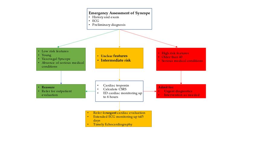
occurred within 15 days of the index syncope [56]. Figure 3 outlines our recommended
out-of-hospital arrhythmias over a 30-day period. Cardiac rhythm monitoring should be
approach toefficiently
the syncope patient
utilized in the ED.as 92% of arrhythmic events occurred within 15 days
in this population,
of the index syncope [56]. Figure 3 outlines our recommended approach to the syncope
patient in the ED.
Figure 3. The authors recommended approach to syncope patients in the ED based on history and
physical exam, ECG, ED diagnosis, and CSRS.
Figure 3. The7.authors recommended approach to syncope patients in the ED based on history and
Conclusions
physical exam, ECG,
Clinical diagnosis,
ED judgement and CSRS.
based on proper history, ECG findings, and the most likely ED
diagnosis remains the primary tool to approach the syncope patient and guide further
management. Low-risk patients can safely be discharged with proper follow up when
needed, while the high-risk patients with clear need for intervention benefit from further in-
hospital management. When in doubt, the clinician may benefit from implementing other
validated clinical decision tools and additional ED testing and monitoring; specifically, the
CSRS appears to perform better than older scores in classifying patient risk. The Basel IX
ECG ALERT-CS tool to identify high- and low-risk groups appears promising. Hospital-
ization, however, does not seem to offer meaningful diagnostic benefits, and unnecessary
hospitalizations can be avoided by efficient outpatient cardiac referral and testing.
Funding: This research received no external funding.
Institutional Review Board Statement: Not applicable.
Informed Consent Statement: Not applicable.
Conflicts of Interest: The authors declare no conflict of interest.
References
1. Ganzeboom, K.S.; Mairuhu, G.; Reitsma, J.B.; Linzer, M.; Wieling, W.; Van Dijk, N. Lifetime Cumulative Incidence of Syncope
in the General Population: A Study of 549 Dutch Subjects Aged 35?60 Years. J. Cardiovasc. Electrophysiol. 2006, 17, 1172–1176.
[CrossRef] [PubMed]
2. Serletis, A.; Rose, S.; Sheldon, A.G.; Sheldon, R.S. Vasovagal syncope in medical students and their first-degree relatives. Eur.
Heart J. 2006, 27, 1965–1970. [CrossRef] [PubMed]
3. Chen, L.Y.; Shen, W.-K.; Mahoney, D.W.; Jacobsen, S.J.; Rodeheffer, R.J. Prevalence of Syncope in a Population Aged More Than
45 Years. Am. J. Med. 2006, 119, 1088.e1–1088.e7. [CrossRef] [PubMed]Medicina 2021, 57, 514 9 of 11
4. Sandhu, R.K.; Raj, S.R.; Thiruganasambandamoorthy, V.; Kaul, P.; Morillo, C.A.; Krahn, A.D.; Guzman, J.C.; Sheldon, R.S.;
Banijamali, H.S.; Macintyre, C.; et al. Canadian Cardiovascular Society Clinical Practice Update on the Assessment and
Management of Syncope. Can. J. Cardiol. 2020, 36, 1167–1177. [CrossRef]
5. Sandhu, R.K.; Sheldon, R.S. Syncope in the Emergency Department. Front. Cardiovasc. Med. 2019, 6, 5. [CrossRef]
6. Sandhu, R.K.; Tran, D.T.; Sheldon, R.S.; Kaul, P. A Population-Based Cohort Study Evaluating Outcomes and Costs for Syncope
Presentations to the Emergency Department. JACC Clin. Electrophysiol. 2018, 4, 265–273. [CrossRef]
7. Sun, B.C. Quality-of-Life, Health Service Use, and Costs Associated With Syncope. Prog. Cardiovasc. Dis. 2013, 55, 370–375.
[CrossRef]
8. Toarta, C.; Mukarram, M.; Arcot, K.; Kim, S.-M.; Gaudet, S.; Sivilotti, A.; Marco, L.; Rowe, B.H.; Thiruganasambandamoorthy, V.
Syncope Prognosis Based on Emergency Department Diagnosis: A Prospective Cohort Study. Acad. Emerg. Med. 2018, 25, 388–396.
[CrossRef]
9. Solbiati, M.; Casazza, G.; Dipaola, F.; Rusconi, A.M.; Cernuschi, G.; Barbic, F.; Montano, N.; Sheldon, R.S.; Furlan, R.; Costantino, G.
Syncope recurrence and mortality: A systematic review. Europace 2015, 17, 300–308. [CrossRef]
10. Costantino, G.; Perego, F.; Dipaola, F.; Borella, M.; Galli, A.; Cantoni, G.; Dell’Orto, S.; Dassi, S.; Filardo, N.; Duca, P.G.; et al.
Short- and Long-Term Prognosis of Syncope, Risk Factors, and Role of Hospital Admission. J. Am. Coll. Cardiol. 2008, 51, 276–283.
[CrossRef] [PubMed]
11. Kaul, P.; Tran, D.T.; Sandhu, R.K.; Solbiati, M.; Costantino, G.; Sheldon, R.S. Lack of benefit from hospitalization in patients with
syncope: A propensity analysis. J. Am. Coll. Emerg. Physicians Open 2020, 1, 716–722. [CrossRef]
12. Bartoletti, A.; Fabiani, P.; Bagnoli, L.; Cappelletti, C.; Cappellini, M.; Nappini, G.; Gianni, R.; Lavacchi, A.; Santoro, G.M. Physical
injuries caused by a transient loss of consciousness: Main clinical characteristics of patients and diagnostic contribution of carotid
sinus massage. Eur. Heart J. 2008, 29, 618–624. [CrossRef] [PubMed]
13. Jorge, J.G.; Pournazari, P.; Raj, S.R.; Maxey, C.; Sheldon, R.S. Frequency of injuries associated with syncope in the prevention of
syncope trials. EP Eur. 2020, 22, 1896–1903. [CrossRef] [PubMed]
14. Jorge, J.G.; Raj, S.R.; Teixeira, P.S.; Teixeira, J.A.C.; Sheldon, R.S. Likelihood of injury due to vasovagal syncope: A systematic
review and meta-analysis. EP Eur. 2021. (epub ahead of print). [CrossRef]
15. Ng, J.; Sheldon, R.S.; Ritchie, D.; Raj, V.; Raj, S.R. Reduced quality of life and greater psychological distress in vasovagal syncope
patients compared to healthy individuals. Pacing Clin. Electrophysiol. 2019, 42, 180–188. [CrossRef] [PubMed]
16. Carvalho, M.S.; Reis Santos, K.; Carmo, P.; Cavaco, D.; Parreira, L.; Morgado, F.; Adragão, P. Prognostic Value of a Very Prolonged
Asystole during Head-Up Tilt Test. Pacing Clin. Electrophysiol. 2015, 38, 973–979. [CrossRef]
17. Ricci, F.; Fedorowski, A.; Radico, F.; Romanello, M.; Tatasciore, A.; Di Nicola, M.; Zimarino, M.; De Caterina, R. Cardiovascular
morbidity and mortality related to orthostatic hypotension: A meta-analysis of prospective observational studies. Eur. Heart J.
2015, 36, 1609–1617. [CrossRef]
18. Brignole, M.; Moya, A.; De Lange, F.J.; Deharo, J.-C.; Elliott, P.M.; Fanciulli, A.; Fedorowski, A.; Furlan, R.; Kenny, R.A.;
Martín, A.; et al. Practical Instructions for the 2018 ESC Guidelines for the diagnosis and management of syncope. Eur. Heart J.
2018, 39, e43–e80. [CrossRef]
19. Sheldon, R. How to Differentiate Syncope from Seizure. Cardiol. Clin. 2015, 33, 377–385. [CrossRef]
20. Sheldon, R.; Rose, S.; Ritchie, D.; Connolly, S.J.; Koshman, M.-L.; Lee, M.A.; Frenneaux, M.; Fisher, M.; Murphy, W. Historical
criteria that distinguish syncope from seizures. J. Am. Coll. Cardiol. 2002, 40, 142–148. [CrossRef]
21. Brignole, M.; Moya, A.; de Lange, F.J.; Deharo, J.C.; Elliott, P.M.; Fanciulli, A.; Fedorowski, A.; Furlan, R.; Kenny, R.A.;
Martin, A.; et al. 2018 ESC Guidelines for the diagnosis and management of syncope. Eur. Heart J. 2018, 39, 1883–1948. [CrossRef]
[PubMed]
22. Shen, W.K.; Sheldon, R.S.; Benditt, D.G.; Cohen, M.I.; Forman, D.E.; Goldberger, Z.D.; Grubb, B.P.; Hamdan, M.H.; Krahn, A.D.;
Link, M.S.; et al. 2017 ACC/AHA/HRS Guideline for the Evaluation and Management of Patients With Syncope A Report of the
American College of Cardiology/American Heart Association Task Force on Clinical Practice Guidelines and the Heart Rhythm
Society. J. Am. Coll. Cardiol. 2017, 70, e39–e110. [CrossRef] [PubMed]
23. Van Dijk, N.; Boer, K.R.; Colman, N.; Bakker, A.; Stam, J.; Van Grieken, J.J.M.; Wilde, A.A.M.; Linzer, M.; Reitsma, J.B.; Wieling, W.
High Diagnostic Yield and Accuracy of History, Physical Examination, and ECG in Patients with Transient Loss of Consciousness
in FAST: The Fainting Assessment Study. J. Cardiovasc. Electrophysiol. 2008, 19, 48–55. [CrossRef]
24. Berecki-Gisolf, J.; Sheldon, A.; Wieling, W.; Van Dijk, N.; Costantino, G.; Furlan, R.; Shen, W.-K.; Sheldon, R. Identifying Cardiac
Syncope Based on Clinical History: A Literature-Based Model Tested in Four Independent Datasets. PLoS ONE 2013, 8, e75255.
[CrossRef] [PubMed]
25. Alboni, P.; Brignole, M.; Menozzi, C.; Raviele, A.; Del Rosso, A.; Dinelli, M.; Solano, A.; Bottoni, N. Diagnostic value of history in
patients with syncope with or without heart disease. J. Am. Coll. Cardiol. 2001, 37, 1921–1928. [CrossRef]
26. Sheldon, R.; Hersi, A.; Ritchie, D.; Koshman, M.-L.; Rose, S. Syncope and Structural Heart Disease: Historical Criteria for
Vasovagal Syncope and Ventricular Tachycardia. J. Cardiovasc. Electrophysiol. 2010, 21, 1358–1364. [CrossRef] [PubMed]
27. Calkins, H.; Shyr, Y.; Frumin, H.; Schork, A.; Morady, F. The value of the clinical history in the differentiation of syncope due to
ventricular tachycardia, atrioventricular block, and neurocardiogenic syncope. Am. J. Med. 1995, 98, 365–373. [CrossRef]Medicina 2021, 57, 514 10 of 11
28. D’Ascenzo, F.; Biondi-Zoccai, G.; Reed, M.J.; Gabayan, G.Z.; Suzuki, M.; Costantino, G.; Furlan, R.; Del Rosso, A.; Sarasin, F.P.;
Sun, B.C.; et al. Incidence, etiology and predictors of adverse outcomes in 43,315 patients presenting to the Emergency Department
with syncope: An international meta-analysis. Int. J. Cardiol. 2013, 167, 57–62. [CrossRef] [PubMed]
29. Quinn, J.V.; Stiell, I.G.; Mcdermott, D.A.; Sellers, K.L.; Kohn, M.A.; Wells, G.A. Derivation of the San Francisco Syncope Rule to
predict patients with short-term serious outcomes. Ann. Emerg. Med. 2004, 43, 224–232. [CrossRef]
30. Probst, M.A.; Gibson, T.; Weiss, R.E.; Yagapen, A.N.; Malveau, S.E.; Adler, D.H.; Bastani, A.; Baugh, C.W.; Caterino, J.M.;
Clark, C.L.; et al. Risk Stratification of Older Adults Who Present to the Emergency Department With Syncope: The FAINT Score.
Ann. Emerg. Med. 2020, 75, 147–158. [CrossRef]
31. Colivicchi, F. Development and prospective validation of a risk stratification system for patients with syncope in the emergency
department: The OESIL risk score. Eur. Heart J. 2003, 24, 811–819. [CrossRef]
32. Del Rosso, A.; Ungar, A.; Maggi, R.; Giada, F.; Petix, N.R.; De Santo, T.; Menozzi, C.; Brignole, M. Clinical predictors of cardiac
syncope at initial evaluation in patients referred urgently to a general hospital: The EGSYS score. Heart 2008, 94, 1620–1626.
[CrossRef]
33. Martin, T.P.; Hanusa, B.H.; Kapoor, W.N. Risk Stratification of Patients with Syncope. Ann. Emerg. Med. 1997, 29, 459–466.
[CrossRef]
34. Quinn, J.; Mcdermott, D. Electrocardiogram Findings in Emergency Department Patients with Syncope. Acad. Emerg. Med. 2011,
18, 714–718. [CrossRef] [PubMed]
35. Thiruganasambandamoorthy, V.; Hess, E.P.; Turko, E.; Tran, M.-L.; Wells, G.A.; Stiell, I.G. Defining abnormal electrocardiography
in adult emergency department syncope patients: The Ottawa Electrocardiographic Criteria. CJEM 2012, 14, 248–258.
36. Thiruganasambandamoorthy, V.; Kwong, K.; Stiell, I.G.; Swampillai, J.; Toarta, C.; Sumner, G.L.; Kuriachan, V.P.; Mukarram, M.;
Taljaard, M.; Hazra, S.; et al. Short-term risk of arrhythmias among emergency department syncope patients with non-sinus
rhythm. Int. J. Cardiol. 2015, 189, 12–14. [CrossRef] [PubMed]
37. Pérez-Rodon, J.; Martínez-Alday, J.; Barón-Esquivias, G.; Martín, A.; García-Civera, R.; Del Arco, C.; Cano-Gonzalez, A.; Moya-
Mitjans, À. Prognostic value of the electrocardiogram in patients with syncope: Data from the Group for Syncope Study in the
Emergency Room (GESINUR). Heart Rhythm 2014, 11, 2035–2044. [CrossRef]
38. Nishijima, D.K.; Lin, A.L.; Weiss, R.E.; Yagapen, A.N.; Malveau, S.E.; Adler, D.H.; Bastani, A.; Baugh, C.W.; Caterino, J.M.;
Clark, C.L.; et al. ECG Predictors of Cardiac Arrhythmias in Older Adults With Syncope. Ann. Emerg. Med. 2018, 71, 452–461.
[CrossRef]
39. White, J.L.; Chang, A.M.; Hollander, J.E.; Su, E.; Weiss, R.E.; Yagapen, A.N.; Malveau, S.E.; Adler, D.H.; Bastani, A.;
Baugh, C.W.; et al. QTc prolongation as a marker of 30-day serious outcomes in older patients with syncope presenting to the
Emergency Department. Am. J. Emerg. Med. 2019, 37, 685–689. [CrossRef]
40. Balasubramaniyam, N.; Palaniswamy, C.; Aronow, W.S.; Khera, S.; Balasubramanian, G.; Harikrishnan, P.; Doshi, J.V.; Nabors, C.;
Peterson, S.J.; Sule, S. Association of corrected QT interval with long-term mortality in patients with syncope. Arch. Med Sci. 2013,
6, 1049–1054. [CrossRef]
41. Aggarwal, A.; Sherazi, S.; Levitan, B.; Lakshmanadoss, U.; Choudhary, N.; Shah, A.; Hsi, D. Corrected QT interval as a predictor
of mortality in elderly patients with syncope. Cardiol. J. 2011, 18, 395–400. [PubMed]
42. Bartczak, A.; Lelonek, M. Early Repolarization Variant in Syncopal Patients Referred to Tilt Testing. Pacing Clin. Electrophysiol.
2013, 36, 456–461. [CrossRef] [PubMed]
43. Thiruganasambandamoorthy, V.; Kwong, K.; Wells, G.A.; Sivilotti, M.L.A.; Mukarram, M.; Rowe, B.H.; Lang, E.; Perry, J.J.;
Sheldon, R.; Stiell, I.G.; et al. Development of the Canadian Syncope Risk Score to predict serious adverse events after emergency
department assessment of syncope. Can. Med. Assoc. J. 2016, 188, E289–E298. [CrossRef] [PubMed]
44. Zimmermann, T.; Du Fay De Lavallaz, J.; Walter, J.E.; Strebel, I.; Nestelberger, T.; Joray, L.; Badertscher, P.; Flores, D.; Widmer, V.;
Geigy, N.; et al. Development of an electrocardiogram-based risk calculator for a cardiac cause of syncope. Heart 2020. [CrossRef]
45. Quinn, J.; Mcdermott, D.; Stiell, I.; Kohn, M.; Wells, G. Prospective Validation of the San Francisco Syncope Rule to Predict
Patients With Serious Outcomes. Ann. Emerg. Med. 2006, 47, 448–454. [CrossRef] [PubMed]
46. Sun, B.C.; Mangione, C.M.; Merchant, G.; Weiss, T.; Shlamovitz, G.Z.; Zargaraff, G.; Shiraga, S.; Hoffman, J.R.; Mower, W.R.
External validation of the San Francisco Syncope Rule. Ann. Emerg. Med. 2007, 49, 420–427. [CrossRef] [PubMed]
47. Thiruganasambandamoorthy, V.; Hess, E.P.; Alreesi, A.; Perry, J.J.; Wells, G.A.; Stiell, I.G. External validation of the San Francisco
Syncope Rule in the Canadian setting. Ann. Emerg. Med. 2010, 55, 464–472. [CrossRef]
48. Birnbaum, A.; Esses, D.; Bijur, P.; Wollowitz, A.; Gallagher, E.J. Failure to validate the San Francisco Syncope Rule in an
independent emergency department population. Ann. Emerg. Med. 2008, 52, 151–159. [CrossRef]
49. de Lavallaz, J.D.; Badertscher, P.; Nestelberger, T.; Isenrich, R.; Miro, O.; Salgado, E.; Geigy, N.; Christ, M.; Cullen, L.;
Than, M.; et al. Prospective validation of prognostic and diagnostic syncope scores in the emergency department. Int. J.
Cardiol. 2018, 269, 114–121. [CrossRef]
50. Dipaola, F.; Costantino, G.; Perego, F.; Borella, M.; Galli, A.; Cantoni, G.; Barbic, F.; Casella, F.; Duca, P.G.; Furlan, R. San Francisco
Syncope Rule, Osservatorio Epidemiologico sulla Sincope nel Lazio risk score, and clinical judgment in the assessment of
short-term outcome of syncope. Am. J. Emerg. Med. 2010, 28, 432–439. [CrossRef] [PubMed]Medicina 2021, 57, 514 11 of 11
51. Costantino, G.; Casazza, G.; Reed, M.; Bossi, I.; Sun, B.; Del Rosso, A.; Ungar, A.; Grossman, S.; D’Ascenzo, F.; Quinn, J.; et al.
Syncope Risk Stratification Tools vs Clinical Judgment: An Individual Patient Data Meta-analysis. Am. J. Med. 2014, 127,
e13–e1126. [CrossRef]
52. Solbiati, M.; Quinn, J.V.; Dipaola, F.; Duca, P.; Furlan, R.; Montano, N.; Reed, M.J.; Sheldon, R.S.; Sun, B.C.; Ungar, A.; et al.
Personalized risk stratification through attribute matching for clinical decision making in clinical conditions with aspecific
symptoms: The example of syncope. PLoS ONE 2020, 15, e0228725. [CrossRef] [PubMed]
53. Thiruganasambandamoorthy, V.; Sivilotti, M.L.A.; Le Sage, N.; Yan, J.W.; Huang, P.; Hegdekar, M.; Mercier, E.; Mukarram, M.;
Nemnom, M.-J.; Mcrae, A.D.; et al. Multicenter Emergency Department Validation of the Canadian Syncope Risk Score. JAMA
Intern. Med. 2020, 180, 737. [CrossRef] [PubMed]
54. Chan, J.; Ballard, E.; Brain, D.; Hocking, J.; Yan, A.; Morel, D.; Hunter, J. External validation of the Canadian Syncope Risk Score
for patients presenting with undifferentiated syncope to the emergency department. Emerg. Med. Australas. 2021, 33, 418–424.
[CrossRef] [PubMed]
55. de Lavallaz, J.D.; Badertscher, P.; Nestelberger, T.; Zimmermann, T.; Miro, O.; Salgado, E.; Christ, M.; Geigy, N.; Cullen, L.;
Than, M.; et al. B-Type Natriuretic Peptides and Cardiac Troponins for Diagnosis and Risk-Stratification of Syncope. Circulation
2019, 139, 2403–2418. [CrossRef] [PubMed]
56. Thiruganasambandamoorthy, V.; Rowe, B.H.; Sivilotti, M.L.A.; Mcrae, A.D.; Arcot, K.; Nemnom, M.-J.; Huang, L.; Mukarram, M.;
Krahn, A.D.; Wells, G.A.; et al. Duration of Electrocardiographic Monitoring of Emergency Department Patients With Syncope.
Circulation 2019, 139, 1396–1406. [CrossRef]
57. Thiruganasambandamoorthy, V.; Ramaekers, R.; Rahman, M.O.; Stiell, I.G.; Sikora, L.; Kelly, S.-L.; Christ, M.; Claret, P.-G.;
Reed, M.J. Prognostic value of cardiac biomarkers in the risk stratification of syncope: A systematic review. Intern. Emerg. Med.
2015, 10, 1003–1014. [CrossRef]
58. Thiruganasambandamoorthy, V.; Mcrae, A.D.; Rowe, B.H.; Sivilotti, M.L.A.; Mukarram, M.; Nemnom, M.-J.; Booth, R.A.;
Calder, L.A.; Stiell, I.G.; Wells, G.A.; et al. Does N-Terminal Pro–B-Type Natriuretic Peptide Improve the Risk Stratification of
Emergency Department Patients With Syncope? Ann. Intern. Med. 2020, 172, 648–655. [CrossRef]
59. Probst, M.; Gibson, T.; Weiss, R.; Yagapen, A.; Malveau, S.; Adler, D.; Bastani, A.; Baugh, C.; Caterino, J.; Clark, C.; et al. Predictors
of Clinically Significant Echocardiography Findings in Older Adults with Syncope: A Secondary Analysis. J. Hosp. Med. 2018, 13,
823–828. [CrossRef]
60. Probst, M.A.; Su, E.; Weiss, R.E.; Yagapen, A.N.; Malveau, S.E.; Adler, D.H.; Bastani, A.; Baugh, C.W.; Caterino, J.M.;
Clark, C.L.; et al. Clinical Benefit of Hospitalization for Older Adults With Unexplained Syncope: A Propensity-Matched
Analysis. Ann. Emerg. Med. 2019, 74, 260–269. [CrossRef]
61. Krishnan, R.J.; Mukarram, M.; Ghaedi, B.; Sivilotti, M.L.A.; Le Sage, N.; Yan, J.W.; Huang, P.; Hegdekar, M.; Mercier, E.;
Nemnom, M.-J.; et al. Benefit of hospital admission for detecting serious adverse events among emergency department patients
with syncope: A propensity-score–matched analysis of a multicentre prospective cohort. Can. Med Assoc. J. 2020, 192, E1198–E1205.
[CrossRef]
62. Canzoniero, J.V.; Afshar, E.; Hedian, H.; Koch, C.; Morgan, D.J. Unnecessary Hospitalization and Related Harm for Patients With
Low-Risk Syncope. JAMA Intern. Med. 2015, 175, 1065. [CrossRef] [PubMed]
63. Cook, O.G.; Mukarram, M.A.; Kim, S.-M.; Arcot, K.; Nemnom, M.-J.; Taljaard, M.; Sivilotti, M.L.A.; Rowe, B.H.; Thiruganasam-
bandamoorthy, V. Application of outpatient cardiac testing among emergency department patients with syncope. Emerg. Med. J.
2018, 35, 486–491. [CrossRef] [PubMed]You can also read