Modifying Effects of Glucose and Insulin/Insulin-Like Growth Factors on Colon Cancer Cells
←
→
Page content transcription
If your browser does not render page correctly, please read the page content below

ORIGINAL RESEARCH
                                                                                                                                                published: 05 July 2021
                                                                                                                                        doi: 10.3389/fonc.2021.645732

                                              Modifying Effects of Glucose
                                              and Insulin/Insulin-Like Growth
                                              Factors on Colon Cancer Cells
                                              Şeyda Berk 1,2, Joseph A. M. J. L. Janssen 2, Peter M. van Koetsveld 2, Fadime Dogan 2,
                                              Naci Değerli 1, Servet Özcan 3,4, Fahrettin Kelestimur 3,5 and Leo J. Hofland 2*
                                              1 Department of Molecular Biology and Genetics, Faculty of Science, Sivas Cumhuriyet University, Sivas, Turkey,
                                              2 Department of Internal Medicine, Division of Endocrinology, Erasmus Medical Center, Rotterdam, Netherlands,
                                              3 Genome and Stem Cell Center (GENKOK), Erciyes University, Kayseri, Turkey, 4 Department of Biology, Faculty of

                                              Sciences, Erciyes University, Kayseri, Turkey, 5 Yeditepe University, Faculty of Medicine, Istanbul, Turkey

                                              There are only a few experimental studies which have investigated effects of glucose alone,
                                              and glucose in combination with insulin/insulin-like growth factors (IGF) on the growth of colon
                                              cancer. In the present study, we studied in vitro in human colorectal cancer cells originating
                                              from four Dukes’ stages of colorectal cancer the effects of glucose, insulin and IGFs on
                      Edited by:
                                              proliferation, migration, cell cycle progression and gene expression of the IGF system. Growth
                      Shixiu Wu,              of colon cancer cells originating from a Dukes’ stage A was glucose-dependent, whereas
   Hangzhou Cancer Hospital, China
                                              growth of cancer cells from Dukes’ stage B, C and D was glucose-independent. Stimulatory
                         Reviewed by:
                                              effects of insulin and IGFs on cell growth were observed only in colon cancer cells originating
                     Antonino Belfiore,
            University of Catania, Italy      from Dukes’ stage C and D. IGF-II stimulated migration in Dukes’ stage B cells only. The
                        Jeff M. P. Holly,     growth stimulatory effects in Dukes’ stage C and D colorectal cancer cells were accompanied
 University of Bristol¸ United Kingdom
                                              by G2/M arrest and associated with an increased IGF-IR/IGF-II receptor ratio. In conclusion,
                   *Correspondence:
                       Leo J. Hofland
                                              our in vitro data suggest that the stimulating effects of glucose, IGFs and insulin on proliferation
              l.hofland@erasmusmc.nl           differ between colorectal cancer cells from early and late Dukes’ stages. Stimulatory effects of
                                              glucose on proliferation appear predominantly present in stage Dukes’ stage A colorectal
                   Specialty section:
                                              cancer cells, while in contrast growth factor-mediated stimulation of cell proliferation is more
         This article was submitted to
             Gastrointestinal Cancers,        pronounced in Dukes’ late stage (metastasized) colorectal cancer cells. Moreover, our study
               a section of the journal       suggests that a stringent glucose control may be important to control tumor growth in early
                 Frontiers in Oncology
                                              stages of colorectal cancer, while inhibition of the endocrine actions of the IGFs and insulin
      Received: 23 December 2020
          Accepted: 08 June 2021
                                              become more important in the late (metastasized) stages of colorectal cancer to restrain
          Published: 05 July 2021             growth of colon cancer cells.
                               Citation:
                                              Keywords: colorectal cancer, glucose, insulin, insulin-like growth factors, IGF-IR, IR
               Berk Ş, Janssen JAMJL,
         van Koetsveld PM, Dogan F,
     Değerli N, Özcan S, Kelestimur F
                 and Hofland LJ (2021)
         Modifying Effects of Glucose
                                              INTRODUCTION
                 and Insulin/Insulin-Like
                     Growth Factors on
                                              Colorectal cancer and type 2 diabetes mellitus are main causes of morbidity and mortality
                   Colon Cancer Cells.        worldwide (1–3). Epidemiologic evidence suggests an association between diabetes mellitus and
              Front. Oncol. 11:645732.        an increased risk of colorectal cancer (4–7). Several studies have suggested that hyperinsulinemia
     doi: 10.3389/fonc.2021.645732            and elevated glucose levels increase the risk for colorectal cancer (4, 7–15). Hyperinsulinemia may

Frontiers in Oncology | www.frontiersin.org                                          1                                          July 2021 | Volume 11 | Article 645732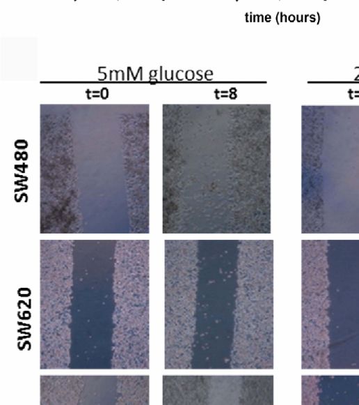
Berk et al.                                                                                                    Glucose, IGFs, Insulin, and Colon Cancer

stimulate insulin receptors (IR) and/or insulin-like growth factor          CO2 and 37°C. Cells were harvested with trypsin solution
receptors (IGFIR/IGFIIR) and thereby induce cell proliferation              (0.05%)-EDTA (0.53 mM) (Gibco, Ref. No: 15400-054). Cells
and suppress apoptosis (16–18).                                             were processed through a semi-automated image-based cell
    The IGF signaling pathway has been considered to be                     analyzer (Cedex XS Innovatis, Roche, The Netherlands), to
involved in the proliferation and differentiation of epithelial             determine cell concentration and viability based on the Trypan
cells of the colon, breast, lung and prostate (19). The IGF                 Blue exclusion method.
system is complex and consists of at least two insulin-like
growth factor receptors (IGF-IR and IGF-IIR), two polypeptide               Drugs and Reagents
ligands (IGF-I and IGF-II) and six binding proteins (IGFBP-1-6)             IGF-I (Sigma, I3769) and IGF-II (Sigma, I2526) were purchased
(20). In addition, a large group of insulin growth factor binding           from Sigma-Aldrich (Zwijndrecht, The Netherlands). Human
protein proteases may cleave IGFBPs and increase levels of free             recombinant insulin was obtained from Sanofi-Aventis
IGF-I and IGF-II (21). It is thought that both IGF-I/II and the             (Insuman Rapid U100). IGF-I and IGF-II were stored at -20°C;
IGF-IR by inducing cellular proliferation, growth and                       insulin was stored at 4°C. Stock solutions of IGF-I and IGF-II
immunosuppression play important roles in tumorigenesis                     were constituted in 0.01 M of acetic acid, insulin was constituted
(22–25). Elevated circulating levels of IGF-I and IGF-II have               in distilled water, all according to the manufacturer’s instruction.
been associated with an increased risk on colorectal cancer (25,
26). IGF-I, IGF-II and its receptor (IGF-IR) are frequently                 Cell Proliferation Assay
overexpressed in many types of tumors including colorectal                  For each cell line the optimal cell number plating density in
cancer (26–29). Overexpression of IGF-II mRNA has been                      medium containing 5 mmol/L glucose with different fetal calf
observed in colorectal cancer tumors compared with normal                   serum (FCS) concentrations (0.1%, 0.5%, 1%, 10%) was
tissue (30).                                                                determined. The cells were plated in 24 well plates at
    Studies investigating expression of the insulin- and IGF                increasing cell density (3.125, 6.250, 12.500, 25.000, 50.000,
receptors in colon cancer cells during the different stages of              100.000 cells/mL per well). For each cell line the minimum
cancer, as well as direct effects of glucose, IGFs and insulin on           FCS concentration was determined to study the effects of IGFs
colon cancer cells may help to better understand the role of the            and insulin on colorectal cancer cell proliferation. Except for cell
insulin-IGF pathway in the pathogenesis of colon cancer.                    line SW620, the growth of the other three cell lines was relatively
Therefore, in the present study we investigated the potential               FCS independent and grew even in medium containing very
effects of glucose, insulin and IGFs on proliferation, migration,           low FCS (0.1%). To compare all data under equal conditions
cell cycle progression and gene expression in colorectal cancer cells       0.5% FCS was selected as plating FCS concentration. After
originating from four different Dukes’ stages (A, B, C and D).              determination of optimal minimum FCS concentration for
                                                                            growth of cell lines, the optimal cell number density was
                                                                            determined after 3 days for each cell line in medium with 0.5%
MATERIALS AND METHODS                                                       FCS (this was for SW1116: 200.000 cells/mL; SW620: 50.000
                                                                            cells/mL; SW480: 50.000 cell/mL; COLO205: 80.000 cells/mL)
Cell Lines and Culture Conditions                                           and after 7 days (this was for SW1116: 100.000 cells/mL; SW620:
SW1116 (ATCC, CCL-233, Lot# 59816766), SW480 (ATCC,                         12.500 cells/mL; SW480: 12.500 cell/mL; COLO205: 40.000 cells/
CCL-228, Lot# 59932372), SW620, (ATCC, CCL-227, Lot#                        mL) (data not shown).
61867814) and COLO205 (ATCC, CCL-222, Lot# 618686374)                           After trypsinization, the cells were plated in 1 mL of medium in
colorectal cancer cell lines were purchased from the American               24 well plates at previously obtained optimal cell density. Plates were
Type Culture Collection (ATCC; Manassas, VA, U.S.A.). The                   placed in a 37°C, 5% CO2 incubator and cells were allowed to attach
details of each cell line are listed in Supplementary Table 1.              overnight. The next day, after washing the plates two times, the
Initially, all cell lines were cultured in medium recommended by            culture medium was replaced with 1 mL/well medium containing
ATCC. According to ATCC, SW1116, SW480 and SW620 were                       0.5% FCS with 5 mmol/L, 12.5 mmol/L or 25 mmol/L glucose to
cultured in medium consisting of Leibovitz’s L-15 (Gibco, Ref               evaluate the effect of different glucose concentration on colorectal
No: 11414-049) supplemented with 10% FCS (Sigma, F7524, Lot                 cancer cell proliferation. For experiments testing the effects of IGFs
No: 060M3396) and penicillin (Natrium-penicillin G; 1x10⁵ U/                and insulin, cells were allowed to attach for one day in medium with
liter) in an incubator at 37°C without CO2. COLO205 was                     0.5% FCS, washed twice as described above, after which the culture
cultured in medium consisting of RPMI-1640 (Gibco, Ref. No:                 medium was replaced with 1 mL/well medium containing 0.5%
11879-020) supplemented with 10% FCS (Sigma, F7524) and                     FCS, 5 mmol/L or 25 mmol/L glucose and increasing
penicillin (1x10⁵ U/liter) in an incubator at 5% CO2 and 37°C.              concentrations of IGF-I, IGF-II or insulin (0.01 nmol/L, 0.1
The cells were first adapted to the medium as recommended by                 nmol/L, 1 nmol/L, 5 nmol/L, 10 nmol/L). After 3 and 7 days
the ATCC for one week, then all the colorectal cancer cell lines            (medium and compounds refreshed at day 3), the cells were
were cultured in a common medium to compare all data under                  harvested for DNA measurement. Measurement of total DNA
the same conditions. For all cell lines, the experimental culture           contents was performed using the bisbenzimide fluorescent dye
medium consisted of DMEM medium (Gibco, Ref No: 11966-                      (Hoechst 33258; Sigma, #B2883) (Boehring Diagnostics, La Jolla,
025) supplemented with 10% FCS (Sigma, F7524), penicillin                   CA) as previously reported (31). The experiments were repeated
(1x10⁵ U/liter) and glucose (5 mmol/L) in an incubator at 5%                twice, and each experiment was performed in quadruplicate.

Frontiers in Oncology | www.frontiersin.org                             2                                      July 2021 | Volume 11 | Article 645732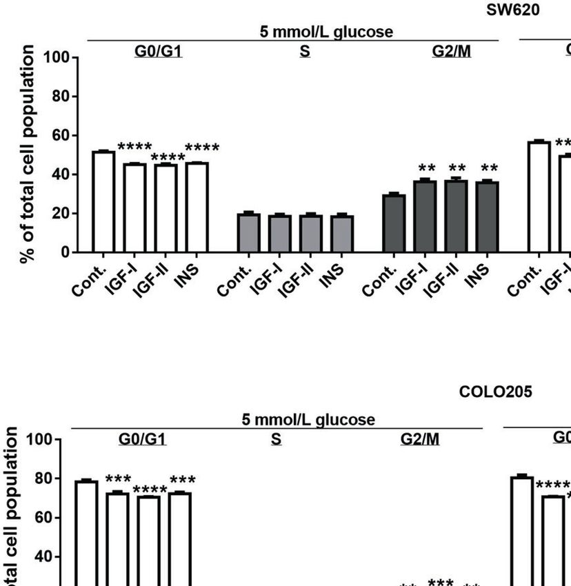
Berk et al.                                                                                                 Glucose, IGFs, Insulin, and Colon Cancer

Cell Cycle Assay                                                           according to the manufacturer’s protocol. PCR conditions were
For cell cycle analysis, the colorectal cancer cell lines SW620 and        95°C for 10 min, followed by 45 cycles of 95°C for 15s and 60°C for
COLO205 were treated with 10 nmol/L IGF-I, IGF-II or insulin.              1 min. Hypoxanthine-guanine phosphoribosyl transferase-1
After 3 days, cells were harvested, washed with NaCl, fixed with            (HPRT1; Sigma-Aldrich), glucuronidase beta (GUSB; Thermo
ice-cold 70% EtOH, and stored at -20°C until analysis. Analyses            Fisher Scientific) and beta-actin (ACTB; Thermo Fisher
were performed using the Muse cell cycle assay kit using a Muse            Scientific) were used to normalize mRNA levels. PCR efficiencies
cell analyzer (Merck Millipore) according to the manufacturer’s            (E) were calculated for the primer–probe combinations used
instructions. The experiments were repeated twice, and each                (Supplementary Table 2) and the relative expression of genes
experiment was performed in triplicate.                                    was calculated using the comparative threshold method, 2-DCt. For
                                                                           accurate RT-qPCR expression profiling of the colorectal cancer
                                                                           cell lines, HPRT, GUSB, and ACTB were determined to normalize
Cell Migration Assay                                                       mRNA levels using the method as previously described (34). The
The in vitro cell migration was measured by the scratch assay
                                                                           sequence of the used primers and probes are listed in
method as previously described (32), with some modifications.
                                                                           Supplementary Table 2. Ct values >35 did not yield consistent
After trypsinization, cells were plated in 2 mL of medium
                                                                           results and were considered below the detection limit of the assay.
containing 10% FCS, 5 mmol/L or 25 mmol/L glucose in poly-
lysine (10 µg/mL) coated 12-wells plates and placed in a 37°C, 5%
CO2 incubator. Cells were grown until a confluent monolayer                 Statistical Analysis
was formed. With a 200 mL pipet tip a scratch was made in the              All the experiments were carried out at least twice. Repeated
cell monolayer. The debris was removed by washing the cells                experiments gave comparable results. For the statistical analysis
with 1 mL of growth medium containing 0.5% FCS, 5 mmol/L or                statistical software GraphPad Prisim 6.0 (GraphPad software, San
25 mmol/L glucose. Thereafter, 2 mL of medium containing 0.5%              Diego, CA) was used. Comparative statistical evaluation among
FCS, 5 mmol/L or 25 mmol/L glucose and the different compounds             groups was performed by a one- -way ANOVA test. When
of interest (IGF-I, IGF-II and insulin at concentration 10 nmol/L)         statistically significant differences were found, a comparison
were added in triplicate. The ability of cells to migrate into the         between groups was made using the Newman-Keuls test. In all
scratch area (wound coverage) was assessed after 2, 4 and 8-hours          analysis, a value of p
Berk et al.                                                                                                               Glucose, IGFs, Insulin, and Colon Cancer

 FIGURE 1 | The effect of glucose on cell proliferation after 7 days. SW1116, SW480, SW620 and COLO205 cells were incubated for 7 days with increasing
 concentrations of glucose (5 - 25 mmol/L). Results are expressed as DNA content/well and as the percentage of the DNA content of 5 mmol/L glucose (black bars).
 Values represent the mean ± SEM of at least two independent experiments in quadruplicate. *p < 0.05, ****p < 0.0001 versus control.

significantly stimulated cell growth at the lowest doses tested (0.01-               glucose (Figure 4B). Unfortunately, due to their low
0.1 nM), whereas IGF-II stimulated cell growth at slightly higher                   attachment, it was impossible to measure migration in SW1116
concentrations (≥ 1 nmol/L; Figure 2, lower panels). At 25 mmol/L                   cells (Dukes A). All three remaining cell lines showed a relative
a slightly different pattern of cell growth was observed in SW620                   low migration rate and SW480 cells (Dukes B) displayed a higher
cells (Dukes C), and stimulation by IGF-II appeared more potent                     migration rate compared to SW620 (Dukes C) and COLO205
compared to IGF-I (Supplementary Figure 1).                                         cells (Dukes D) (Figures 4A–C). When cells were cultured in 5
                                                                                    mmol/L glucose and 25 mmol/L glucose, the percentage of
Effects of IGFs and Insulin on Colorectal                                           wound closure after 8 hours was 16.25% and 14.82% for the
Cancer Cell Cycle Progression                                                       SW480 cells; 7.49% and 5.45% for SW620 cells; and 8.66% and
As SW620 (Dukes C) and COLO205 cells (Dukes D) were the                             10.43% for COLO205 cells, respectively.
only two cell lines that in our hands were stimulated by IGFs and                       Furthermore, we only observed in SW480 cells a significant
insulin, we also analyzed the effects of these compounds on cell                    increase in migration after 10 nmol/L IGF-II in 5, but not in 25
cycle progression (Figure 3). The percentage of SW620 cells in                      mmol/L glucose (Figures 4D, E and Supplementary Figure 2,
G2/M phase significantly increased b Both at 5 and 25 mmol/L                         respectively). No statistically significant stimulatory effects on
glucose, when stimulated by 10 nmol/L IGF-I (to 36.3%, p
Berk et al.                                                                                                                   Glucose, IGFs, Insulin, and Colon Cancer

                                      A

                                      B

                                      C

                                      D

 FIGURE 2 | Dose dependent effect of 7-days treatment with increasing concentrations (range 0.01-10 nmol/L) of IGF-I (open bars), IGF-II (grey bars) or insulin (black
 bars) on SW1116 (A), SW480 (B), SW620 (C) and COLO205 (D) cell proliferation in 5 mmol/L glucose. Results are expressed as DNA content/well and as the
 percentage of untreated control. Values represent the mean ± SEM of at least two independent experiments in quadruplicate. *p < 0.05, **p < 0.01, ***p < 0.001,
 ****p < 0.0001 versus control.

COLO205) (Figure 5B). Moreover, SW620 cells had the lowest                            expression in COLO205 cells was relatively high compared to
expression levels of IGF-IIR, IR-A, IR-B, compared to the other                       the three other studied cell lines, whereas SW620 cells had very
cell lines (Figures 5D–F). In COLO205 cells, mRNA expression                          low IGFBP-2 and IGFBP-3 expression (Figure 6).
of the IGF-IR, IGF-IIR, as well as IR-A and IR-B was relatively
high, whereas the mRNA expression of IGF-I and IGF-II was
undetectable and very low, respectively (Figures 5A–F). The                           DISCUSSION
ratio IGF-IR/IGF-IIR progressively increased from cells derived
from Dukes’ stage A and B tumors to cells derived from Dukes’                         To the best of our knowledge, there are no previous studies
stage C and D tumors. The IR-A/IR-B ratio was lowest in                               which systematically have studied 1] whether cell proliferation,
SW1116 (Dukes’ stage A) and highest in SW480 (Dukes’ stage                            cell cycle progression and migration in cells from different stages
B) tumor cells (at 5 mmol/L glucose; Table 1; at 25 mmol/L                            of colon cancer are modified by glucose and 2] whether effects of
glucose; Supplementary Table 3). IGFBP-2 and IGFBP-3                                  IGF-I, IGF-II and insulin on cell proliferation, cell cycle

Frontiers in Oncology | www.frontiersin.org                                       5                                            July 2021 | Volume 11 | Article 645732
Berk et al.                                                                                                                   Glucose, IGFs, Insulin, and Colon Cancer

                          A

                          B

 FIGURE 3 | Effect of 3-days treatment with the growth factors IGF-I, IGF-II and insulin (INS) on cell cycle distribution in SW620 (A) and COLO205 (B) cell lines. Two
 different conditions were tested: medium containing 0.5% FCS with 5 mmol/L glucose (5 mmol/L) or 0.5% FCS with 25 mmol/L glucose (25 mmol/L). The cells were
 incubated for 3 days with 10 nmol/L IGF-I, IGF-II or insulin, respectively. Values are expressed as the percentage of untreated control and represent the mean ± SEM
 of at least two independent experiments in triplicate. **p < 0.01, ***p < 0.001, ****p < 0.0001 versus control.

progression and migration change during different stages of                           switch leads to increased proliferation and migration (37).
colon cancer.                                                                         Although thus the exact mechanism is not clear at the
    In this study we investigated four well-characterized                             moment, our study suggests that hyperglycemia influences
colorectal cancer cell lines originating from Dukes’ stages A to                      growth in the early stages of colorectal cancer.
D. We found that proliferation of SW1116 cells, originating from                          In our study stimulating effects of IGF-I, IGF-II and insulin
Dukes’ stage A, progressively increased at higher glucose                             on cell proliferation were only found in the SW620 and
concentrations. In contrast, glucose did not modify growth of                         COLO205 cell lines. Both cell lines originate from
the three other studied cell lines, which originated from Dukes’                      (metastasized) advanced stages of colon cancer (Dukes’ stage C
stages B to D. These results suggest that glucose is an important                     and D). The stimulating effects of IGF-I, IGF-II and insulin on
growth factor in the early stage of colon tumor growth. Although                      proliferation were not significantly modified by glucose. In
in a meta-analysis it has been found that diabetes mellitus is                        contrast, the IGFs and insulin did not influence proliferation of
associated with an increased risk of colorectal cancer, it is still                   the SW1116 and SW480 cell lines (which originate from Dukes’
unclear whether glucose has a role in the initiation of colon                         stage A and B, respectively).
cancer independent of hyperinsulinemia (5). High glucose may                              Our results suggest that IGFs and insulin are especially
trigger several direct and indirect mechanisms that cooperate to                      important for growth of advanced colorectal cancer cells which
promote cancer cell proliferation. For example, high glucose may                      have metastasized, but they seem not to play an important role in
favor anabolic metabolism and thereby fuel tumor growth (35).                         the early growth of colon cancer cells localized and confined to
A link between high glucose levels and cancer was already                             the bowel. In our study we only studied growth properties of our
proposed more than 50 years ago by Warburg (36). He                                   cell lines in monolayer cultures. In the study of growth properties
suggested that cancer cells use the glycolysis pathway for                            of cancer cell lines, 3D cell cultures might provide additional
respiration and cell division rather than oxidative                                   information compared to monolayer cell cultures. Therefore, it
phosphorylation (36). Hyperglycemia may enhance WNT                                   remains to be studied whether similar differential responses are
signaling and thereby proliferation (35). In addition,                                observed when cells are cultured in 3D cell cultures.
hyperglycemia may also change IGF-IR signaling. Clemmons                                  We observed that IGF-I, IGF-II and insulin induced a G2/M
et al. found that, following exposure to hyperglycemia, cells                         arrest in both SW620 (Dukes’ stage C) and COLO205 cells
undergo a signaling switch leading to an entirely different                           (Dukes’ stage D). This suggests that the IGFs and insulin may
mechanism to activate both the “metabolic” (PI-3 kinase) and                          positively regulate cell-cycle progression and thereby growth of
“mitogenic” (MAP) pathways of the IGF-IR (37). This signaling                         colon cancer cells.

Frontiers in Oncology | www.frontiersin.org                                       6                                            July 2021 | Volume 11 | Article 645732
Berk et al. Glucose, IGFs, Insulin, and Colon Cancer
A
D
B
E
C
FIGURE 4 | Panel (A) and (B): Cell migration of SW480 (light grey bars), SW620 (grey bars) and COLO205 (black bars) cells at 2, 4, 8 hours after scratch in
medium containing 5 mmol/L glucose (A) or 25 mmol/L glucose (B). The percentage of non-recovered wound area was calculated by dividing the non-recovered
area after 8 hours by the initial wound area at time 0 hours. (C) Representative pictures of the scratch at 0 time and 8 hours after scratch in SW480, SW620 and
COLO205 cells in medium containing 5 mmol/L glucose (left panel) or 25 mmol/L glucose (right panel). Original magnification is x50. (D) Percentage of cell migration
of SW480, SW620 and COLO205 cells after 8 hours of incubation with 10 nmol/L IGF-I, IGF-II or insulin, respectively, in medium containing 5 mmol/L glucose.
(E) Representative pictures of the scratch of SW480, SW620 and COLO205 cells at t = 0 and t = 8 hours of incubation with 10 nmol/L IGF-I, IGF-II or insulin,
respectively, in medium containing 5 mmol/L glucose. Original magnification x50. Values are expressed as the percentage of wound closure compared to t=0 and
represent the mean ± SEM of at least two independent experiments performed in triplicate. *p < 0.05, **p < 0.01, ****p < 0.0001 versus control.
Numerous epidemiologic studies have found increased cancer IGF-IR and the IR-A, but to other factors like the real number of
risk associated with high circulating IGF-I levels, IGF-IR and IR-A receptors at the cellular surface and the affinity
hyperinsulinemia or both (19, 38). Although these associations of ligand-receptor interactions.
do not prove causation there are experimental data to support a Our results suggest that local (autocrine and paracrine)
role of the IGFs and insulin in the development of cancer. production of the IGF-II (but not IGF-I) may play a role in
Lowering levels of circulating IGF-I in mice has been shown to promoting (local) tumor growth in the early non-metastasized
inhibit the growth of colon cancer xenografts and to reduce stages of colon cancer. Our results further suggest that the
metastatic spread to the liver (39). endocrine IGF/insulin system becomes more important for
We are not aware of any previous studies comparing mRNA growth in the late stages of (metastasized) colorectal cancer,
expression of parameters of the insulin/IGF system in different suggesting that a difference in response to insulin/IGFs may be a
Dukes’ stages of colon cancer. Expression of IGF-II mRNA property acquired with metastatic spread of colon cancer cells to
progressively decreased with advanced Dukes’ stages while the secondary sites. Nevertheless, our results are based on cell line
expression of IGF-IR and IGF-IIR was highest in the most models from colon cancer tissue specimens in vitro. Since these
advanced COLO205 cells (Dukes’ stage D). The proliferating models do not perfectly mimic in vivo conditions, our results
response of the cells to insulin/IGFs was very similar in SW620 should be interpreted with caution. In addition, we only
and COLO205 cells despite the latter cells having much higher measured mRNAs but not the actual protein levels of the IGF/
expression of IGF-IR and IR-A, the two potentially most insulin system.
important mitogenic receptors. This suggests that growth of Previous evidence suggests that insulin may induce mitosis of
these cells is not directly related to mRNA expression of the normal colorectal epithelial cells, possibly by increasing their
Frontiers in Oncology | www.frontiersin.org 7 July 2021 | Volume 11 | Article 645732Berk et al. Glucose, IGFs, Insulin, and Colon Cancer
A B
C D
E F
FIGURE 5 | mRNA expression levels of the IGF-I (A), IGF-II (B), IGF-IR (C), IGF-IIR (D), >IR-A (E) and IR-B (F) (expressed as relative mRNA expression, normalized to the
house-keeping genes HPRT, GUSB and ACTB) in four human colorectal cancer cell lines. Two different conditions were tested: medium containing 5 mmol/L glucose (open
bars) or 25 mmol/L glucose (black bars). Values represent the mean ± SEM of two independent experiments. *p < 0.05, **p < 0.01, ***p < 0.001. nd, not detectable.
energetic metabolism (40). There are two insulin receptors IGF-IR/IR-B by IGF-I predominantly results in metabolic
formed in vivo by alternative splicing: IR-A, which misses exon signaling. Frasca et al. have reported an IR-A relative
11, and IR-B which contains exon 11 (18). Insulin binds both to abundance in colorectal cancer cells compared to normal
the IR-A and the IR-B. However, insulin can also bind to the colonic epithelial cells, with median values for the IR-A
IGF-IR. This occurs with much lower affinity than to the IR. ranging from 68–73% in cancer to 35–43% in normal tissue
The IRs may also form hybrids with the IGF-IR (18). IGF-I (41). Furthermore, it has been found that IR-A is the
binds to the IGF-IR, hybrids or IR, but has a much lower affinity predominant isoform of the IRs in both the undifferentiated
for the IR than IGF-IR while IGF-II binds to the IGF-IR, IR-A intestinal epithelial stem cells and in the rapidly dividing
and the IGF-IR/IR-A hybrid receptor. Stimulation of the IGF-IR progenitors of the crypt (42).
and the IGF-IR/IR-A hybrid receptor by IGF-I or IGF-II Recent data have elucidated molecular mechanisms how the
predominantly induces proliferation (41). Stimulation of the IRs may be involved in cancer (43). The IRs and especially the
IR-A by insulin or IGF-II also predominantly induces IR-A is overexpressed in several human malignancies. In our
proliferation whereas stimulation of the IR-B by insulin or the hands the mRNA IR-A/IR-B ratio was also higher in advanced
Frontiers in Oncology | www.frontiersin.org 8 July 2021 | Volume 11 | Article 645732Berk et al. Glucose, IGFs, Insulin, and Colon Cancer
A B
C D
FIGURE 6 | mRNA expression levels of the IGFBP-1 (A), IGFBP-2 (B), IGFBP-3 (C) and IGFBP-6 (D) (expressed as relative mRNA expression, normalized to the
house-keeping genes HPRT, GUSB and ACTB) in four human colorectal cancer cell lines. Two different conditions were tested: medium containing 5 mmol/L
glucose (open bars) or 25 mmol/L glucose (black bars). Values represent the mean ± SEM of two independent experiments. *p < 0.05, **p < 0.01, ***p < 0.001.
Dukes’ stages, but IR-A mRNA expression did not significantly for IGF-II on SW480 cells. In addition, we observed in the three
correlate with Dukes’ stages. studied cell lines no difference between 5 mmol/L and 25 mmol/L
We also evaluated in vitro cell migration with the scratch assay glucose on growth factor stimulated migration. These results
method. By analyzing the growth factor stimulated migration suggest only a limited effect of the IGF/insulin system on
within 8 hours after scratch in normal and high glucose migration in the early stages of colon cancer.
conditions we minimalized potential effects of cell proliferation The activities of IGFs may be modulated by the IGFBPs: they
on migration. Only stimulating effects on migration were observed may potentiate or inhibit IGFs action, but also mediate IGF-
TABLE 1 | Summarizing table of all data of cells in 5 mmol/L glucose condition.
Column1 SW1116 SW480 SW620 COLO205
Duke stage A B C D
Glucose dependency yes no no no
IGF-I proliferation no no yes yes
IGF-II proliferation no no yes yes
INS proliferation no no yes yes
Relative mRNA expression
IGF-I 0,00013 ± 0,0001 N.D. N.D. N.D.
IGF-II 1,397 ± 0,271 0,041 ± 0,018 0,009 ± 0,001 0,0003 ± 0,0003
IG-IR 0,012 ± 0,003 0,012 ± 0,001 0,008 ± 0,001 0,044 ± 0,006
IGF-IIR 0,085 ± 0,011 0,110 ± 0,019 0,033 ± 0,004 0,146 ± 0,022
Ratio IGF-IR/IGF-IIR 0,137 0,105 0,256 0,301
IR-A 0,013 ± 0,002 0,025 ± 0,008 0,007 ± 0,001 0,040 ± 0,006
IR-B 0,005 ± 0,002 0,002 ± 0,001 0,001 ± 0,0003 0,007 ± 0,001
Ratio IR-A/IR-B 2,460 10,686 5,180 5,435
IGFBP-1 0,040 ± 0,033 0,003 ± 0,002 0,008 ± 0,001 0,041 ± 0,008
IGFBP-2 0,220 ± 0,037 0,251 ± 0,044 0,013 ± 0,001 0,304 ± 0,005
IGFBP-3 0,026 ± 0,009 0,037 ± 0,004 0,002 ± 0,0005 0,096 ± 0,025
IGFBP-6 0,058 ± 0,008 0,265 ± 0,105 0,142 ± 0,014 0,007 ± 0,001
N.D., not detectable.
Frontiers in Oncology | www.frontiersin.org 9 July 2021 | Volume 11 | Article 645732Berk et al. Glucose, IGFs, Insulin, and Colon Cancer
independent biological effects (44, 45). We observed in our study stage colorectal cancer stringent glucose control may be important
the highest IGFBP-1, IGFBP-2 and IGFBP-3 mRNA expression for tumor progression, while in advanced stages of colon cancer
in Dukes’ stage D colorectal cancer cells, having the lowest level inhibition of the endocrine actions of the IGFs and insulin are
IGFBP-6. Interestingly, a recent study showed in vitro a dose- more important to restrain growth of colon cancer cells.
dependent inhibitory role of IGFBP-6 on proliferation, invasion
and migration of colorectal cancer cells (46). However, although
IGFBPs may modify effects of IGFs, their modifying effects are
through a variety of mechanisms, highly cell type specific and
DATA AVAILABILITY STATEMENT
dependent on environment. As such, the relative contributions of The original contributions presented in the study are included in the
the evaluated IGFBPs on colorectal cell growth and migration are article/Supplementary Material, further inquiries can be directed
difficult to disentangle. to the corresponding author.
There are several limitations to our study. First, we measured
only mRNA and not the actual protein levels of the components
IGF/insulin system. Since mRNA levels do not necessarily reflect
protein levels, further studies on this are required. In addition, in AUTHOR CONTRIBUTIONS
order to better understand the mechanisms underlying the
SB, JJ, LH, PK, and SO contributed to designing the experiments
differential responses of the cell lines to glucose and IGF/
and analysis of the data. SB, PK, and FD executed and analyzed
insulin, it is worthwhile study the expression of glucose
the experiments. SB, JJ, ND, SO FK, and LH contributed to the
transporters and downstream signaling by assessment of the
writing and critically revised the manuscript. All authors
phosphorylation of IR and IGF-1R and their main effectors such
contributed to the article and approved the submitted version.
as IRS1, AKT and ERK.
In conclusion, our data suggest that there is a dissociation
between the effects of glucose, IGF-I, IGF-II and insulin on cell
proliferation between early and late stage colorectal cancer cells. SUPPLEMENTARY MATERIAL
Stimulatory effects of glucose appear to be present only in Dukes’
stage A colorectal cancer cells while growth factor-mediated cell The Supplementary Material for this article can be found online
proliferative responses seem to be more prominently present in at: https://www.frontiersin.org/articles/10.3389/fonc.2021.
late Dukes’ stage cells. Moreover, our study suggests that in early 645732/full#supplementary-material
REFERENCES 11. Kaaks R, Toniolo P, Akhmedkhanov A, Lukanova A, Biessy C, Dechaud H,
et al. Serum C-peptide, Insulin-Like Growth Factor (IGF)-I. IGF-binding
1. Engelgau MM, Geiss LS, Saaddine JB, Boyle JP, Benjamin SM, Gregg EW, Proteins, and Colorectal Cancer in Women. J Natl Cancer Inst (Bethesda)
et al. The Evolving Diabetes Burden in the United States. Ann Intern Med (2000) 92:1592–600. doi: 10.1093/jnci/92.19.1592
(2004) 140:945–50. doi: 10.7326/0003-4819-140-11-200406010-00035 12. Palmqvist R, Stattin P, Rinaldi S, Biessy C, Stenling R, Riboli E, et al. Plasma
2. Jemal A, Murray T, Ward E, Samuels A, Tiwari RC, Ghafoor A, et al. Cancer Insulin, IGF-binding Proteins-1 and -2 and Risk of Colorectal Cancer: A
Statistics. CA Cancer J Clin (2005) 55:10–30. doi: 10.3322/canjclin.55.1.10 Prospective Study in Northern Sweden. Int J Cancer (2003) 107:89–93.
3. Parkin DM, Bray F, Ferlay J, Pisani P. Global Cancer Statistics, 2002. CA doi: 10.1002/ijc.11362
Cancer J Clin (2005) 55:74–108. doi: 10.3322/canjclin.55.2.74 13. Saydah SH, Platz EA, Rifai N, Pollak MN, Brancati FL, Helzlsouer KJ.
4. Nilsen LT, Vatten L. Prospective Study of Colorectal Cancer Risk and Physical Association of Markers of Insulin and Glucose Control With Subsequent
Activity, Diabetes, Blood Glucose and BMI: Exploring the Hyperinsulinemia Colorectal Cancer Risk. Cancer Epidemiol Biomarkers Prev (2003) 12:412–8.
Hypothesis. Br J Cancer (2001) 84:417–22. doi: 10.1054/bjoc.2000.1582 14. Gunter MJ, Hoover DR, Yu H, Wassertheil-Smoller S, Rohan TE, Manson JE,
5. Larsson SC, Orsini N, Wolk A. Diabetes Mellitus and Risk of Colorectal et al. Insulin, Insulin-Like Growth Factor-I, Endogenous Estradiol, and Risk of
Cancer: A Meta-Analysis. J Natl Cancer Inst (2005) 97:1679–87. doi: 10.1093/ Colorectal Cancer in Postmenopausal Women. Cancer Res (2008) 68:329–37.
jnci/dji375 doi: 10.1158/0008-5472.CAN-07-2946
6. Yang YX, Hennessy S, Lewis JD. Type 2 Diabetes Mellitus and the Risk of 15. Kabat GC, Kim MY, Strickler HD, Shikany JM, Lane D, Luo J, et al. A
Colorectal Cancer. Clin Gastroenterol Hepatol (2005) 3:587–94. doi: 10.1016/ Longitudinal Study of Serum Insulin and Glucose Levels in Relation to
s1542-3565(05)00152-7 Colorectal Cancer Risk Among Postmenopausal Women. Br J Cancer
7. Limburg PJ, Vierkant RA, Fredericksen ZS, Leibson CL, Rizza RA, Gupta AK, (2012) 106:227–32. doi: 10.1038/bjc.2011.512
et al. Clinically Confirmed Type 2 Diabetes Mellitus and Colorectal Cancer 16. Le Roith D, Parrizas M, Blakesley VA. The Insulin-Like Growth Factor-I
Risk: A Populationbased, Retrospective Cohort Study. Am J Gastroenterol Receptor and Apoptosis. Implications for the Aging Progress. Endocrine
(2006) 101:1872–9. doi: 10.1111/j.1572-0241.2006.00725.x (1997) 7:103–5. doi: 10.1007/BF02778074
8. Levine W, Dyer AR, Shekelle RB, Schoenberger JA, Stamler J. Post-Load 17. Pandini G, Frasca F, Mineo R, Sciacca L, Vigneri R, Belfiore A. Insulin/
Plasma Glucose and Cancer Mortality in Middle-Aged Men and Women. Am Insulin-Like Growth Factor I Hybrid Receptors Have Different Biological
J Epidemiol (1990) 131:254–62. doi: 10.1093/oxfordjournals.aje.a115495 Characteristics Depending on the Insulin Receptor Isoform Involved. J Biol
9. Platz EA, Hankinson SE, Rifai N, Colditz GA, Speizer FE, Giovannucci E. Chem (2002) 277:39684–95. doi: 10.1074/jbc.M202766200
Glycosylated Hemoglobin and Risk of Colorectal Cancer and Adenoma. 18. Belfiore A, Frasca F, Pandini G, Sciacca L, Vigneri R. Insulin Receptor
Cancer Causes Control (1999) 10:379–86. doi: 10.1023/a:1008953611657 Isoforms and Insulin Receptor/Insulin-Like Growth Factor Receptor
10. Schoen RE, Tangen CM, Kuller LH, Burke GL, Cushman M, Tracy RP, et al. Hybrids in Physiology and Disease. Endocr Rev (2009) 30:586–623.
Increased Blood Glucose and Insulin, Body Size, and Incident Colorectal doi: 10.1210/er.2008-0047
Cancer. J Natl Cancer Inst (Bethesda) (1999) 91:1147–54. doi: 10.1093/jnci/ 19. Pollak M. Insulin and Insulin-Like Growth Factor Signalling in Neoplasia. Nat
91.13.1147 Rev Cancer (2008) 8:915–28. doi: 10.1038/nrc2536
Frontiers in Oncology | www.frontiersin.org 10 July 2021 | Volume 11 | Article 645732Berk et al. Glucose, IGFs, Insulin, and Colon Cancer
20. Annunziata M, Granata R, Ghigo E. The IGF System. Acta Diabetol (2011) Geometric Averaging of Multiple Internal Control Genes. Genome Biol (2002)
48:1–9. doi: 10.1007/s00592-010-0227-z 3:RESEARCH0034. doi: 10.1186/gb-2002-3-7-research0034
21. Yu H, Rohan T. Role of the Insulin-Like Growth Factor Family in Cancer 35. Garcia-Jimenez C, Garcia-Martinez JM, Chocarro-Calvo A, de la Vieja A. A
Development and Progression. J Natl Cancer Inst (2000) 18:1472–89. New Link Between Diabetes and Cancer: Enhanced WNT/Beta-
doi: 10.1007/s00592-010-0227-z Cateninsignaling by High Glucose. J Mol Endocrinol (2014) 52:R51–66.
22. Kawamoto K, Onodera H, Kondo S, Kan S, Ikeuchi D, Maetani S, et al. doi: 10.1530/JME-13-0152
Expression of Insulin-Like Growth Factor-2 can Predict the Prognosis of 36. Warburg O. On the Origin of Cancer Cells. Science (1956) 123:309–14.
Human Colorectal Cancer Patients: Correlation With Tumor Progression, doi: 10.1126/science.123.3191.309
Proliferative Activity and Survival. Oncology (1998) 55:242– 8. doi: 10.1159/ 37. Clemmons D, Maile L, Xi G, Shen X, Radhakrishnan Y. IGF-I Signalling in
000011858 Response to Hyperglycemia and the Development of Diabetic Complications.
23. Kawamoto K, Onodera H, Kan S, Kondo S, Imamura M. Possible Paracrine Curr Diabetes Rev (2011) 7:235–45. doi: 10.2174/157339911796397848
Mechanism of Insulin-Like Growth Factor-2 in the Development of Liver 38. Gallagher EJ, LeRoith D. Minireview: IGF, Insulin, and Cancer. Endocrinology
Metastases From Colorectal Carcinoma. Cancer (Phila) (1999) 85:18–25. (2011) 152:2546–51. doi: 10.1210/en.2011-0231
doi: 10.1002/(sici)1097-0142(19990101)85:13.0.co;2-4 39. Wu Y, Yakar S, Zhao L, Hennighausen L, LeRoith D. Circulating Insulin-Like
24. Peters G, Gongoll S, Langner C, Mengel M, Piso P, Klempnauer J, et al. IGF-1R, Growth Factor-I Levels Regulate Colon Cancer Growth and Metastasis.
IGF-1 and IGF-2 Expression as Potential Prognostic and Predictive Markers in Cancer Res (2002) 62:1030–5.
Colorectalcancer. Virchows Arch (2003) 443:139–45. doi: 10.1007/s00428-003- 40. Vigneri PG, Tirrò E, Pennisi MS, Massimino M, Stella S, Romano C, et al. The
0856-5 Insulin/IGF System in Colorectal Cancer Development and Resistance to
25. Durai R, Yang W, Gupta S, Seifalian AM, Winslet MC. The Role of Insulin- Therapy. Front Oncol (2015) 5:230. doi: 10.3389/fonc.2015.00230
Like Growth Factor System in Colorectal Cancer: Review of Current 41. Frasca F, Pandini G, Scalia P, Sciacca L, Mineo R, Costantino A, et al. Insulin
Knowledge. Int J Colorectal Dis (2005) 20:203–20. doi: 10.1007/s00384-004- Receptor Isoform A, a Newly Recognized, High-Affinity Insulin-Like Growth
0675-4 Factor II Receptor in Fetal and Cancer Cells. Mol Cell Biol (1999) 19:3278–88.
26. Michell NP, Langman MJ, Eggo MC. Insulin-Like Growth Factors and Their doi: 10.1128/mcb.19.5.3278
Binding Proteins in Human Colonocytes: Preferential Degradation of Insulin- 42. Andres SF, Simmons JG, Mah AT, Santoro MA, Van Landeghem L, Lund PK.
Like Growth Factor Binding Protein 2 in Colonic Cancers. Br J Cancer (1997) Insulin Receptor Isoform Switching in Intestinal Stem Cells, Progenitors,
76:60–6. doi: 10.1038/bjc.1997.337 Differentiated Lineages and Tumors: Evidence That IR-B Limits Proliferation.
27. Hakam A, Yeatman TJ, Lu L, Mora L, Marcet G, Nicosia SV, et al. Expression J Cell Sci (2013) 126:5645–56. doi: 10.1242/jcs.132985
of Insulin-Like Growth Factor-I Receptor in Human Colorectal Cancer. Hum 43. Frasca F, Pandini G, Sciacca L, Pezzino V, Squatrito S, Belfiore A, et al. The
Pathol (1999) 30:1128–33. doi: 10.1016/s0046-8177(99)90027-8 Role of Insulin Receptors and IGF-I Receptors in Cancer and Other Diseases.
28. Baserga R, Peruzzi F, Reiss K. The IGF-1 Receptor in Cancer Biology. Int J Arch Physiol Biochem (2008) 114:23–37. doi: 10.1080/13813450801969715
Cancer (2003) 107:873–7. doi: 10.1002/ijc.11487 44. Jones JI, Clemmons DR. Insulin-Like Growth Factors and Their Binding
29. Zhang R, Xu GL, Li Y, He LJ, Chen LM, Wang GB, et al. The Role of Insulin- Proteins: Biological Actions. Endocr Rev (1995) 16:3–34. doi: 10.1210/edrv-
Like Growth Factor 1 and its Receptor in the Formation and Development of 16-1-3
Colorectal Carcinoma. Int J Med Res (2013) 41:1228–35. doi: 10.1177/ 45. Slater T, Haywood NJ, Matthews C, Cheema H, Wheatcroft SB. Insulin-Like
0300060513487631 Growth Factor Binding Proteins and Angiogenesis: From Cancer to
30. Freier S, Weiss O, Eran M, Flyvbjerg A, Dahan R, Nephesh I, et al. Expression Cardiovascular Disease. Cytokine Growth Factor Rev (2019) 46:28–35.
of the Insulin-Like Growth Factors and Their Receptors in Adenocarcinoma doi: 10.1016/j.cytogfr.2019.03.005
of the Colon. Gut (1999) 44:704–8. doi: 10.1136/gut.44.5.704 46. Qiu F, Gao W, Wang B. Correlation of IGFBP-6 Expression With Apoptosis
31. Hofland LJ, van Koetsveld PM, Lamberts SW. Percoll Density Gradient and Migration of Colorectal Carcinoma Cells. Cancer Biomark (2018) 21:893–
Centrifugation of Rat Pituitary Tumor Cells: A Study of Functional 8. doi: 10.3233/CBM-170947
Heterogeneity Within and Between Tumors With Respect to Growth Rates,
Prolactin Production and Responsiveness to the Somatostatin Analog SMS Conflict of Interest: The authors declare that the research was conducted in the
201-995. Eur J Cancer (1990) 26:37–44. doi: 10.1016/0277-5379(90)90254-q absence of any commercial or financial relationships that could be construed as a
32. Liang C, Park AY, Guan J. In Vitro Scratch Assay: A Convenient and potential conflict of interest.
Inexpensive Method for Analysis of Cell Migration In Vitro. Nat Protoc
(2007) 2:329–33. doi: 10.1038/nprot.2007.30 Copyright © 2021 Berk, Janssen, van Koetsveld, Dogan, Değ erli, Özcan, Kelestimur
33. De Martino MC, van Koetsveld PM, Feelders RA, de Herder WW, Dogan F, and Hofland. This is an open-access article distributed under the terms of the Creative
Janssen JAMJL, et al. IGF and mTOR Pathway Expression and In Vitro Effects Commons Attribution License (CC BY). The use, distribution or reproduction in other
of Linsitinib and mTOR Inhibitors in Cancer. Endocr Relat Cancer (2019) forums is permitted, provided the original author(s) and the copyright owner(s) are
64:673–84. doi: 10.1007/s12020-019-01869-1 credited and that the original publication in this journal is cited, in accordance with
34. Vandesompele J, De Preter K, Pattyn F, Poppe B, Van Roy N, De Paepe A, accepted academic practice. No use, distribution or reproduction is permitted which
et al. Accurate Normalization of Real-Time Quantitative RT-PCR Data by does not comply with these terms.
Frontiers in Oncology | www.frontiersin.org 11 July 2021 | Volume 11 | Article 645732You can also read