Exploring Pavlova pinguis chemical diversity: a potentially novel source of high value compounds - Nature
←
→
Page content transcription
If your browser does not render page correctly, please read the page content below
www.nature.com/scientificreports

 OPEN Exploring Pavlova pinguis chemical
 diversity: a potentially novel source
 of high value compounds
 Tomásia Fernandes1, Antera Martel 2
 & Nereida Cordeiro 1,3*

 To uncover the potential of Pavlova pinguis J.C. Green as a natural source of value added compounds,
 its lipophilic extracts were studied before and after alkaline hydrolysis using gas chromatography-
 mass spectrometry (GC-MS). The GC-MS analysis of the lipophilic extracts showed a wide chemical
 diversity including 72 compounds distributed by fatty acids (29), sterols (14), fatty alcohols (13) and
 other lipophilic compounds (16). Fatty acids represented the main class of identified compounds
 presenting myristic, palmitic, palmitoleic and eicosapentaenoic acids as its main components. Through
 the ∑ω6/∑ω3 ratio (0.25) and sterol composition it was possible to observe that P. pinguis is a valuable
 source of ω3 fatty acids and stigmasterol (up to 43% of total sterols). After alkaline hydrolysis, fatty
 acids and fatty alcohols content increased by 32 and 14% respectively, in contrast to, monoglycerides
 which decreased by 84%. The long chain alcohols content enables the exploitation of this microalga
 as a source of these bioactive compounds. Smaller amounts of sugars and other compounds were also
 detected. The present study is a valuable reference to the metabolite characterization of P. pinguis and
 shows the potential of this microalga for nutraceutical and pharmaceutical industries.

 The search for natural products with pharmaceutical and industrial applications has driven the attention of the
 scientific community towards the marine environment1,2. The large spectrum of marine organisms combined
 with their intrinsic chemical variability make these organisms a huge source from which to isolate new mole-
 cules with a broad range of applications3. From the marine organisms, microalgae have emerged as versatile cell
 factories to produce high-value compounds due to their rich biodiversity, growth rate, phenotypic plasticity and
 simple nutrient requirements1.
 The richness of microalgal biodiversity is often underestimated in the biotechnological field, being restricted
 to few species of Chlorophytes and Cyanophytes that dominate the market4. This fact represents a constraint for
 the full development of microalgae based industries once it overshadows the diversity of compounds amongst
 microalgae taxa5. Therefore, to exploit the potential of microalgae as versatile cell factories the following chal-
 lenges are found: microalgal strain selection, cultivation optimization and downstream biomass extraction4,6.
 These can be overcome through a detailed phytochemical characterization and identification of the high-value
 components of microalgal extracts6.
 Microalgal cell components have been recognized as precious sources of health promoting phyto-
 chemicals that can prevent and/or improve cardiovascular diseases, hyper-tension, and arthritis and act as
 anti-inflammatory, anticarcinogenic and antitumoral agents7,8. Included in the health beneficial phytochemicals
 synthesized by microalgae are terpenes, sterols, phenolics, polyunsaturated fatty acids (PUFA), vitamins, carbo-
 hydrates, proteins among other compounds9,10.
 Although much research has focused on the aquaculture potential of several Pavlova species11–13, only specific
 algal compounds (e.g. fatty acids and sterols) have been analyzed to determine their biological activity, nutritional
 value and applicability14. This target analysis restricts the detection of compounds that are present in low quan-
 tities which, in turn, makes difficult the inclusion of unknowns in microalgal extract analysis15. From the classes
 of widespread natural products, the composition of long-chain aliphatic alcohols (LC-alcohols), steryl glycosides
 and monoglycerides in microalgae are poorly studied9.

 1
 LB3, Faculty of Sciences and Engineering, University of Madeira, Campus Universitário da Penteada, 9020-105,
 Funchal, Portugal. 2Banco Español de Algas (BEA), Instituto de Oceanografía y Cambio Global (IOCAG), Universidad
 de Las Palmas de Gran Canaria, Las Palmas de Gran Canaria, Spain. 3CIIMAR - Interdisciplinary Centre of Marine
 and Environmental Research, University of Porto, 4450-208, Matosinhos, Portugal. *email: ncordeiro@staff.uma.pt

Scientific Reports | (2020) 10:339 | https://doi.org/10.1038/s41598-019-57188-y 1
www.nature.com/scientificreports/ www.nature.com/scientificreports

 In the Haptophyta Pavlova pinguis J. C. Green16 only specific classes of compounds have been analyzed to
 assess its potential as food for larval hatcheries in aquaculture and as ecological biomarkers13,17,18. For instance,
 Milke, et al.11 and Parrish, et al.12 assessed the ability of P. pinguis and other Pavlova species to sustain postlarval
 sea scallop growth focusing on its proximate, fatty acid and sterol composition. In this microalga the complete
 characterization of lipid components (simple and complex lipids) is still largely unexplored6,9. Thus, in the present
 study the analysis of the lipophilic fraction of P. pinguis was performed in order to identify its lipophilic features
 before and after alkaline hydrolysis through gas chromatography–mass spectrometry (GC–MS) and evaluate its
 prospects for further improvement in bioactive compounds.

 Materials and Methods
 Growth and culture conditions. The haptophyta Pavlova pinguis (RCC 1539) was obtained from the
 Roscoff Culture Collection (RCC). The microalgal cultures were made by inoculating starter cultures into
 1L of sterile f/2 – Si medium with pH adjusted to 7.0 under 70 μmol m−2 s−1 light intensity with 16:8 h (light:
 dark cycles) at 25 °C. At the end of the logarithmic phase, the medium was centrifuged for 7 min. at 3720 g and
 the pellets washed. Microalgae growth was monitored daily with a Neubauer–improved counting chamber
 (Marienfield–Superior) and a light microscope (Olympus BX41) with a 40x magnification. The specific growth
 rate was determined as described in Fernandes, et al.19.

 Solvent extraction. The extraction of non-polar phases was made as described by Ma et al.20, with some
 modifications. To 0.10 g of microalgal freeze dried biomass an aqueous solution (methanol:water in a 1:1 ratio)
 and chloroform in 1:1 ratio were added. After homogenization, the mixture was left stirring for 15 min. and cen-
 trifuged at 4430 g for 10 min. The organic layer was carefully removed and transferred into pre-weighted tubes.
 The insoluble residue was washed three times with chloroform and dried in Na2SO4 filters. The extracts were
 evaporated in a nitrogen atmosphere and the amount of extractable substances was gravimetrically quantified and
 expressed as a percentage by weight of the freeze dried biomass (dry weight, dw). The extractable substances are
 presented as an average of at least three replicates.

 Fourier transform infrared (FTIR) spectroscopy. FTIR with attenuated total reflectance (ATR) was used
 to identify the major functional groups in the raw microalga and chloroform extracts. FTIR-ATR spectra were
 collected on a Perkin–Elmer Spectrum Two instrument coupled with a Diamond ATR accessory (DurasamplIR
 II, Smiths Detection, UK) scanning over the wavenumber range of 4000–650 cm−1 at a resolution of 4 cm−1 and
 36 scans.

 Alkaline hydrolysis. The alkaline hydrolysis was performed in two aliquots of the chloroform extracts to
 detect molecules in their esterified forms according to Santos, et al.9. To each extract, 10 mL of 0.5 M NaOH in
 aqueous methanol was added and the mixtures were heated at 100 °C for 1 h in a nitrogen atmosphere. Then,
 the samples were allowed to cool, prior to the acidification of the mixtures to pH 2 with 1 M HCl. Following this
 step the hydrolyzed samples were extracted with dichloromethane. The solvent was evaporated to dryness under
 nitrogen.

 Gas chromatography–mass spectrometry (GC-MS) analysis. Prior to GC–MS analysis, the extracts
 were silylated accordingly to Santos, et al.9. Two aliquots of each dried extract (before and after alkaline hydrol-
 ysis) and an accurate amount of internal standard (tetracosane, 0.30 and 0.40 mg) were dissolved in 250 μL of
 pyridine, 250 μL of N,O-bis(trimethylsilyl)trifluoroacetamide and 50 μL of trimethylclorosilane. Then, the
 mixture was kept at 70 °C for 30 min to proceed to the conversion of the hydroxyl and/or carboxyl groups into
 trimethylsilyl (TMS) ethers and/or esters, respectively. TMS were analyzed in a gas chromatographer (Agilent
 HP 6890) equipped with a mass selective detector (Agilent 5973) and a ValcoBon 17704 capillary column VB1
 (30 m × 0.25 mm inner diameter, 0.25 µm film thickness). The chromatographic conditions were as follows: oven
 initial temperature was 80 °C for 5 min.; increasing 4 °C min−1 until reach the 208 °C; followed by 2 °C min−1 to
 260 °C; and 5 °C min−1 until reaching the final temperature of 300 °C for 4 min. The temperature of the injector
 was 250 °C; the transfer line, 290 °C; and the split ratio was 33:1. Helium was used as the carrier gas at a constant
 flow of 1.0 mL min−1. The identification of the extracted compounds as TMS derivatives was made by comparison
 of the mass spectra fragmentation to those in the GC-MS spectral library (Wiley-NIST Mass Spectral Library
 1999), literature data21–24 or by injection of standards. For semi-quantitative analysis, GC–MS was calibrated with
 pure reference compounds (mannose, trans-ferulic acid, nonadecan-1-ol, eicosan-1-ol, 5α–Chlolestane, choles-
 terol, stigmasterol, hexadecanoic, and nonadecanoic acids) relative to tetracosane.

 Antioxidant activity. The 2,2-diphenyl-1-picrylhydrazyl (DPPH) radical scavenging activity was deter-
 mined according to Maadane, et al.25 with some modifications. Stock solutions of butylated hydroxytolu-
 ene (BHT, 1 mg mL−1) and extract (3 mg mL−1) were prepared in methanol and dimethyl sulfoxide, respectively.
 The stock solutions were added to 1300 µL of DPPH radical solution (83 µM). Then the absorbance of samples
 was measured at 520 nm with a UV/Vis spectrometer Lambda 25 (Perkin Elmer), after 30 min in the dark at room
 temperature. The DPPH scavenging effect was calculated by the Equation (1):
 Scavenging effect (%) = [1 − (A sample − A sample blank )/Acontrol] × 100

 where Asample is the absorbance of DPPH solution with the sample or standard, Asample blank is the absorbance of
 sample without DPPH and Acontrol is the absorbance of DPPH solution without sample.

Scientific Reports | (2020) 10:339 | https://doi.org/10.1038/s41598-019-57188-y 2
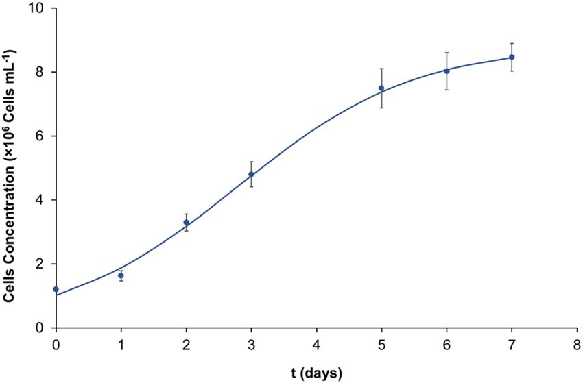
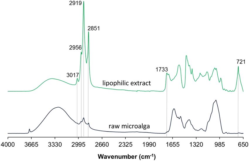
www.nature.com/scientificreports/ www.nature.com/scientificreports

 Figure 1. Growth curve of P. pinguis in f/2 growth medium.

 Figure 2. FTIR–ATR spectra of the lipophilic extractives and the raw marine microalga P. pinguis, the
 characteristic bands of the lipophilic extracts are highlighted.

 Statistical analysis. Statistical analysis of the data was carried out using the software IBM SPSS Statistics
 24. Differences between treatments were assessed with Student’s t-test, p-values < 0.05 were considered to be
 statistically significant.

 Results and Discussion
 Growth and extraction yield. The overall yield of a specific desired product is dependent on the microal-
 gal growth rate and on the product content26. Thus, when exploring the potential of a microalgal strain for further
 improvement in high valued compounds, the microalgal growth should be considered. The growth rate deter-
 mined for P. pinguis was 0.8 day−1 and the maximum cell concentration, reached by this microalga, was 8.46 × 10⁶
 cells mL−1, Fig. 1. According to Steinrucken, et al.27 this microalgal strain can be considered as a high growth rate
 strain since its growth rate is ≥0.7 day−1. The average dry biomass production observed at the end of the batch
 cultivation was 368 mg L−1 achieved in 7 days, this value being close to the dry weight (dw) estimated by Mansour,
 et al.18 of 390 mg L−1.
 The yields of the chloroform extractable substances, in P. pinguis, accounted for 11.92% dw. This yield was
 higher than that previously reported by Mansour, et al.18 for this microalga cultivated in the fE and GSe growth
 media and extracted with a modified Bligh and Dyer, in which, the chloroform extracts accounted 7.6% and 3.5%
 dw, respectively. The total chloroform extractable substances obtained in this study (5.17 pg cell−1) were also
 higher than that found for other P. pinguis strains (2 pg cell−1) grown in f/2 medium and extracted with chloro-
 form through modified Bligh and Dyer22.

 FTIR analysis. The FTIR-ATR was employed to P. pinguis biomass and chloroform extracts as a first approach
 to perform a qualitative analysis of the extractable substances of P. pinguis (Fig. 2). Through the FTIR spectrum
 of the raw microalga, it is possible to visualize three main regions, that relate to the main macromolecular pools:
 (i) the carbohydrate region between 1200–900 cm−1 (νC–O–C of carbohydrates); (ii) the protein bands at 1655
 cm−1 and 1545 cm−1 (νC-O of amide II and δN–H of amide I, respectively); (iii) and the lipid associated peaks at

Scientific Reports | (2020) 10:339 | https://doi.org/10.1038/s41598-019-57188-y 3
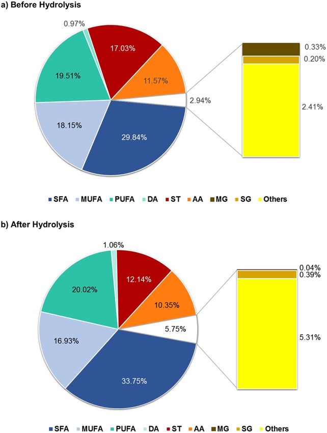
www.nature.com/scientificreports/ www.nature.com/scientificreports

 1740 (νC=O of the ester functional groups) and 3050–2800 cm−1 28. This observation indicates the co-presence of
 lipids, proteins and polysaccharides in the microalgal biomass. In the FTIR spectrum of the chloroform extract it
 is possible to visualize that the signals often attributed to the characteristic functional groups of lipids increased
 their intensity, namely those in the 3050–2800 cm−1 region (C-H stretch), which indicates that lipids are the major
 component of the lipophilic extract. Moreover, the presence of peaks at around 720, 1745 and 3010 cm−1, that are
 related to the CH2 rocking, C=O and C-H stretch, respectively, indicates the presence of unsaturated hydrocar-
 bons in the lipophilic extract29.

 GC-MS analysis. To screen microalgae for commercial purposes, different aspects have to be covered: the
 first is to explore the chemical diversity of microalgae; the second is to evaluate the quality of these extracts by
 searching for known bioactive compounds30.
 The microalgal strain under study presented a wide chemical diversity in the extract: 29 fatty acids, 14 sterols,
 13 fatty alcohols, and 16 other compounds. Through Table 1 it is possible to observe qualitative and quantitative
 differences, in the P. pinguis chloroform extracts, before and after hydrolysis. Before hydrolysis 71% of the extract-
 able substances were quantified while after hydrolysis this percentage increased to 88%.
 Alkaline hydrolysis is often used for the analysis of compounds in their esterified forms31. In the chloroform
 extracts, submitted to alkaline hydrolysis, total fatty acids increased by 32% being the major increase verified
 for the unsaturated fatty acids (46%). This slight increase might be explained from poor extraction, such that
 only the smaller lipids will be available for derivatization and analysis, including volatile compounds. Moreover,
 these observations reveal that 32% of total fatty acids were in complex forms. Additionally, the absence of sig-
 nificant differences (p < 0.05) in sterols amounts, in contrast to, sugars and monoglycerides levels, after alkaline
 hydrolysis, indicates that the major complex forms present in the chloroform extracts of P. pinguis where mainly
 glycolipids and fatty acids esterified with glycerol (mono, di and triglycerides). The chromatogram obtained for
 the derivatized chloroform extract of P. pinguis after alkaline hydrolysis is displayed in Fig. 3. According to Milke
 et al.11 the levels of free fatty acids in Pavlova spp., Chaetoceros muelleri and Placopecten magellanicus samples
 ranged 0.30 and 3.10%. In the P. pinguis studied, fatty acids before hydrolysis accounted 5.71% of microalgal dry
 weight. These differences might be explained by several factors such as: (i) the strain-to-strain variation; (ii) the
 cultivation conditions; (iii) the methodology applied.

 Fatty acids. In Fig. 4 it are represented the main families identified in the P. pinguis chloroform extractable sub-
 stances before (Fig. 4a) and after (Fig. 4b) alkaline hydrolysis. Through this figure it is possible to visualize that
 fatty acids were the main family present in the chloroform extracts accounting up to 72% of the total compounds
 identified (Fig. 4b). The major fatty acids found in P. pinguis were palmitoleic (C16:1ω7 – PAA), myristic (C14:0
 – MA), palmitic (C16:0 – PA) and eicosapentaenoic (C20:5ω3 – EPA) acids which together accounted over 30%
 of the total identified compounds before and after hydrolysis.
 Microalgae are the primary producers of essential fatty acids that cannot be synthesized by humans, which, in
 turn, must obtain them through their diet32. These are linoleic (C18:2ω6 - LA) and α-linolenic (C18:3ω3 - ALA)
 acids which represent the omega-6 LC-PUFA and the omega-3 LC-PUFA, respectively32. P. pinguis presented a
 high content of LA (1.86 mg g−1 dw) and minor amounts of ALA (0.89 mg g−1 dw). As main precursor of the ω3
 fatty acid synthesis, the minor amounts of ALA can be explained by the high levels of stearidonic (SA; 2.82 mg g−1
 dw), EPA (3.49 mg g−1 dw) and docosahexaenoic (2.01 mg g−1 dw) acids8,33. These results are consistent with those
 in literature that point P. pinguis as a high value omega-3 LC-PUFA producing strain14.
 Docosahexaenoic acid (C22:6ω3 – DHA) is one of the main components of the structural lipids of the brain,
 whereas EPA display an important role in cardiovascular and immunological health32. The government health
 agencies worldwide recommend a dietary intake of DHA and EPA ranging from 200 and 670 mg day−1 34. In P.
 pinguis, DHA and EPA accounted a total of 5.50 mg g−1 dw, which means that 122 g of dry microalga represents
 the highest dietary reference value.
 The ω6 fatty acids (main precursors of pro-inflammatory mediators) and the ω3 fatty acids (major precursors
 of anti-inflammatory molecules) compete for the same enzyme sets when metabolized35. Therefore, a balanced
 intake of ω6:ω3 fatty acids close to 1:1 is recommended. Western diets are characterized by high levels of ω6 Fatty
 acids and an unbalanced Σω6/Σω3 fatty acids ratio of 20:1 promoting the pathogenesis of various diseases32. This
 trend might be inverted by decreasing the intake of ω6 rich sources and increasing the intake of ω3 rich sources32.
 P. pinguis presented high amounts of ω3 fatty acids (13.36 mg g−1 dw) and a low Σω6/Σω3 fatty acids ratio (1:4)
 which, in turn, makes it suitable for dietary supply of ω3 fatty acids. This ratio was close to that obtained by
 Slocombe, et al.14 (1:3) for P. pinguis.

 Fatty alcohols. Fatty alcohols have been studied for their antibacterial activity and cholesterol-lowering abil-
 ity36,37. The biological properties of these biomolecules are linked with the carbon chain length that is thought to
 determine their antibacterial activity and mode of action in biological systems36. Fatty alcohols accounted up to
 12% of the total compounds identified (Fig. 4a). The major fatty alcohols found in P. pinguis were hexadecanol
 (C16-OH), octadece-9-nol (C18:1-OH) and octadecanol (C18-OH) which together accounted up to 40% of the
 total fatty alcohols. The mass fragmentation of C18:1-OH is displayed in Fig. 5a. Through this figure it is possible
 to observe three key fragment ions from alcohols: the base peak at m/z 75 [(CH3)2SiOH]+, the m/z 325 [M − 15]+
 and the molecular ion [M]+ at m/z 340.
 In P. pinguis, it was identified two very long chain alcohols: octacosanol (C28-OH) and dotriacontanol
 (C32-OH). The consumption of 5–20 mg day−1 of very long aliphatic chain alcohols is known to decrease the
 low-density lipoprotein (LDL) cholesterol9,37. Therefore, 3.04–12.15 g of microalgal biomass and 0.36–1.44 g of
 microalgal extract are the quantities needed to fulfill these requirements (Table 1). Moreover, alcohols such as

Scientific Reports | (2020) 10:339 | https://doi.org/10.1038/s41598-019-57188-y 4
www.nature.com/scientificreports/ www.nature.com/scientificreports

 Content (mg g−1 of microalgal dw) Content (mg g−1 of extract)
 N° Identified Compounds MW BH AH BH AH
 Fatty acids 57.34 ± 1.33a 75.60 ± 4.49b 482.95 ± 26.06a 631.77 ± 23.20b
 Saturated 24.99 ± 0.67a 35.55 ± 2.60b 210.60 ± 12.07a 297.02 ± 14.12b
 2 Octanoic acid 216 n.d. 1.03 ± 0.00 n.d. 8.65 ± 0.27
 4 Nonanoic acid 230 0.77 ± 0.00a 1.05 ± 0.01b 6.49 ± 0.23a 8.78 ± 0.20b
 6 Decanoic acid 244 n.d. 1.03 ± 0.01 n.d. 8.62 ± 0.34
 13 Dodecanoic acid 272 0.78 ± 0.01a 1.05 ± 0.01b 6.54 ± 0.28a 8.79 ± 0.27b
 17 Myristic acid 300 7.43 ± 0.41a 9.43 ± 0.54b 62.62 ± 5.37a 78.86 ± 4.03b
 20 Pentadecanoic acid 314 1.06 ± 0.02a 1.38 ± 0.03b 8.91 ± 0.45a 11.53 ± 0.25b
 28 Palmitic acid (PA) 328 7.32 ± 0.22a 9.97 ± 1.66b 61.71 ± 3.62a 83.13 ± 11.56b
 18 Hexadecanoic methyl ester 270 0.85 ± 0.01a 1.05 ± 0.01b 7.14 ± 0.34a 8.79 ± 0.36b
 30 Heptadecanoic acid a
 342 2.54 ± 0.01a
 3.26 ± 0.02 b
 21.37 ± 0.58 a
 27.29 ± 0.71b
 39 2-Octyl-Cyclopropaneheptanoic acid 354 0.82 ± 0.00a
 1.09 ± 0.01 b
 6.89 ± 0.23 a
 9.10 ± 0.31b
 38 Octadecanoic acid 356 1.81 ± 0.03a
 3.07 ± 0.50 b
 15.24 ± 0.67 a
 25.61 ± 3.40b
 46 Eicosanoic acid 384 0.79 ± 0.00a
 1.07 ± 0.01 b
 6.69 ± 0.24 a
 8.93 ± 0.20b
 53 Docosanoic acid 412 0.83 ± 0.02a
 1.07 ± 0.01 b
 7.00 ± 0.24 a
 8.94 ± 0.23b
 Monounsaturated 15.20 ± 0.54a 17.83 ± 1.00b 128.12 ± 8.53a 149.07 ± 5.78b
 24 Palmitoleic acida 326 10.97 ± 0.39a 13.06 ± 0.78b 92.46 ± 6.15a 109.19 ± 5.00b
 25 7-Hexadecenoic acid 326 0.92 ± 0.02a 1.17 ± 0.01b 7.75 ± 0.37a 9.79 ± 0.31b
 37 Vaccenic acid 354 1.27 ± 0.08a 1.72 ± 0.16b 10.68 ± 0.98a 14.33 ± 0.91b
 36 Oleic acid 354 1.25 ± 0.08a 1.89 ± 0.08b 10.55 ± 0.93a 15.76 ± 0.28b
 44 Gondoic acid 382 0.79 ± 0.00 n.d. 6.68 ± 0.22 a
 n.d.
 Polyunsaturated 16.34 ± 0.29a 21.09 ± 0.92b 137.38 ± 5.69a 176.31 ± 4.50b
 22 4,7,10,13-Hexadecatetraenoic acid 322 0.82 ± 0.01a
 1.08 ± 0.01 b
 6.91 ± 0.27 a
 9.01 ± 0.29b
 26 Methyl-4,7,10,13-hexadecatetraenoate 262 0.85 ± 0.01a
 1.05 ± 0.00 b
 6.86 ± 0.27 a
 8.80 ± 0.28b
 34 Linoleic acid (LA) a
 352 1.86 ± 0.03a
 2.58 ± 0.03 b
 15.65 ± 0.71 a
 21.59 ± 0.81b
 35 α-Linolenic acid (ALA) 350 0.89 ± 0.01a
 1.18 ± 0.02 b
 7.49 ± 0.30 a
 9.90 ± 0.29b
 33 Stearidonic acid (SA) 348 2.82 ± 0.09a 3.04 ± 0.41a 23.80 ± 1.47a 25.39 ± 2.67a
 32 3,6,9,12,15-Octadecapentaenoic acid 346 0.90 ± 0.01a 1.18 ± 0.02b 7.60 ± 0.30a 9.84 ± 0.33b
 42 Eicosapentaenoic acid (EPA) 374 3.49 ± 0.09a 4.73 ± 0.26b 29.43 ± 1.44a 39.54 ± 1.51b
 48 7,10,13,16,19-Docosapentaenoic acid 402 1.59 ± 0.08a 2.05 ± 0.08b 13.36 ± 0.68a 17.14 ± 0.51b
 49 4,7,10,13,16-Docosapentaenoic acid 402 1.11 ± 0.02a 1.53 ± 0.06b 9.37 ± 0.32a 12.80 ± 0.32b
 47 Docosahexaenoic acid (DHA) 400 2.01 ± 0.10a
 2.67 ± 0.15 b
 16.91 ± 0.76 a
 22.30 ± 0.95b
 Diacids 0.81 ± 0.01 a
 1.12 ± 0.03 b
 6.86 ± 0.27 a
 9.37 ± 0.33b
 50 Octadecenedioic acid 456 0.81 ± 0.01a
 1.12 ± 0.03 b
 6.86 ± 0.27 a
 9.37 ± 0.33b
 Fatty Alcohols 9.69 ± 0.26 a
 10.90 ± 0.39 b
 81.54 ± 0.32 a
 91.14 ± 0.90b
 1 1-Octanol 202 n.d. 1.20 ± 0.08 n.d. 10.05 ± 0.35
 7 1-Undecanol 244 0.63 ± 0.01a 0.83 ± 0.00b 5.31 ± 0.24a 6.93 ± 0.23b
 12 1-Dodecanol 258 0.63 ± 0.00a 0.83 ± 0.01b 5.28 ± 0.18a 6.99 ± 0.25b
 14 1-Tridecanol 272 0.64 ± 0.01a 0.83 ± 0.01b 5.36 ± 0.26a 6.98 ± 0.25b
 16 1-Tetradecanol 286 0.67 ± 0.01a 0.88 ± 0.01b 5.61 ± 0.26a 7.36 ± 0.17b
 21 1-Hexadecanol 314 1.50 ± 0.09a 1.21 ± 0.08b 12.63 ± 0.39a 10.07 ± 0.42b
 31 1-Octadecanol 342 1.18 ± 0.06a 1.17 ± 0.07a 9.94 ± 0.16a 9.81 ± 0.31a
 29 Octadece-9-nol 340 1.46 ± 0.12a
 1.21 ± 0.12 b
 12.29 ± 0.62 a
 10.10 ± 0.72b
 43 1-Eicosanol 368 0.69 ± 0.01 n.d. 5.77 ± 0.15 a
 n.d.
 51 1-Docosanol 396 0.65 ± 0.01a 0.85 ± 0.00b 5.48 ± 0.13a 7.10 ± 0.19b
 58 1-Octacosanol 482 0.97 ± 0.02a
 1.02 ± 0.06a
 8.13 ± 0.18 a
 8.54 ± 0.25b
 71 1-Dotriacontanol 538 0.68 ± 0.00a
 0.86 ± 0.01b
 5.73 ± 0.15 a
 7.21 ± 0.21b
 Sterols 14.26 ± 1.04a 12.79 ± 1.01a 120.26 ± 11.78a 106.86 ± 6.16a
 57 22-Stigmasten-3-one 412 0.26 ± 0.02 n.d. 2.15 ± 0.20a n.d.
 59 Stigmastane-3,6-dione 428 0.34 ± 0.06a 0.19 ± 0.01b 2.84 ± 0.58a 1.56 ± 0.09b
 60 Campesterol 472 0.57 ± 0.02a 0.56 ± 0.03a 4.81 ± 0.29a 4.71 ± 0.26a
 61 Stigmasterol 484 6.14 ± 0.52a 5.07 ± 0.29b 51.75 ± 5.69a 42.38 ± 1.99b
 62 24-Ethyl-δ(22)-coprostenol 486 0.75 ± 0.10a
 0.63 ± 0.04a
 6.31 ± 0.92a
 5.25 ± 0.31a
 63 β-Sitosterol 486 0.78 ± 0.03a
 0.90 ± 0.21a
 6.53 ± 0.31a
 7.46 ± 1.55a
 64 4α,24-Dimethyl-5α-cholestan-3β-ol 488 1.09 ± 0.04a
 0.95 ± 0.11a
 9.22 ± 0.57a
 7.97 ± 0.72b

 Continued

Scientific Reports | (2020) 10:339 | https://doi.org/10.1038/s41598-019-57188-y 5
www.nature.com/scientificreports/ www.nature.com/scientificreports

 Content (mg g−1 of microalgal dw) Content (mg g−1 of extract)
 N° Identified Compounds MW BH AH BH AH
 66 4α-methyl,24-ethyl-5α-cholest-22E-en-3β-ol 500 0.77 ± 0.04a 0.70 ± 0.10b 6.52 ± 0.54a 5.82 ± 0.71a
 68 4α-methyl-24-ethyl-5α-cholestan-3-ol 502 n.d. 0.34 ± 0.03 n.d. 2.86 ± 0.23
 69 4α,24β-dimethyl-5α-cholestan-3β,4β-diol 504* 0.37 ± 0.04a 0.32 ± 0.02a 3.10 ± 0.44a 2.65 ± 0.2a
 72 4α-methyl-24β-ethyl-5α-cholestan-3β,4β-diol 518* 0.71 ± 0.09a 0.58 ± 0.05b 6.04 ± 0.93a 4.88 ± 0.32a
 67 Unidentified C30 Sterol 1.96 ± 0.12a 1.64 ± 0.18b 16.48 ± 1.35a 13.66 ± 1.12b
 70 Unidentified C30 Sterol 0.17 ± 0.01a 0.21 ± 0.01b 1.45 ± 0.08a 1.76 ± 0.12b
 65 Unidentified C30 Sterol 0.36 ± 0.02a 0.71 ± 0.07b 3.06 ± 0.26a 5.90 ± 0.46b
 Monoglycerides 0.27 ± 0.01a 0.04 ± 0.01b 2.31 ± 0.12a 0.37 ± 0.05b
 40 1-Monotridecanoin 432 0.06 ± 0.00a 0.04 ± 0.01b 0.52 ± 0.05a 0.37 ± 0.05b
 45 Monomyristin 446 0.09 ± 0.01 n.d. 0.72 ± 0.05a n.d.
 52 Monopalmitin 474 0.07 ± 0.01 n.d. 0.59 ± 0.07a n.d.
 55 Monostearin 502 0.06 ± 0.01 n.d. 0.48 ± 0.07a n.d.
 Sugars 0.17 ± 0.01a 0.41 ± 0.02b 1.42 ± 0.10a 3.64 ± 0.02b
 19 Rhamnose 452 n.d. 0.21 ± 0.01 n.d. 1.85 ± 0.02
 27 Deoxyglucose 452 n.d. 0.20 ± 0.01 n.d. 1.79 ± 0.00
 56 Glucosamine 612 0.17 ± 0.01 n.d. 1.42 ± 0.10a n.d.
 Others 2.02 ± 0.02a 5.60 ± 0.07b 17.05 ± 0.83a 46.84 ± 1.29b
 3 2,4,6,8-Tetramethyl-1-undecene 210 0.03 ± 0.00 n.d. 0.21 ± 0.04a n.d.
 5 2-Methyltetradecane 212 0.05 ± 0.01 n.d. 0.46 ± 0.07a n.d.
 8 2-Methyl-4-nonadecene 280 0.03 ± 0.00 n.d. 0.24 ± 0.04a n.d.
 10 3-Methyl-4-nonadecene 280 0.04 ± 0.01a 0.05 ± 0.01a 0.32 ± 0.07a 0.41 ± 0.10a
 9 2,6-bis(1,1-Dimethylethyl)phenol 278 0.99 ± 0.02a
 1.56 ± 0.08b
 8.43 ± 0.43a
 13.02 ± 0.28b
 15 Methylsuccinic acid 276 0.05 ± 0.01 n.d. 0.45 ± 0.04a
 n.d.
 11 3-Methoxycinnamic acid 250 n.d. 1.18 ± 0.01 n.d. 9.88 ± 0.38
 41 Dehydroabietic acid 372 0.18 ± 0.02a 0.66 ± 0.05b 1.49 ± 0.13a 5.55 ± 0.49b
 54 Pinoresinol 502 n.d. 1.30 ± 0.03 n.d. 10.85 ± 0.55
 23 Mannitol 614 0.65 ± 0.00a 0.85 ± 0.01b 5.46 ± 0.21a 7.13 ± 0.29b
 Total Identified 83.75 ± 2.00a 105.34 ± 5.90b 705.52 ± 37.96a 880.41 ± 29.68b

 Table 1. Compounds detected in the lipophilic extracts of P. pinguis before (BH) and after (AH) alkaline
 hydrolysis. Values (means ± SD of four replicates) in the same row, not sharing a common superscript
 are significantly different (p < 0.05). Compounds are numbered by their elution order (see Fig. 2). All the
 compounds containing hydroxyl and/or carboxyl groups are identified as the correspondent TMS derivatives.
 MW – Molecular weight of compounds after silylation. aContains the iso- and anteiso-isomers; bContains cis
 and trans isomers; *Identified as the mono-TMS ether; n.d. – non detected; dw – dry weight.

 Figure 3. Chromatogram of the derivatized P. pinguis lipophilic extract after alkaline hydrolysis. Peak
 identification as in Table 1. IS – Internal Standard (Tetracosane, 0.40 mg).

Scientific Reports | (2020) 10:339 | https://doi.org/10.1038/s41598-019-57188-y 6
www.nature.com/scientificreports/ www.nature.com/scientificreports

 Figure 4. Main families identified in P. pinguis lipophilic extracts (a) before and (b) after alkaline hydrolysis, in
 percentage of the total identified compounds.

 undecanol (C11-OH), dodecanol (C12-OH) and tridecanol (C13-OH), have been pointed in previous studies by
 their bactericidal activity36. After alkaline hydrolysis the fatty alcohols increased by 12%, with the highest increase
 verified for the C12-OH (32%).
 The detected docosanol (C22-OH) is known by its antiproliferative effect of chinese hamster ovary cells K1
 (CHO-K1) and human melanoma (CRL-1974TM) cell lines38. Investigations concerning the effects of long chain
 alcohols are often performed with long chain alcohols isolated from sugarcane where these compounds make
 up 0.10–0.30% of its mass39. In the present study this class of compounds comprised 0.16 and 0.19% of P. pinguis
 dried biomass, before and after alkaline hydrolysis respectively.

 Sterols. Microalgae are recognized for their wide diversity of sterols that are often used for chemotaxonomic and
 phylogenetic comparisons7. This diversity along with the high sterol content make microalgae promising sources
 of novel sterols with potential novel activities40.
 Sterols accounted up to 17% of the total identified compounds (Fig. 4a). The major sterols were stigmasterol
 (40–43%), 4α, 24-Dimethyl-5α.cholestan-3β-ol (7–8%) and an unidentified sterol (13–14%), which together
 accounted over 50% of total sterols (Table 1). The dominance of stigmasterol across Pavlova species namely P. pin-
 guis have been reported in studies targeting aquaculture and chemotaxonomy11,22. β-sitosterol, campesterol and
 stigmasterol - in their non-esterified forms, have been subject to the Food and Drug Admnistration (FDA) health
 claim for reduced risk of coronary heart disease40. P. pinguis presented high levels of stigmasterol which alone rep-
 resented up to 7% of the total identified compounds in the lipophilic profile. Campesterol and β-sitosterol were
 also identified in the P. pinguis chloroform extracts accounting 0.57 and 0.78 mg g−1 of microalgal biomass and
 4.81 and 6.53 mg g−1 of extract, respectively (Table 1). In contrast to Milke, et al.11 stigmastanol and cholesterol
 were not detected in P. pinguis lipophilic extracts.

Scientific Reports | (2020) 10:339 | https://doi.org/10.1038/s41598-019-57188-y 7
www.nature.com/scientificreports/ www.nature.com/scientificreports

 Figure 5. Mass spectra of some assigned peaks as trimethylsilyl (TMS) ethers and/or esters: (a) Octadece-9-nol
 (peak 29), (b) Monomyristin (peak 45), (c) Pinoresinol (peak 54) and (d) 4α-methyl-24β-ethyl-5α-cholestan-
 3β,4β-diol as mono TMS ether (peak 72).

 Pavlova species are often recognized by their unusual dihydroxylated sterols called Pavlovols17. In P. pin-
 guis two dihydroxylated sterols were identified, 4α-methyl-24β-ethyl-5α-cholestan-3β, 4β-diol (ethylpavlovol)
 and 4α, 24β-dimethyl-5α-cholestan-3β, 4β-diol (methylpavlovol), and a structurally isomeric form of dinos-
 terol, 4α-methyl-24-ethyl-5α-cholest-22E-en-3β-ol. As with fatty acids, the sterol profiles of microalgae are
 species-specific and often used as chemotaxonomic markers17.
 Despite in Milke, et al.11 the pavlovols have not been found for Pavlova species, the authors recognize that they
 can constitute the sterol composition of this microalga specie. Moreover, Volkman, et al.22 found the existence of
 these unusual sterols in the composition of two other P. pinguis strains and pointed pavlovols as chemotaxonomic
 markers of Pavlovales.
 Figure 5b shows the mass spectrum of the compound identified as ethylpavlovol. Through comparison of
 the obtained mass spectrum with the one obtained previously by Volkman, et al.22, it was possible to identify the
 ethylpavlovol as its mono TMS ether. The assignment was done by the presence of the following mass fragments:
 m/z 43, 487, 503 and 518, as well as, the base peak at m/z 371 [M-147(C3H6O2TMS)]+.
 Phytosterols are playing a key role in nutraceutic and pharmaceutical industries, as precursors of some bioac-
 tive molecules7,40. Moreover, it is estimated that the dietary intake of phytosterols is in the range of 150 to 400 mg
 day−1. P. pinguis can contribute to the intake of around 143 mg of free sterols per 100 g of microalgal dry weight.
 After alkaline hydrolysis it were not verified significant differences (p < 0.05) in the amounts of sterols (Table 1).
 This observation indicates that sterols were non-esterified and were as free sterols.

 Other compounds. In the classes of monoglycerides, sugars and other components, compositional differences
 before and after hydrolysis were verified. In Table 1 it is possible to observe that after alkaline hydrolysis the mono-
 glycerides: monomyristin, monopalmitin and monostearin, were not detected. Sugars and the other components
 classes were those who presented the highest increase after alkaline hydrolysis, 3 and 5 times higher respectively.
 The increase of the sugar content suggests the presence of polar lipids, namely glycolipids incorporating rham-
 nose and deoxyglucose, in the chloroform extracts of P. pinguis. The sulfoquinovosyl diacylglycerols (SQDGs) are
 one of the most abundant glycolipids found in microalgal cells41. SQDGs are constituted by a 6-sulfoquinovose
 unit, which, in turn, is constituted by a sulphur group attached to the quinovose (6-deoxyglucose)42. Moreover, it
 has been reported that microalgal glycolipids may contain other sugar moieties than galactose such as mannose
 and rhamnose43.
 The mass fragmentation of monomyristin is presented in Fig. 5c. Although in the mass spectrum it is not pos-
 sible to visualize the molecular ion, the presence of the following ions: m/z 73, 103, 147, 205 347 indicates that this
 compound is monomyristin. The fragment m/z 347 corresponds to the mass fragment [M-103 (CH2OTMS)]+
 and the m/z 103, 147 and 205 are associated to the silylated glycerol backbone.
 The detection of smaller amounts of 3-methocycinnamic acid and pinoresinol 1.12 and 1.23% of total
 identified compounds, respectively, was observed after alkaline hydrolysis. According to Klejdusa, et al.44 the

Scientific Reports | (2020) 10:339 | https://doi.org/10.1038/s41598-019-57188-y 8
www.nature.com/scientificreports/ www.nature.com/scientificreports

 Figure 6. 2,2-diphenyl-1-picrylhydrazyl (DPPH) radical scavenging activity (%) of P. pinguis chloroform
 extracts (µg mL−1). Butylated hydroxytoluene (BHT) was used as a reference compound and its EC50 value is
 9.79 g mL−1.

 cinnamic acid derivatives are precursors in the phenyl-propanoid pathway for the synthesis of polyphenols,
 which indicates that it is possible that this microalgal strain have other phenols that could be extracted by polar
 solvents. In Fig. 5d it is possible to visualize the fragmentation pattern of obtained for pinoresinol, namely the
 base peak m/z 73 [(CH3)3Si]+, the m/z 487 resultant from the loss of a methyl group [M-15]+ and the molec-
 ular ion [M]+ m/z 502. Phenols are natural products that are recognized by their antioxidant, antimicrobial
 and antiviral activities44.
 P. pinguis chloroform extracts presented a strong concentration-dependent DPPH radical scavenging activity
 with a determination coefficient close to 1 (R2 = 0.99), Fig. 6. The presence of phenols in the lipophilic fraction
 after alkaline hydrolysis suggest that this ability might be resultant from polyphenol-associated lipids. The esti-
 mated EC50 for P. pinguis chloroform extracts was of 1 057 µg mL−1.
 Besides nutraceutics and pharmaceutics the rich composition verified for P. pinguis as for other species of the
 genus Pavlova45 can also be used in aquaculture for animal consumption as dietetic supply and in food industry as
 additive and/or nutritional supplements attributing to consumer a higher level of bioactive compounds.

 Conclusions
 The need for naturally derived health promoting phytochemicals instead of the chemically derived drugs have
 prompted the search for new sources of natural products. Pavlova pinguis presented a manifold range of metab-
 olites which demonstrated its versatility and potential as a source of high value compounds. The high content of
 unsaturated fatty acids, long chain aliphatic alcohols and stigmasterol, demonstrate the potential of this microalga
 not only for aquaculture but also for nutraceutics and pharmaceutics uses. To fully exploit the phytochemical
 features of microalgae for commercial purposes, a non-targeted approach should be taken in order to uncover
 whole extract chemical diversity.

 Received: 17 June 2019; Accepted: 19 December 2019;
 Published: xx xx xxxx

 References
 1. Falaise, C. et al. Antimicrobial Compounds from Eukaryotic Microalgae against Human Pathogens and Diseases in Aquaculture.
 Mar. Drugs 14, https://doi.org/10.3390/md14090159 (2016).
 2. Shannon, E. & Abu-Ghannam, N. Antibacterial Derivatives of Marine Algae: An Overview of Pharmacological Mechanisms and
 Applications. Mar. Drugs 14, https://doi.org/10.3390/md14040081 (2016).
 3. Talero, E. et al. Bioactive Compounds Isolated from Microalgae in Chronic Inflammation and Cancer. Mar. Drugs 13, 6152–6209,
 https://doi.org/10.3390/md13106152 (2015).
 4. Barra, L., Chandrasekaran, R., Corato, F. & Brunet, C. The challenge of ecophysiological biodiversity for biotechnological
 applications of marine microalgae. Mar. Drugs 12, 1641–1675, https://doi.org/10.3390/md12031641 (2014).
 5. Stengel, D. B., Connan, S. & Popper, Z. A. Algal chemodiversity and bioactivity: sources of natural variability and implications for
 commercial application. Biotechnol. Adv. 29, 483–501, https://doi.org/10.1016/j.biotechadv.2011.05.016 (2011).
 6. Yao, L., Gerde, J. A., Lee, S. L., Wang, T. & Harrata, K. A. Microalgae lipid characterization. J. Agric. Food Chem. 63, 1773–1787,
 https://doi.org/10.1021/jf5050603 (2015).
 7. Francavilla, M., Trotta, P. & Luque, R. Phytosterols from Dunaliella tertiolecta and Dunaliella salina: a potentially novel industrial
 application. Bioresour. Technol. 101, 4144–4150, https://doi.org/10.1016/j.biortech.2009.12.139 (2010).
 8. Fernandes, T., Fernandes, I., Andrade, C. A. P. & Cordeiro, N. Changes in fatty acid biosynthesis in marine microalgae as a response
 to medium nutrient availability. Algal Res. 18, 314–320, https://doi.org/10.1016/j.algal.2016.07.005 (2016).
 9. Santos, S. A. et al. Chlorophyta and Rhodophyta macroalgae: a source of health promoting phytochemicals. Food Chem. 183,
 122–128, https://doi.org/10.1016/j.foodchem.2015.03.006 (2015).
 10. Katiyar, R. et al. Microalgae: An emerging source of energy based bio-products and a solution for environmental issues. Renew. Sust.
 Energ. Rev. 72, 1083–1093, https://doi.org/10.1016/j.rser.2016.10.028 (2017).

Scientific Reports | (2020) 10:339 | https://doi.org/10.1038/s41598-019-57188-y 9
www.nature.com/scientificreports/ www.nature.com/scientificreports

 11. Milke, L. M., Bricelj, V. M. & Parrish, C. C. Biochemical characterization and nutritional value of three Pavlova spp. in unialgal and
 mixed diets with Chaetoceros muelleri for postlarval sea scallops, Placopecten magellanicus. Aquaculture 276, 130–142, https://doi.
 org/10.1016/j.aquaculture.2008.01.040 (2008).
 12. Parrish, C. C., Milke, L. M. & Bricelj, V. M. Characterisation of 4α-methyl sterols in Pavlova spp. and postlarval sea scallops,
 Placopecten magellanicus. Aquaculture 311, 261–262, https://doi.org/10.1016/j.aquaculture.2010.11.003 (2011).
 13. Ponis, E. et al. Nutritional value of six Pavlovophyceae for Crassostrea gigas and Pecten maximus larvae. Aquaculture 254, 544–553,
 https://doi.org/10.1016/j.aquaculture.2005.11.017 (2006).
 14. Slocombe, S. P. et al. Unlocking nature’s treasure-chest: screening for oleaginous algae. Scientific reports 5, 9844, https://doi.
 org/10.1038/srep09844 (2015).
 15. Azizan, A. et al. Metabolite Profiling of the Microalgal Diatom Chaetoceros Calcitrans and Correlation with Antioxidant and Nitric
 Oxide Inhibitory Activities via (1)H NMR-Based Metabolomics. Mar. Drugs 16, https://doi.org/10.3390/md16050154 (2018).
 16. Green, J. C. A new species of Pavlova from Madeira. British Phycological Bulletin 3, 299–303, https://doi.
 org/10.1080/00071616700650181 (1967).
 17. Volkman, J. K. In The Physiology of Microalgae, Developments in Applied Phycology Vol. 6 (eds. Borowitzka, M. A., Beardall, J. &
 Raven, J. A.) (Springer, 2016).
 18. Mansour, M. P., Frampton, D. M. F., Nichols, P. D., Volkman, J. K. & Blackburn, S. I. Lipid and fatty acid yield of nine stationary-
 phase microalgae: Applications and unusual C24–C28 polyunsaturated fatty acids. J. Appl. Phycol. 17, 287–300, https://doi.
 org/10.1007/s10811-005-6625-x (2005).
 19. Fernandes, T., Fernandes, I., Andrade, C. A. P. & Cordeiro, N. Marine microalgae growth and carbon partitioning as a function of
 nutrient availability. Bioresour. Technol. 214, 541–547, https://doi.org/10.1016/j.biortech.2016.05.001 (2016).
 20. Ma, N. L., Teh, K. Y., Lam, S. S., Kaben, A. M. & Cha, T. S. Optimization of cell disruption methods for efficient recovery of bioactive
 metabolites via NMR of three freshwater microalgae (chlorophyta). Bioresour. Technol. 190, 536–542, https://doi.org/10.1016/j.
 biortech.2015.03.036 (2015).
 21. Christie, W. W. The Lipid Web, http://www.lipidhome.co.uk (2018).
 22. Volkman, J. K., Farmer, C. L., Barrett, S. M. & Sikes, E. L. Unusual dihydroxysterols as chemotaxonomic markers for microalgae
 from the order Pavlovales (Haptophyceae). J. Phycol. 33, 1016–1023 (1997).
 23. Gutiérrez, A., Rodríguez, I. M. & del Río, J. C. Chemical composition of lipophilic extractives from sisal (Agave sisalana) fibers.
 Industrial Crops and Products 28, 81–87, https://doi.org/10.1016/j.indcrop.2008.01.008 (2008).
 24. Silvério, F. O., Barbosa, L. C. A., Silvestre, A. J. D., Piló-Veloso, D. & Gomide, J. L. Comparative study on the chemical composition
 of lipophilic fractions from three wood tissues of Eucalyptus species by gas chromatography-mass spectrometry analysis. Journal of
 Wood Science 53, 533–540, https://doi.org/10.1007/s10086-007-0901-0 (2007).
 25. Maadane, A. et al. Antioxidant activity of some Moroccan marine microalgae: Pufa profiles, carotenoids and phenolic content.
 Journal of biotechnology 215, 13–19, https://doi.org/10.1016/j.jbiotec.2015.06.400 (2015).
 26. Sukenic, A. Ecophysiological considerations in the optimization of Eicosapentaenoic acid production by Nannochloropsis sp.
 (Eustigmatophyceae). Bioresour. Technol. 35, 263–269 (1991).
 27. Steinrucken, P., Erga, S. R., Mjos, S. A., Kleivdal, H. & Prestegard, S. K. Bioprospecting North Atlantic microalgae with fast growth
 and high polyunsaturated fatty acid (PUFA) content for microalgae-based technologies. Algal Res. 26, 392–401, https://doi.
 org/10.1016/j.algal.2017.07.030 (2017).
 28. Driver, T. et al. Metabolic responses of eukaryotic microalgae to environmental stress limit the ability of FT-IR spectroscopy for
 species identification. Algal Res. 11, 148–155, https://doi.org/10.1016/j.algal.2015.06.009 (2015).
 29. Forfang, K., Zimmermann, B., Kosa, G., Kohler, A. & Shapaval, V. FTIR Spectroscopy for Evaluation and Monitoring of Lipid
 Extraction Efficiency for Oleaginous Fungi. PloS one 12, 1–17, https://doi.org/10.1371/journal.pone.0170611 (2017).
 30. Martin, A. C. et al. Evaluating solvent extraction systems using metabolomics approaches. RSC Adv. 4, 26325–26334 (2014).
 31. Yu, S. et al. Characterization of steryl glycosides in marine microalgae by gas chromatography-triple quadrupole mass spectrometry
 (GC-QQQ-MS). J. Sci. Food Agric. 98, 1574–1583, https://doi.org/10.1002/jsfa.8629 (2018).
 32. Simopoulos, A. P. An Increase in the Omega-6/Omega-3 Fatty Acid Ratio Increases the Risk for Obesity. Nutrients 8, 128, https://
 doi.org/10.3390/nu8030128 (2016).
 33. Huerlimann, R. et al. Effects of growth phase and nitrogen starvation on expression of fatty acid desaturases and fatty acid
 composition of Isochrysis aff. galbana (TISO). Gene 545, 36–44, https://doi.org/10.1016/j.gene.2014.05.009 (2014).
 34. Ryan, A. S. et al. Effects of long-chain polyunsaturated fatty acid supplementation on neurodevelopment in childhood: a review of
 human studies. Prostaglandins Leukot. Essent. Fatty Acids 82, 305–314, https://doi.org/10.1016/j.plefa.2010.02.007 (2010).
 35. Das, U. N. Essential fatty acids: biochemistry, physiology and pathology. Biotechnol. J. 1, 420–439, https://doi.org/10.1002/
 biot.200600012 (2006).
 36. Togashi, N. et al. Antibacterial Activity of Long-Chain Fatty Alcohols against Staphylococcus aureus. Molecules 12, 139–148 (2007).
 37. Hargrove, J. L., Greenspan, P. & Hartle, D. K. Nutritional Significance and Metabolism of Very Long Chain Fatty Alcohols and Acids
 from Dietary Waxes. Exp. Biol. Med. 229, 215–226 (2004).
 38. Vergara, M., Olivares, A. & Altamirano, C. Antiproliferative evaluation of tall-oil docosanol and tetracosanol over CHO-K1 and
 human melanoma cells. Electronic Journal of Biotechnology 18, 291–294, https://doi.org/10.1016/j.ejbt.2015.05.004 (2015).
 39. Jackson, M. A. & Eller, F. J. Isolation of long-chain aliphatic alcohols from beeswax using lipase-catalyzed methanolysis in
 supercritical carbon dioxide. The Journal of Supercritical Fluids 37, 173–177, https://doi.org/10.1016/j.supflu.2005.08.008 (2006).
 40. Luo, X., Su, P. & Zhang, W. Advances in Microalgae-Derived Phytosterols for Functional Food and Pharmaceutical Applications.
 Mar. Drugs 13, 4231–4254, https://doi.org/10.3390/md13074231 (2015).
 41. da Costa, E., Silva, J., Mendonca, S. H., Abreu, M. H. & Domingues, M. R. Lipidomic Approaches towards Deciphering Glycolipids
 from Microalgae as a Reservoir of Bioactive Lipids. Marine drugs 14, https://doi.org/10.3390/md14050101 (2016).
 42. Domonkos, I., Kis, M. & Gombos, Z. Versatile roles of lipids and carotenoids in membranes. Acta Biologica Szegediensis 59, 83–104
 (2015).
 43. Guschinak, I. A., Harwood, J. L. In Algae for Biofuels and Energy (ed. Borowitzka, M. A., Moheimani, N. R.) 17–36 (Springer, 2013).
 44. Klejdusa, B., Kopecký, J., Benešová, L. & Vacek, J. Solid-phase/supercritical-fluid extraction for liquid chromatography of phenolic
 compounds in freshwater microalgae and selected cyanobacterial species. J. Chromatogr. A 1216, 763–771, https://doi.org/10.1016/j.
 chroma.2008.11.096 (2009).
 45. Guihéneuf, F., Mimouni, V., Ulmann, L. & Tremblin, G. Combined effects of irradiance level and carbon source on fatty acid and
 lipid class composition in the microalga Pavlova lutheri commonly used in mariculture. Journal of Experimental Marine Biology and
 Ecology 369, 136–143, https://doi.org/10.1016/j.jembe.2008.11.009 (2009).

 Acknowledgements
 This work was partially supported by the European Territorial Cooperation Programme PCT-MAC 2014–2020
 through project REBECA (MAC/1.1a/060). Tomásia Fernandes was financially supported by a doctoral grant
 from ARDITI (Regional Agency for Development of Research, Technology and Innovation of Madeira), Project
 M1420-09-5369-FSE-000001.

Scientific Reports | (2020) 10:339 | https://doi.org/10.1038/s41598-019-57188-y 10
www.nature.com/scientificreports/ www.nature.com/scientificreports

 Author contributions
 T.F. and N.C. contributed on the conception and design of the study. T.F. executed the experiment, analyzed and
 interpreted the data, and wrote the manuscript. N.C. and A.M. made possible the experiment execution with
 administrative and financial support, supervised the experimental and made the critical revision for important
 intellectual content of the manuscript. All the authors read and final approved the manuscript.

 Competing interests
 The authors declare no competing interests.

 Additional information
 Correspondence and requests for materials should be addressed to N.C.
 Reprints and permissions information is available at www.nature.com/reprints.
 Publisher’s note Springer Nature remains neutral with regard to jurisdictional claims in published maps and
 institutional affiliations.
 Open Access This article is licensed under a Creative Commons Attribution 4.0 International
 License, which permits use, sharing, adaptation, distribution and reproduction in any medium or
 format, as long as you give appropriate credit to the original author(s) and the source, provide a link to the Cre-
 ative Commons license, and indicate if changes were made. The images or other third party material in this
 article are included in the article’s Creative Commons license, unless indicated otherwise in a credit line to the
 material. If material is not included in the article’s Creative Commons license and your intended use is not per-
 mitted by statutory regulation or exceeds the permitted use, you will need to obtain permission directly from the
 copyright holder. To view a copy of this license, visit http://creativecommons.org/licenses/by/4.0/.

 © The Author(s) 2020

Scientific Reports | (2020) 10:339 | https://doi.org/10.1038/s41598-019-57188-y 11
You can also read