Altered asymmetries of the structural networks comprising the fronto limbic brain circuitry of preterm infants - Nature
←
→
Page content transcription
If your browser does not render page correctly, please read the page content below
www.nature.com/scientificreports
OPEN Altered asymmetries
of the structural networks
comprising the fronto‑limbic brain
circuitry of preterm infants
Joo Young Lee1,5, Yong‑Ho Choi2,5, Jong Ho Cha1, Ji Young Lee3, Young‑Jun Lee3,
Bo‑Hyun Kim2, Il‑Kewon Kim4, Jong‑Min Lee2* & Hyun Ju Lee 1,4*
This study aimed to elaborate upon prior findings suggestive of the altered lateralization of structural
connectivity in the developing preterm brain by using diffusion tensor imaging tractography to
explore how network topological asymmetries in fronto-limbic neural circuitry are altered at
36–41 weeks, postmenstrual age in 64 preterm infants without severe brain injury and 33 term-
born infants. We compared the pattern of structural connectivity and network lateralization of the
betweenness centrality in the medial fronto-orbital gyrus, superior temporal gyrus, amygdala, and
hippocampus—the structures comprising the fronto-limbic brain circuit—between preterm and term
infants. Global efficiency, local efficiency, and small-world characteristics did not differ significantly
between the two hemispheres in term-born infants, suggesting that integration and segregation are
balanced between the left and right hemispheres. However, the preterm brain showed significantly
greater leftward lateralization of small-worldness (P = 0.033); the lateralization index of the
betweenness centrality revealed that the medial fronto-orbital gyrus (P = 0.008), superior temporal
gyrus (P = 0.031), and hippocampus (P = 0.028) showed significantly increased leftward asymmetry in
preterm infants relative to term-infants independent of sex, age at imaging, and bronchopulmonary
dysplasia. The altered lateralization of fronto-limbic brain circuitry might be involved in the early
development of social–emotional disorders in preterm infants.
Significant recent improvements in the capacity of the neonatal intensive care unit to administer preterm care
have engendered a burgeoning interest in the neurodevelopment of premature infants. Accumulating evidence
indicates that such children suffer from multiple neurodevelopmental disorders; indeed, this population features
75% and 7% prevalence of developmental disabilities and autism spectrum disorders (ASD), r espectively1–3.
Moreover, why some preterm birth induces adverse long-term neurodevelopmental effects despite exhibiting
no measurable focal brain injury on magnetic resonance imaging (MRI) remains unclear.
Recent studies using advanced neuroimaging techniques have revealed meaningful differences in white matter
microstructure and structural brain networks between preterm infants and term-born neonates. Specifically, pre-
term infants were observed to exhibit aberrant structural connectivity, including global efficiency, local efficiency,
and small-worldness, in major tracts connecting the corticospinal tracts, commissural fibres, and c erebellum4.
However, while we have previously shown that preterm infants exhibit significantly lower global and local effi-
ciency, increases in small-worldness indicate an enhanced resilience against prematurity-related p athology5.
Cerebral lateralization refers to the functional specialization of the two cerebral hemispheres: e.g., lan-
guage production and motor functions are lateralized to the left cerebral hemisphere and emotional process-
ing to the right. This spatial segregation of dynamic brain networks is reportedly a critical process in early
neurodevelopment6, the impairment of which is associated with premature birth even in the absence of focal
brain injury7. While Kwon et al.8 demonstrated that preterm neonates show a significant lack of lateralization in
1
Department of Pediatrics, Hanyang University Hospital, Hanyang University College of Medicine, 17
Haengdang‑dong, Seongdong‑gu, Seoul 133‑792, Republic of Korea. 2Department of Biomedical Engineering,
Hanyang University, Seoul, Republic of Korea. 3Department of Radiology, Hanyang Hospital, Hanyang University
College of Medicine, Seoul, Republic of Korea. 4Division of Neonatology and Developmental Medicine, Seoul
Hanyang University Hospital, Seoul, Republic of Korea. 5These authors contributed equally: Joo Young Lee and
Yong-Ho Choi. *email: ljm@hanyang.ac.kr; blesslee77@hanmail.net
Scientific Reports | (2021) 11:1318 | https://doi.org/10.1038/s41598-020-79446-0 1
Vol.:(0123456789)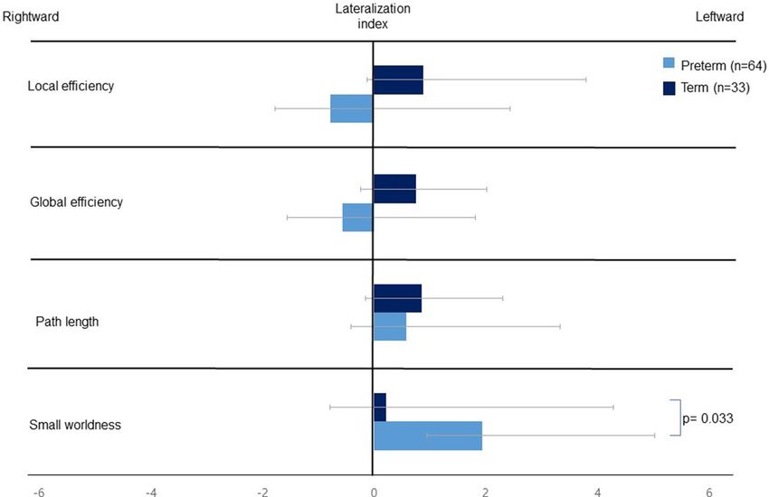
www.nature.com/scientificreports/
regions involving both receptive and expressive language, the mechanisms mediating social–emotional process-
ing remain poorly characterized.
The aberrant network connectivity of the fronto-limbic circuit has been implicated in emotion, social com-
munication, psychiatric, and neurodevelopmental d isorders9,10. A previous study of 6-month-old healthy infants
demonstrated that the rightward lateralization of the fronto-limbic brain development of white matter micro-
structure helps to support the development of social communication skills11. Other research performed across
the past decade suggests that altered lateralization of the neural networks comprising the fronto-limbic circuit
contributes to social and emotional problems in children and a dults12–14. To date, however, no study has inves-
tigated cerebral lateralization in the fronto-limbic connection of the structural brain networks of preterm neo-
nates. To address this dearth in the literature, we studied the asymmetry of both global and local networks of
the fronto-limbic circuit in the neonatal brain using diffusion tensor imaging (DTI) tractography and structural
network analysis. We hypothesized that preterm birth results in architectural changes in the global network or
specific fronto-limbic connections at near-term age. Furthermore, we speculated that the altered lateralization
of preterm brain in fronto-limbic connection might be related to social–emotional scores at 18 months of cor-
rected age in the developing preterm brain.
Results
Table 1 presents the prenatal and neonatal data of both groups. Compared to the full-term infants, preterm
infants had a significantly lower gestational age (30.08 ± 3.94 vs. 38.39 ± 1.22, P < 0.001) and lower birth weight
(1423.61 ± 588.44 vs. 3202.63 ± 326.40, P < 0.001). Preterm infants exhibited significantly lower 1-min and 5-min
Apgar scores. With respect to comorbidities, grade 1 intraventricular haemorrhages (P = 0.054), culture-proven
sepsis (P = 0.102), and bronchopulmonary dysplasia (P < 0.001) were more frequent among preterm infants. Neu-
rodevelopmental outcome data were available at 18 months of age (corrected for the prematurity) for 57 out of 64
(89%) of the preterm infants and for 21 out of 33 (64%) of the term infants. The preterm infants had significantly
lower mean composite scores on cognitive (95.47 ± 14.47 vs. 102.38 ± 8.74, P = 0.013), motor (95.38 ± 16.41 vs.
105.04 ± 11.03, P = 0.015) and social–emotional (97.28 ± 17.39 vs. 108.57 ± 11.95, P = 0.008) domains compared
with term-born infants. The incidence of language delay was significantly higher among preterm infants than
term infants (42.1% vs. 10%). Thirteen (13/57, 22.8%) preterm infants had a Bayley-SE score of < 85—i.e., > 1 SD
below the standardized mean for the assessment. No term infant had a Bayley-SE score of < 85.
We compared the differences in local efficiency, clustering coefficient, global efficiency, path length, and the
small-worldness between preterm infants and term-born neonates. These measures were comparable between
the two groups (Table 2; all P > 0.05). The small worldness tended to be higher in preterm infants than term-born
infants. We quantified the betweenness centrality for the fronto-limbic connections: the medial fronto-orbital
gyrus (mFOG), superior temporal gyrus (STG), amygdala, and hippocampus. However, the betweenness central-
ity of the mFOG (0.019 ± 0.024 vs. 0.010 ± 0.014, P = 0.169), STG (0.048 ± 0.042 vs. 0.046 ± 0.042, P = 0.599), and
hippocampus (0.007 ± 0.009 vs. 0.005 ± 0.007, P = 0.082) in the left hemisphere tended to be higher in preterm
infants than in term-born infants independent of age at MRI and bronchopulmonary dysplasia.
The results of the statistical analysis of the lateralization index of the global network metrics are summarized
in Fig. 1. While the left hemisphere tended to be more efficient with respect to global and local efficiency in
term-born infants, the right hemispheric predirection of global and local efficiency was observed in preterm
infants. However, a left-hemispheric predominance in the small-world properties was observed in both groups.
When comparing the lateralization index of the small-worldness between the 2 groups, preterm infants exhibited
significantly more leftward lateralization than did the full-term group, suggesting the prominence of small-world
properties in the left hemispheres of preterm infants (1.942 ± 4.488 and 0.212 ± 4.076, P = 0.033).
Figure 2 presents the comparison of the betweenness centrality in the fronto-limbic network of both preterm
and full-term infants; in the latter group, the mFOG was associated with a leftward asymmetry of between-
ness centrality, while the STG and hippocampus were associated with a rightward asymmetry. However, the
mFOG of preterm infants exhibited a significantly greater leftward asymmetry than did their full-term coun-
terparts (36.823 ± 26.562 vs. 10.855 ± 28.167, P = 0.008). The rightward predominance of the betweenness cen-
trality in the STG and hippocampus of term-born controls was not observed in preterm infants. When com-
paring the lateralization index of the fronto-limbic cortex between preterm and full-term infants, the mFOG
(36.823 ± 26.562 vs. 10.855 ± 28.167, P = 0.008), STG (2.001 ± 25.551 vs. − 16.820 ± 21.777, P = 0.031), and hip-
pocampus (21.636 ± 30.623 vs. − 6.167 ± 32.312, P = 0.028) of preterm infants was observed to exhibit significantly
greater leftward asymmetry after correcting for sex, age at MRI, and bronchopulmonary dysplasia.
Our assessment of how the age at MRI scan affects the asymmetry of the global network, mFOG, STG,
amygdala and hippocampus at 36–41 weeks of corrected age revealed no correlation between age at MRI scan
and global network or fronto-limbic asymmetries in all infants (n = 97) (Fig. 3). Figure 4 shows the subgroup
analyses for the comparison of network topological asymmetries between 57 preterm infants with abnormal
Bayley-SE (< 1SD below average, n = 13) and normal Bayley-SE (≥ 1SD below average, n = 44). The preterm infants
with abnormal Bayley-SE exhibited greater leftward lateralization of global and local efficiency and small-world
properties than did those with normal Bayley-SE. After controlling for gestational age and age at imaging, we
found significant differences in the leftward asymmetry of the betweenness centrality in the mFOG (P = 0.044)
and hippocampus (P = 0.032) between preterm infant with normal and abnormal Bayley-SE.
Discussion
The present study observed fronto-limbic asymmetry in the structural brain networks of preterm infants by
using DTI and structural network analyses. The preterm infants with no evidence of severe brain injury exhib-
ited significantly more leftward lateralization of small-world properties than did their full-term counterparts.
Scientific Reports | (2021) 11:1318 | https://doi.org/10.1038/s41598-020-79446-0 2
Vol:.(1234567890)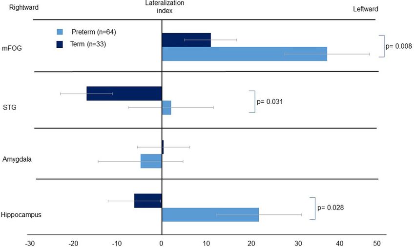
www.nature.com/scientificreports/
Variables Preterm (n = 64) Term (n = 33) P value
Perinatal characteristics
Caesarean section 54/64 (84.4) 9/33 (27.2) 0.001
Antenatal corticosteroids 29/64 (45.3) 0 < 0.001
Preeclampsia 8/64 (12.5) 0 0.102
GDM 5/64 (7.8) 2 (6.1) 0.669
Histologic chorioamnionitis 30/64 (51.5) - NA
Infant characteristics
Gestational age, wk 30.08 ± 3.94 38.39 ± 1.22 < 0.001
Birth weight, g 1423.61 ± 588.44 3202.63 ± 326.40 < 0.001
Male sex 13/64 (39.4) 8/33 (34.8) 0.785
Apgar 1 min 4.02 ± 1.69 6.73 ± 2.12 < 0.001
Apgar 5 min 6.58 ± 1.31 8.40 ± 1.35 < 0.001
PDA 30/64 (46.9) 0 < 0.001
IVH, grade 1 11/64 (17.2) 1 (3.0) 0.054
Culture-proven sepsis 8/64 (12.5) 0 0.102
NEC (requiring surgery) 3/64 (4.7) 0 0.315
ROP (requiring surgery) 6/64 (9.4) 0 0.092
Days on mechanical ventilator 14.26 ± 20.79 3.23 ± 1.25 0.985
Bronchopulmonary dysplasia 29/64 (45.3) 0 < 0.001
Age at MRI scan, wk 37.30 ± 1.33 38.61 ± 0.86 0.171
Maternal characteristics
Maternal age, years 33.38 ± 4.420 33.35 ± 4.33 0.982
Maternal smoker 1 (1.5) 0 1.000
Maternal education 0.379
Less than high school 14 (21.9) 5 (15.2)
High school graduate 49 (76.6) 26 (78.8)
College/university graduate 1 (1.6) 2 (6.1)
Neurodevelopmental follow-up at 18 mo of age
Cognitive composite score, mean (SD) 95.47 ± 14.47 (57/64) 102.38 ± 8.74 (21/33) 0.013
Infants with scores < 85, n (%) 10/57 (17.5) 0 (0) 0.056
Language composite score, mean (SD) 89.21 ± 15.42 (57/64) 93.95 ± 6.68 (21/33) 0.063
Infants with scores < 85, n (%) 24/57 (42.1) 2 (10) 0.012
Motor composite score, mean (SD) 95.38 ± 16.41 (57/64) 105.04 ± 11.03 (21/33) 0.015
Infants with scores < 85, n (%) 11/57 (19.3) 0 (0) 0.057
S–E composite score, mean (SD) 97.28 ± 17.39 (57/64) 108.57 ± 11.95 (21/33) 0.008
Infants with scores < 85, n (%) 13/57 (22.8) 0 (0) 0.017
Table 1. Demographic, clinical characteristics and neurodevelopmental outcome of preterm infants and term
infants. Data are presented as the mean ± SD or number (%). GDM gestational diabetes mellitus, PDA patent
ductus arteriosus, IVH intraventricular, NEC necrotizing enterocolitis, ROP retinopathy of prematurity, MRI
magnetic resonance imaging, S–E social–emotional.
Moreover, we found the STG and hippocampi of preterm infants to feature significantly greater leftward asym-
metries in betweenness centrality, and their mFOG to exhibit greater activation, than those of full-term infants.
DTI has revealed that the hemispheric structural asymmetry in the brains of children involves not only
regional brain volumes but also white matter m icrostructure15,16. Despite evidence indicating a variety of regional
differences in the hemispheric asymmetry of term-born children, patterns of neural network asymmetry in
preterm infants still require elucidation. Ratnarajah et al.17 recently reported structural connectivity asymmetry
in 124 full-term neonates, indicating specific lateralized brain functions in the topology of each cerebral hemi-
sphere. Moreover, leftward asymmetric efficiency at both global and local levels was found in the term-born
brain, which may reflect the early development of the lateralization of neurological functions at birth. While this
finding was consistent with our observation that full-term infants exhibit the leftward lateralization of global
and local efficiency, we further found that preterm infants exhibit the rightward lateralization of global and local
efficiency and a more balanced information propagation with marked small-worldness in the left cerebral hemi-
sphere relative to full-term infants. These findings indicate the possible compensatory development of neural
networks in preterm infants and suggest that the adaptive growth of preterm brain networks was preserved and
enhanced following the stabilization of their clinical status. Recent investigations into the small-world property
have significantly impacted the understanding of the topological organization of the neonatal brain network in
early life18. Furthermore, the present results agree with our previous observation that structural brain networks
Scientific Reports | (2021) 11:1318 | https://doi.org/10.1038/s41598-020-79446-0 3
Vol.:(0123456789)www.nature.com/scientificreports/
Left Right
Variables Preterm (n = 64) Term (n = 33) P value* Preterm (n = 64) Term (n = 33) P value*
Global brain network
Local efficiency 0.778 ± 0.036 0.762 ± 0.049 0.425 0.790 ± 0.042 0.775 ± 0.051 0.587
Global efficiency 0.674 ± 0.029 0.673 ± 0.026 0.519 0.682 ± 0.032 0.684 ± 0.028 0.081
Path length 1.694 ± 0.078 1.704 ± 0.083 0.523 1.675 ± 0.095 1.675 ± 0.084 0.113
Small-worldness 1.219 ± 0.116 1.191 ± 0.107 0.623 1.179 ± 0.108 1.180 ± 0.105 0.717
Betweenness centrality of FLC
mFOG 0.019 ± 0.024 0.010 ± 0.014 0.169 0.006 ± 0.008 0.007 ± 0.011 0.079
STG 0.048 ± 0.042 0.046 ± 0.042 0.599 0.039 ± 0.023 0.047 ± 0.036 0.830
Amygdala 0.001 ± 0.005 0.001 ± 0.001 0.107 0.001 ± 0.002 0.001 ± 0.001 0.570
Hippocampus 0.007 ± 0.009 0.005 ± 0.007 0.082 0.004 ± 0.004 0.008 ± 0.019 0.155
Table 2. Network measures of the left and right hemisphere in the preterm and term infants. Data are
presented as the mean ± SD. FLC fronto-limbic circuit, mFOG medial fronto-orbital gyrus, STG superior
temporal gyrus. *Adjusted for sex, age at MRI scan and bronchopulmonary dysplasia.
Figure 1. The comparison of the lateralization index of the global network metrics between preterm and full-
term infants. We controlled for sex, age at MRI scan, and bronchopulmonary dysplasia when performing the
analyses. P values of ≤ 0.05 were considered to indicate statistical significance. All P values were corrected for the
false-discovery rate.
after preterm birth are reorganized to accelerate the development of small-world property in preterm infants
without apparent brain abnormalities at term-equivalent a ges5.
Despite considerable advances in the survival rate among preterm infants, the mechanisms underlying the
development of neural asymmetry present at or before birth—especially those concerning the alteration of the
lateralization of structural connectivity—have lacked rigorous exploration. The few existing studies on full-term
neonates have focused on the leftward predominance of language and motor functions and the larger gray and
white matter volumes at the areas associated with these functions, as well as early white matter maturation in
emisphere16,19. A recent neuroimaging study of 26 very preterm infants and 25 controls revealed that
the left h
cerebral lateralization in the left language region, including Wernicke’s and Broca’s areas, was impaired in the
preterm group at term-equivalent a ges8.
Our study demonstrated that social–emotional development at 18 months of age, as assessed by the maternal
completion of the Bayley-SE Scale, was diminished in preterm infants relative to term-born neonates. However,
since subtle impairments may not be evident in infancy and can either improve or worsen during preschool
age, evaluations performed at a corrected age of 18 months should be further explored by follow-up studies to
consolidate the present results. This possibility was anticipated by early findings that preterm infants of less than
27 weeks of age were three times more likely to develop ASD than controls after controlling for sex, SGA, and
Scientific Reports | (2021) 11:1318 | https://doi.org/10.1038/s41598-020-79446-0 4
Vol:.(1234567890)www.nature.com/scientificreports/
Figure 2. The comparison of the lateralization index of the betweenness centrality in the fronto-limbic network
of both preterm and full-term infants. We controlled for sex, age at MRI scan, and bronchopulmonary dysplasia
when performing the analyses. P values of ≤ 0.05 were considered to indicate statistical significance. All P values
were corrected for the false-discovery rate. Abbreviations: mFOG, medial froto-orbital gyrus; STG, superior
temporal gyrus.
maternal education20. The study20 further suggested that preterm infants of < 34 weeks of age who were observed
in the neonatal intensive care unit to have potential risk factors such as low-grade intraventricular haemorrhage
or high-frequency ventilation were likely to have a relatively high risk of developing ASD. Notably, the differential
activation of the right hemispheric limbic structures is believed to be determined and established during prena-
tal and postnatal development17, indicating that the right hemisphere of the neonatal brain has its own dense,
efficient network for social and emotional processing. Ratnarajah et al.17 suggested that limbic structures, such
as the cingulate gyrus and hippocampus, of term-born infants show a rightward asymmetry in the betweenness
centrality of emotional processing, suggesting that brain regions involved in emotional processes play crucial
roles in efficient communication in the right hemisphere. However, few studies have considered the correlation
between the pathophysiology of socioemotional processing and social–emotional delay among preterm infants
using DTI-based connectomes.
Frontotemporolimbic connections are important interactive pathways involving the “social brain” via con-
nections between the mFOG, STG, amygdala, and hippocampus. This network lateralization to the right hemi-
sphere at the neonatal stage could be associated with social information processing and emotional regulation
in later neurodevelopment. The presently observed rightward asymmetry of betweenness centrality of the STG
and hippocampus in term-born controls is consistent with previous fi ndings17,21. Altered lateralization in these
regions favoring the left hemisphere rather than the right might contribute substantially to the early onset of
social–emotional disorders in preterm i nfants9.
Our results suggest that the leftward lateralization of the STG and hippocampus in preterm infants may
contribute to the maladaptive development of social communication by altering the network connectivity of the
fronto-limbic circuit and thus compromising normative social–emotional processing. This conclusion is partially
supported by research on the asymmetry of FA values of the uncinate fasciculus: the largest white matter associa-
tion tract connecting the prefrontal cortex, superior temporal cortex, and limbic s ystem11. Specifically, the right
uncinated fasciculus connecting the mFOG, amygdala, and STG at 6 months was found to be a strong predictor
of the emergence of joint attention at 9 months of a ge11. The asymmetry of the mFOG and STG is considered to
be associated with a rightward shift according to the infant’s maturation pattern, which should be lateralized to
the right at term-equivalent ages in n eonates11,22. However, we observed an excessive leftward asymmetry that
involved redundant leftward lateralization of the mFOG in preterm infants relative to their full-term counter-
parts; this finding agrees with previous observations on functional MRI of reduced activation in the right mFOG
of patients with ASD relative to c ontrols23.
To date, no study has reported significant differences in the lateralization of the fronto-limbic circuit between
preterm and term-born infants. This study is, to the best of our knowledge, the first to identify the altered later-
alization of betweenness centrality in the efficiency of the networks of the fronto-limbic circuit with DTI per-
formed at the term-equivalent age in preterm infants. We further addressed these connections in relation to the
Bayley-SE scale scores of infants without brain injuries at 18 month of corrected age. However, the present study
is subject to several limitations: (1) The relatively small sample size in term-born neonates may have resulted
in insufficient power to detect possible associations. This is partially due to the high cost of data collection and
the substantial lack of term-born neonates for follow-up. (2) The confirmation of an association between socio-
emotional development and fronto-limbic asymmetry was limited by our small number of preterm infants with
Scientific Reports | (2021) 11:1318 | https://doi.org/10.1038/s41598-020-79446-0 5
Vol.:(0123456789)www.nature.com/scientificreports/
Figure 3. The features of global network asymmetries and fronto-limbic asymmetry were plotted against age
at which all infants underwent the MRI scan (n = 97). P values of ≤ 0.05 were considered to indicate statistical
significance. All P values were corrected for the false-discovery rate. Abbreviations: mFOG, medial fronto-
orbital gyrus; STG, superior temporal gyrus.
Scientific Reports | (2021) 11:1318 | https://doi.org/10.1038/s41598-020-79446-0 6
Vol:.(1234567890)www.nature.com/scientificreports/
Figure 4. Comparison of network topological asymmetries between 57 preterm infants with normal scores on
the Bayley-III socio-emotional scale (≥ 1SD below average, n = 44) and abnormal scores on the socio-emotional
scale (< 1SD below average, n = 13). We controlled for gestational age and age at MRI scan when performing the
analyses. An asterisk indicates statistical significance. P values of ≤ 0.05 were considered to indicate statistical
significance. All P values were corrected for the false-discovery rate. Abbreviations: mFOG, medial fronto-
orbital gyrus; STG, superior temporal gyrus.
Scientific Reports | (2021) 11:1318 | https://doi.org/10.1038/s41598-020-79446-0 7
Vol.:(0123456789)www.nature.com/scientificreports/
low socio-emotional scores and by the preterm infants not having been able to undergo more assessments for
the subdomain of behavior at older ages. (3) We analyzed the development of the central nervous system in
children at an age when it is undergoing constant change, and its correlation with clinical development is highly
dynamic. Hence, the development or alterations of the nervous systems of the included preterm infants may
have been delayed at the point of observation—despite our having collected data at a corrected age of 18 months
after birth. The value of the correlative analysis between anatomical and functional findings should be assessed
by larger prospective cohort studies.
The present study benefitted from recruited a large number of preterm infants with available 3 T MRI imag-
ing data at near-term age, allowing for the exploration of distinct white matter tracts, rather than the WM
microstructure in its entirety, in relation to socio-emotional development in children born preterm. We focused
on infants without significant brain injuries, using network topological asymmetries in fronto-limbic neural
circuitry. Our results may implicate the alteration of fronto-limbic brain circuitry in the early development of
social–emotional disorders in preterm infants. The present study further indicates that the altered interhemi-
spheric leftward asymmetries of small-worldness in the global network and fronto-temporo-limbic areas may
help to elucidate the etiology of neurodevelopmental disorders across the long-term follow-up of preterm infants.
Methods
The present study recruited 83 preterm infants from the Hanyang Inclusive Clinic for Developmental Disorders,
Hanyang University Seoul Hospital, between December 2016 and April 2018. Our sample included infants with
no evidence of genetic disorders, congenital infections, or congenital brain abnormalities, and for whom MRI
data obtained at near-term age (GA, 36–41 weeks) was available. We excluded eight infants with (1) intraven-
tricular or intracranial hemorrhage greater than grade I on brain ultrasound at 3 days, 7 days, or 3 weeks of birth;
(2) periventricular leukomalacia or white matter injury on term-equivalent MRI; or (3) intrauterine growth
retardation. Of the 83 eligible preterm children, 75 met our inclusion criteria. 33 term-born infants (GA range,
37–42 weeks) with normal MRI findings and neurodevelopment were also recruited for the term-born control
group. Eleven of the remaining 75 preterm infants were removed due to poor image quality that precluded DTI
processing; hence, the initial sample included 64 datasets. Our final sample of preterm infants and term-born
infants included 64 and 33 datasets, respectively. Images were obtained from the controls and preterm infants
during natural sleep without sedation. The infants underwent follow-up neurosensory and neurodevelopmental
assessments at 4, 9, and 18 months of corrected age. Demographic data included maternal details, GA, birth
weight, sex, and Apgar scores. Neonatal outcomes included patent ductus arteriosus, culture-proven sepsis,
necrotizing enterocolitis, retinopathy of prematurity, and bronchopulmonary dysplasia. This present study pro-
tocol and scanning procedure were approved by the institutional review board of Hanyang University Hospital
(IRB No. 2017-04-004-002). All procedures were performed in accordance with the principles of the Declara-
tion of Helsinki, and informed consent for participation was obtained from the children’s parents prior to their
participation in the study.
Neurodevelopmental assessment. The Bayley Scales of Infant and Toddler Development, Third Edition
(BSID-III)24, were used to perform the neurological and developmental assessments at Hanyang Inclusive Clinic
for Developmental Disorders when the children were 18 months of age (corrected for prematurity). The routine
neurological examination was completed by a developmental pediatrician, while the developmental assessment
was performed by paediatric physiotherapist and neonatologist. Seven of the preterm infants (11%) did not
complete the Bayley-III test at 18 months of corrected age; the parents of three of the infants did not permit the
assessment of their children; two infants dropped out of the study during follow-up, and two of the participants
were withdrawn from the study because of their inability to cooperate with testing.
Developmental outcomes included cognitive, language, motor, and social–emotional composite BSID-III
scores based on the child’s adjusted age at the time of evaluation. Assessments of the cognitive, language, and
motor domains were performed using the items administered to the child, while assessments of social–emotional
domains were informed by the primary caregiver’s responses to a questionnaire. The normative means (standard
deviation [SD]) of each outcome score was 100; scores of less than 85 (1 SD below average) were considered to
indicate neurodevelopmental delay.
MRI. All scans were performed on 3 T MRI (Philips Real-Time Compact Magnet, Achieva 3.0-T Xseries,
16-channel SENSE head coil). The T1-weighted images included sagittal and axial T1 spin-echo sequences
(400/25/2, repetition time [TR]/echo time [TE]/signal intensity averages) and axial T2 Turbo spin-echo
sequences (3000/100/1). DTIs were obtained using a single-shot spin-echo planar sequence with a SENSE fac-
tor of two and an echo-planar imaging factor (TR/TE, 5243/76 ms; voxel size, 1.97 × 1.97 × 2 mm; field-of-view,
150 mm, 45 axial sections). Diffusivities were measured along 15 directions with an electrostatic gradient model
(b = 800). No sedation was used and the infants remained asleep in the scanner by feeding them before the scan.
A pulse oximeter was monitored to ensure stable heart rate and respiration.
Image processing. The diffusion-weighted images were processed using the FMRIB Software Library
(http://www.fmrib.ox.ac.uk/fsl in the public domain). Motion artifacts and eddy current distortions were cor-
rected by normalizing each diffusion-weighted volume to the nondiffusion-weighted volume (b0) with the
FMRIB Linear Image Registration Tool. Each brain mask was created using the Brain Extraction Tool (BET).
Subsequently, DTIs were reconstructed for each voxel using DTIFit in FMRIB’s Diffusion Toolbox. Fractional
anisotropy (FA) and mean diffusivity (MD) values were then calculated. Whole-brain deterministic diffusion
fiber tracking was performed for each infant using quantitative anisotropy in DSI Studio (dsi-studio.labsolver.
Scientific Reports | (2021) 11:1318 | https://doi.org/10.1038/s41598-020-79446-0 8
Vol:.(1234567890)www.nature.com/scientificreports/
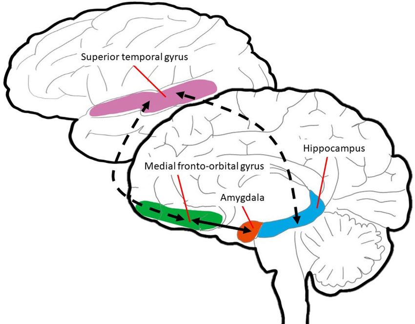
Figure 5. Schematic representation of fronto-temporo-limbic areas connected via medial and lateral cortical
pathways. The brain diagram presented in the top register presents a lateral view, wherein the pink area indicates
the superior temporal gyrus. The lower register presents a medial view of the medial fronto-orbital gyrus,
amygdala, and hippocampus depicted in green, red, and blue, respectively. The circuit linking this structures is
key to the mediation of responses to social–emotional stimuli by the prefrontal, temporal, and limbic systems.
org) and was estimated using a streamlined algorithm in DSI Studio25. Fiber tracking was initiated from the
center of each voxel and proceeded to adjacent voxels by following the direction of water diffusivity. Fiber track-
ing was stopped when the FA value fell below 0.1 and if the streamline turned with a curvature angle of more
than 45°. Every step of image processing was performed in a blind way and MRI images with spurious brain
connections due to possible noise effects on the whole brain network were excluded for network construction
and statistical analyses.
Network construction. Topological organizations of global structural networks were characterized with
the Brain Connectivity Toolbox in MATLAB (The Mathworks, Inc., Natick, Massachusetts, USA)26,27. Network
nodes were defined based on the Johns Hopkins University (JHU) neonate atlas. All neonatal FA images were
nonlinearly transformed to the JHU neonate atlas, and labels in the JHU atlas space were inversely transformed
to FA images using the inverse warping field. The 64 labels in the JHU neonate atlas were defined as nodes
for an individual structure network. We used estimated fiber tracts in the whole brain for edge definition and
quantified connectivity between cortical regions. The connectivity strength was then computed by obtaining
the average FA value along the fiber tracts4. The average FA value was calculated by the arithmetic average of
FA taken over all voxels comprising each fiber and then averaged over all fibers comprising the tract. Finally, we
constructed unweighted 64 × 64 connectivity network matrices for each subject (Fig. 5).
Network analysis. The global and local efficiency, characteristic path length, and small-worldness were
computed to characterize the potential ease with which information could be transferred concurrently across a
network and locally communicated neighborhood. The network integration refers to how efficiently and rapidly
all the nodes integrate discrete specialized information. The most popular network integration measures include
characteristic path length ( Lp) and global efficiency ( Eglob): a shorter path length and increased global efficiency
suggest the potential integration of effective connections. The characteristic path length was defined by the aver-
age shortest path length in a network28.
1
Lp = Lij
N(N − 1) i∈G
j∈G,j�=i
where Lij is the shortest path length between nodes i and j and N is the set of all nodes in the network G . The
global efficiency was defined as the mean of the inverse shortest path length in a network29.
Scientific Reports | (2021) 11:1318 | https://doi.org/10.1038/s41598-020-79446-0 9
Vol.:(0123456789)www.nature.com/scientificreports/
1
Eglob = 1 Lij
N(N − 1) i∈G
j∈G,j�=i
Segregation indicates the relative strength of within-network connections compared to integration. The most
popular measures of segregation include the clustering coefficient and local efficiency. The clustering coefficient
was defined as the ratio between the number of existing edges between neighbors and the number of all possible
connections between neighbors30.
1 2 ∗ Ei
Cp =
N i∈G ki (ki − 1)
where ki is the degree of node i and Ei is the number of edges with the neighbors of node i .
The local efficiency of a node was calculated as the global efficiency of the neighborhood sub-network of
etwork26
this node. The local efficiencies across all nodes were averaged to estimate the local efficiency of the n
1
Elocal = Elocal,i
N i∈G
where Elocal,i is the local efficiency of node i . Reflecting both integrated and segregated information processing,
small-worldness was defined by the clustering coefficient and characteristic path length28.
Lp /Lran
σ=
Cp /Cran
where Cran and Lran are the mean clustering coefficient and characteristic path length of 1—random networks.
All network metrics were calculated using the Brain Connectivity Toolbox (http://www.brain-connec tivit y-toolb
ox.net).
Informed by prior s tudies9,11,31, a regions-of-interest (ROI) connectivity analysis was performed to explore
fronto-temporo-limbic regions, including the bilateral mFOG, STG, amygdala, and hippocampus (Fig. 5). The
mFOG is a key cortical structure that plays an important role in social domains related to emotional regulation
and behavioral suppression. The STG mediates the initial social perception of visual and auditory social cues
in the temporal lobe. The amygdala and hippocampus are the limbic regions linked to social cognition and
emotional processes through the fronto-limbic pathway. ROIs were defined based on the JHU neonate atlas.
Betweenness centrality refers to how important a given node is to the efficient communication between the other
nodes32. The betweenness centrality of a node was defined as the fraction of all shortest paths between any two
nodes a and b that passes through a given node n. We characterized network lateralization of the betweenness
centrality between the left and right structural networks of the medial fronto-orbital gyrus, STG, amygdala, and
hippocampus. We thus represented fronto-limbic brain circuitry in terms of between centrality.
Statistical analyses. SPSS 21.0 (SPSS, Chicago, IL) software was used for statistical calculations. We used
Student’s t-test, Mann–Whitney U-test, Fisher’s exact test, and the chi-square analysis to compare clinical vari-
ables between groups. Based on the prevalences of impaired cognition or motor outcomes among very-low
birth weight newborns (15% among term-born children and 40% among very preterm children)33, the required
sample size to detect differences in prevalence between the study and general populations was 94 for all infants
(52 for each group) with an alpha of 0.05, a desired power of 0.8, and a dropout rate 10%. For the term infants,
the total sample size was smaller than the calculated sample due to lack of follow-up assessment. General linear
models were generated to compare the network parameters and lateralization indices of the network param-
eters between preterm and full-term infants. The analysis of structural connectivity incorporated the global
network matrix, including clustering, local efficiency, path length, global efficiency, and small-worldness, and
the betweenness centrality of the fronto-limbic connections. A correlation analysis was performed to explore
whether the age at MRI scan correlated with the lateralization index of the structural network. The subgroup
analyses for the comparisons of structural network metrics between 57 preterm infants with abnormal Bayley-
SE (< 1SD below average) and normal Bayley-SE (≥ 1SD below average) were performed using clinical variables
such as gestational age and age at scan as covariates.
The asymmetry of the aforementioned network metrics was evaluated with the asymmetry score:
AS = 100 × (X(L) − X(R))/(X(L) + X(R)), where X(R) and X(L) indicate the network parameters of the right and
left hemispheres, respectively17. By incorporating the relative network metrics of both hemispheres into one value,
the AS(X) index permits the assessment of differences between the right and left hemispheres. Ranging between
+ 100 and − 100, the AS(X) is positive when metric X shows prominent rightward asymmetry and negative
when the opposite is the case. Lw measures the overall routing efficiency of the network and is inversely related
to Eglobal. For a network, the higher the Lw, the less efficient the global integration. Hence, positive AS(Lw)
indicates a leftward advantage of global integration, and negative AS(Lw) a rightward asymmetry of global inte-
gration. Continuous variables are represented as the mean ± SD. A P value of ≤ 0.05 was considered to indicate
statistical significance. The resulting P values were corrected for the false discovery rate (FDR); FDR-adjusted P
values of < 0.05 were considered to indicate statistical significance for multiple comparisons.
Received: 4 February 2020; Accepted: 21 October 2020
Scientific Reports | (2021) 11:1318 | https://doi.org/10.1038/s41598-020-79446-0 10
Vol:.(1234567890)www.nature.com/scientificreports/
References
1. Anderson, P. J. & Doyle, L. W. Cognitive and educational deficits in children born extremely preterm. Semin. Perinatol. 32, 51–58.
https://doi.org/10.1053/j.semperi.2007.12.009 (2008).
2. Potharst, E. S. et al. High incidence of multi-domain disabilities in very preterm children at five years of age. J. Pediatr. 159, 79–85.
https://doi.org/10.1016/j.jpeds.2010.12.055 (2011).
3. Agrawal, S., Rao, S. C., Bulsara, M. K. & Patole, S. K. Prevalence of autism spectrum disorder in preterm infants: a meta-analysis.
Pediatrics https://doi.org/10.1542/peds.2018-0134 (2018).
4. Brown, C. J. et al. Structural network analysis of brain development in young preterm neonates. Neuroimage 101, 667–680. https
://doi.org/10.1016/j.neuroimage.2014.07.030 (2014).
5. Lee, J. Y., Park, H. K. & Lee, H. J. Accelerated small-world property of structural brain networks in preterm infants at term-
equivalent age. Neonatology 115, 99–107. https://doi.org/10.1159/000493087 (2019).
6. Song, J. W. et al. Asymmetry of white matter pathways in developing human brains. Cereb. Cortex 25, 2883–2893. https://doi.
org/10.1093/cercor/bhu084 (2015).
7. Scheinost, D. et al. Cerebral lateralization is protective in the very prematurely born. Cereb. Cortex 25, 1858–1866. https://doi.
org/10.1093/cercor/bht430 (2015).
8. Kwon, S. H. et al. Adaptive mechanisms of developing brain: cerebral lateralization in the prematurely-born. Neuroimage 108,
144–150. https://doi.org/10.1016/j.neuroimage.2014.12.032 (2015).
9. Perez, D. L. et al. Frontolimbic neural circuit changes in emotional processing and inhibitory control associated with clinical
improvement following transference-focused psychotherapy in borderline personality disorder. Psychiatry Clin. Neurosci. 70,
51–61. https://doi.org/10.1111/pcn.12357 (2016).
10. Bigler, E. D. et al. Superior temporal gyrus, language function, and autism. Dev. Neuropsychol. 31, 217–238. https://doi.
org/10.1080/87565640701190841 (2007).
11. Elison, J. T. et al. Frontolimbic neural circuitry at 6 months predicts individual differences in joint attention at 9 months. Dev. Sci.
16, 186–197. https://doi.org/10.1111/desc.12015 (2013).
12. Toller, G. et al. Right fronto-limbic atrophy is associated with reduced empathy in refractory unilateral mesial temporal lobe
epilepsy. Neuropsychologia 78, 80–87. https://doi.org/10.1016/j.neuropsychologia.2015.09.010 (2015).
13. Zhou, Q. et al. Hemispheric asymmetry of the frontolimbic cortex in young adults with borderline personality disorder. Acta
Psychiatr. Scand. 136, 637–647. https://doi.org/10.1111/acps.12823 (2017).
14. Madsen, K. S., Jernigan, T. L., Vestergaard, M., Mortensen, E. L. & Baare, W. F. C. Neuroticism is linked to microstructural left-
right asymmetry of fronto-limbic fibre tracts in adolescents with opposite effects in boys and girls. Neuropsychologia 114, 1–10.
https://doi.org/10.1016/j.neuropsychologia.2018.04.010 (2018).
15. Sowell, E. R. et al. Mapping sulcal pattern asymmetry and local cortical surface gray matter distribution in vivo: maturation in
perisylvian cortices. Cereb. Cortex 12, 17–26. https://doi.org/10.1093/cercor/12.1.17 (2002).
16. Dubois, J. et al. Structural asymmetries in the infant language and sensori-motor networks. Cereb. Cortex 19, 414–423. https://
doi.org/10.1093/cercor/bhn097 (2009).
17. Ratnarajah, N. et al. Structural connectivity asymmetry in the neonatal brain. Neuroimage 75, 187–194. https://doi.org/10.1016/j.
neuroimage.2013.02.052 (2013).
18. Cao, M. et al. Early development of functional network segregation revealed by connectomic analysis of the preterm human brain.
Cereb. Cortex 27, 1949–1963. https://doi.org/10.1093/cercor/bhw038 (2017).
19. Gilmore, J. H. et al. Regional gray matter growth, sexual dimorphism, and cerebral asymmetry in the neonatal brain. J. Neurosci.
27, 1255–1260. https://doi.org/10.1523/jneurosci.3339-06.2007 (2007).
20. Kuzniewicz, M. W. et al. Prevalence and neonatal factors associated with autism spectrum disorders in preterm infants. J. Pediatr.
164, 20–25. https://doi.org/10.1016/j.jpeds.2013.09.021 (2014).
21. Leroy, F. et al. Early maturation of the linguistic dorsal pathway in human infants. J. Neurosci. 31, 1500–1506. https://doi.
org/10.1523/JNEUROSCI.4141-10.2011 (2011).
22. Sowell, E. R. et al. Longitudinal mapping of cortical thickness and brain growth in normal children. J. Neurosci. 24, 8223–8231.
https://doi.org/10.1523/JNEUROSCI.1798-04.2004 (2004).
23. Carlisi, C. O. et al. Comparison of neural substrates of temporal discounting between youth with autism spectrum disorder and
with obsessive-compulsive disorder. Psychol. Med. 47, 2513–2527. https://doi.org/10.1017/S0033291717001088 (2017).
24. Durrant, C. et al. Developmental trajectories of infants born at less than 30 weeks’ gestation on the Bayley-III Scales. Arch. Dis.
Child. Fetal Neonatal Ed. https://doi.org/10.1136/archdischild-2019-317810 (2020).
25. Yeh, F. C., Verstynen, T. D., Wang, Y., Fernandez-Miranda, J. C. & Tseng, W. Y. Deterministic diffusion fiber tracking improved by
quantitative anisotropy. PLoS ONE 8, e80713. https://doi.org/10.1371/journal.pone.0080713 (2013).
26. Rubinov, M. & Sporns, O. Complex network measures of brain connectivity: uses and interpretations. Neuroimage 52, 1059–1069.
https://doi.org/10.1016/j.neuroimage.2009.10.003 (2010).
27. Oishi, K. et al. Multi-contrast human neonatal brain atlas: application to normal neonate development analysis. Neuroimage 56,
8–20. https://doi.org/10.1016/j.neuroimage.2011.01.051 (2011).
28. Watts, D. J. & Strogatz, S. H. Collective dynamics of “small-world” networks. Nature 393, 440–442. https://doi.org/10.1038/30918
(1998).
29. Latora, V. & Marchiori, M. Efficient behavior of small-world networks. Phys. Rev. Lett. 87, 198701. https://doi.org/10.1103/PhysR
evLett.87.198701 (2001).
30. Onnela, J. P., Saramaki, J., Kertesz, J. & Kaski, K. Intensity and coherence of motifs in weighted complex networks. Phys. Rev. E
Stat. Nonlinear Soft Matter Phys. 71, 065103. https://doi.org/10.1103/PhysRevE.71.065103 (2005).
31. Zilbovicius, M. et al. Autism, the superior temporal sulcus and social perception. Trends Neurosci. 29, 359–366. https://doi.
org/10.1016/j.tins.2006.06.004 (2006).
32. Goh, K. I., Oh, E., Kahng, B. & Kim, D. Betweenness centrality correlation in social networks. Phys. Rev. E Stat. Nonlinear Soft
Matter Phys. 67, 017101. https://doi.org/10.1103/PhysRevE.67.017101 (2003).
33. Erdei, C., Austin, N. C., Cherkerzian, S., Morris, A. R. & Woodward, L. J. Predicting school-aged cognitive impairment in children
born very preterm. Pediatrics 145, 1. https://doi.org/10.1542/peds.2019-1982 (2020).
Acknowledgements
This work was supported by the National Research Foundation of Korea Grant funded by the Korean Govern-
ment MSIT (NRF-2020-R1F1A1048529 and 2020-M3E5D9080787).
Author contributions
H.J.L. and J.Y.L drafted the manuscript and made substantial contributions to design of this study. J.L.,Y.C and
J.H.C participated in data acquisition and performed data analysis. J.Y.L., Y.L., I.K. and B.K. were active in
research conception and critical revision of the manuscript. Joo Young Lee and Yong-Ho Choi contributed to
this work equally.
Scientific Reports | (2021) 11:1318 | https://doi.org/10.1038/s41598-020-79446-0 11
Vol.:(0123456789)www.nature.com/scientificreports/
Competing interests
The authors declare no competing interests.
Additional information
Correspondence and requests for materials should be addressed to J.-M.L. or H.J.L.
Reprints and permissions information is available at www.nature.com/reprints.
Publisher’s note Springer Nature remains neutral with regard to jurisdictional claims in published maps and
institutional affiliations.
Open Access This article is licensed under a Creative Commons Attribution 4.0 International
License, which permits use, sharing, adaptation, distribution and reproduction in any medium or
format, as long as you give appropriate credit to the original author(s) and the source, provide a link to the
Creative Commons licence, and indicate if changes were made. The images or other third party material in this
article are included in the article’s Creative Commons licence, unless indicated otherwise in a credit line to the
material. If material is not included in the article’s Creative Commons licence and your intended use is not
permitted by statutory regulation or exceeds the permitted use, you will need to obtain permission directly from
the copyright holder. To view a copy of this licence, visit http://creativecommons.org/licenses/by/4.0/.
© The Author(s) 2021
Scientific Reports | (2021) 11:1318 | https://doi.org/10.1038/s41598-020-79446-0 12
Vol:.(1234567890)You can also read