Paleoneuroanatomy of the European lambeosaurine dinosaur Arenysaurus ardevoli
←
→
Page content transcription
If your browser does not render page correctly, please read the page content below
Paleoneuroanatomy of the European
                                 lambeosaurine dinosaur Arenysaurus
                                 ardevoli
                                 P Cruzado-Caballero1,2 , J Fortuny3,4 , S Llacer3 and JI Canudo2
                                1 CONICET—Instituto de Investigación en Paleobiologı́a y Geologı́a, Universidad Nacional de Rı́o

                                 Negro, Roca, Rı́o Negro, Argentina
                                2 Área de Paleontologı́a, Facultad de Ciencias, Universidad de Zaragoza, C/Pedro Cerbuna,

                                 Zaragoza, Spain
                                3 Institut Català de Paleontologia Miquel Crusafont, C/Escola Industrial, Sabadell, Spain
                                4 Departament de Resistència de Materials i Estructures a l’Enginyeria, Universitat Politècnica de

                                 Catalunya, Terrassa, Spain

                                     ABSTRACT
                                     The neuroanatomy of hadrosaurid dinosaurs is well known from North America
                                     and Asia. In Europe only a few cranial remains have been recovered that include
                                     the braincase. Arenysaurus is the first European endocast for which the paleoneu-
                                     roanatomy has been studied. The resulting data have enabled us to draw ontogenetic,
                                     phylogenetic and functional inferences. Arenysaurus preserves the endocast and
                                     the inner ear. This cranial material was CT scanned, and a 3D-model was gener-
                                     ated. The endocast morphology supports a general pattern for hadrosaurids with
                                     some characters that distinguish it to a subfamily level, such as a brain cavity that is
                                     anteroposteriorly shorter or the angle of the major axis of the cerebral hemisphere
                                     to the horizontal in lambeosaurines. Both these characters are present in the
                                     endocast of Arenysaurus. Osteological features indicate an adult ontogenetic stage,
                                     while some paleoneuroanatomical features are indicative of a subadult ontogenetic
                                     stage. It is hypothesized that the presence of puzzling mixture of characters that
                                     suggest different ontogenetic stages for this specimen may reflect some degree of
                                     dwarfism in Arenysaurus. Regarding the inner ear, its structure shows differences
Submitted 5 November 2014            from the ornithopod clade with respect to the height of the semicircular canals. These
Accepted 5 February 2015
Published 24 February 2015
                                     differences could lead to a decrease in the compensatory movements of eyes and
                                     head, with important implications for the paleobiology and behavior of hadrosaurid
Corresponding author
P Cruzado-Caballero,                 taxa such as Edmontosaurus, Parasaurolophus and Arenysaurus. The endocranial
pccaballero@unrn.edu.ar              morphology of European hadrosaurids sheds new light on the evolution of this group
Academic editor                      and may reflect the conditions in the archipelago where these animals lived during
Andrew Farke                         the Late Cretaceous.
Additional Information and
Declarations can be found on
page 13                          Subjects Paleontology
                                 Keywords European lambeosaurine, Paleoneurology, Hadrosaurid, Paleobiology, Inner ear,
DOI 10.7717/peerj.802
                                 Dinosauria
    Copyright
2015 Cruzado-Caballero et al.

Distributed under
                                 INTRODUCTION
Creative Commons CC-BY 4.0       Hadrosaurids are the most abundant ornithopod dinosaurs from the Late Cretaceous
                                 of Laurasia, with a very complete record including ontogenetic series, mummies, eggs,
OPEN ACCESS

                                  How to cite this article Cruzado-Caballero et al. (2015), Paleoneuroanatomy of the European lambeosaurine dinosaur Arenysaurus
                                  ardevoli. PeerJ 3:e802; DOI 10.7717/peerj.802
ichnites, etc. (see Lull & Wright, 1942; Horner, Weishampel & Forster, 2004 for reviews).
                                   This rich record also includes natural cranial endocasts or complete skulls allowing the
                                   generation of silicone or latex rubber models of the endocast (Lambe, 1920; Gilmore,
                                   1924; Ostrom, 1961; Serrano-Brañas et al., 2006; Lauters et al., 2013). The endocranial
                                   morphology of hadrosaurids has been studied since the first quarter of the 20th century
                                   (as in the case of Edmontosaurus regalis (Lambe, 1920) or Lambeosaurus Gilmore, 1924).
                                   Nowadays, non-invasive techniques such as CT scans shed new light on the paleoneurology
                                   of dinosaurs and other extinct taxa (Witmer et al., 2008; Evans, Ridgely & Witmer, 2009;
                                   Godefroit, Bolotsky & Lauters, 2012; Lautenschlager & Hübner, 2013). CT scan techniques
                                   are currently common in biology and paleontology as a way of obtaining digital models
                                   of inner regions as in the case of endocranial morphology, where these cavities may
                                   sometimes be filled by matrix. One of the great advantages of the CT scan is also that it
                                   makes it possible to access features without destroying the specimen (by contrast with
                                   very old methods) with minimum manipulation of the specimen and to create 3D models
                                   allowing manipulation or measurement without damage to the specimen. A CT scan
                                   allows a 3D visualization with a high or very high resolution, depending on the type of CT
                                   scan used and the goal of the study.
                                       To date, endocranial morphology is mainly known from North American specimens
                                   (Lull & Wright, 1942; Ostrom, 1961; Hopson, 1979; Evans, Ridgely & Witmer, 2009; Farke
                                   et al., 2013) and to a lesser extent from Asian remains (Young, 1958; Saveliev, Alifanov &
                                   Bolotsky, 2012; Godefroit, Bolotsky & Lauters, 2012; Lauters et al., 2013), including isolated
                                   individuals and ontogenetic series. In Europe, however, the cranial record of hadrosaurids
                                   is very scarce, and no paleoneurological analyses have yet been performed. The European
                                   hadrosaurids with cranial material are Tethyshadros, Telmatosaurus and Arenysaurus
                                   (Nopcsa, 1900; Dalla Vecchia, 2009; Pereda-Suberbiola et al., 2009b). In the case of Tel-
                                   matosaurus, a latex rubber model of poor quality was described historically (Nopcsa, 1900).
                                       Arenysaurus forms part of the rich hadrosaurid fauna from the Iberian Peninsula,
                                   although cranial remains are scarce (Cruzado-Caballero, Pereda Suberbiola & Ruiz-
                                   Omeñaca, 2010; Cruzado-Caballero, Ruiz-Omeñaca & Canudo, 2010; Cruzado-Caballero
                                   et al., 2013; Prieto-Márquez et al., 2013). It was described by Pereda-Suberbiola et al.
                                   (2009b) as the first European lambeosaurine hadrosaurid preserving most of the cranial
                                   elements, including an almost complete and uncrushed braincase (Fig. 1). The Arenysaurus
                                   remains, together with other hadrosaurid and lambeosaurine material, helped to exchange
                                   the vision of a primitive European fauna for one that is more diverse, permitting
                                   osteological comparison with derived hadrosaurid faunas from North America and Asia,
                                   and studies of the phylogenetic relations between them (Company, Galobart & Gaete,
                                   1998; Casanovas et al., 1999; Pereda-Suberbiola et al., 2009a; Cruzado-Caballero, Pereda
                                   Suberbiola & Ruiz-Omeñaca, 2010; Cruzado-Caballero, Ruiz-Omeñaca & Canudo, 2010;
                                   Cruzado-Caballero, 2012). Recently, Cruzado-Caballero et al. (2013) and Prieto-Márquez
                                   et al. (2013) have raised the possibility of a North American influence on the European
                                   lambeosaurine fauna.

Cruzado-Caballero et al. (2015), PeerJ, DOI 10.7717/peerj.802                                                               2/16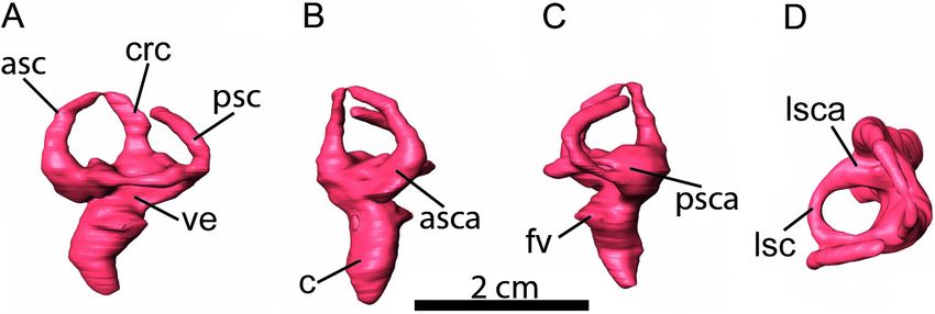
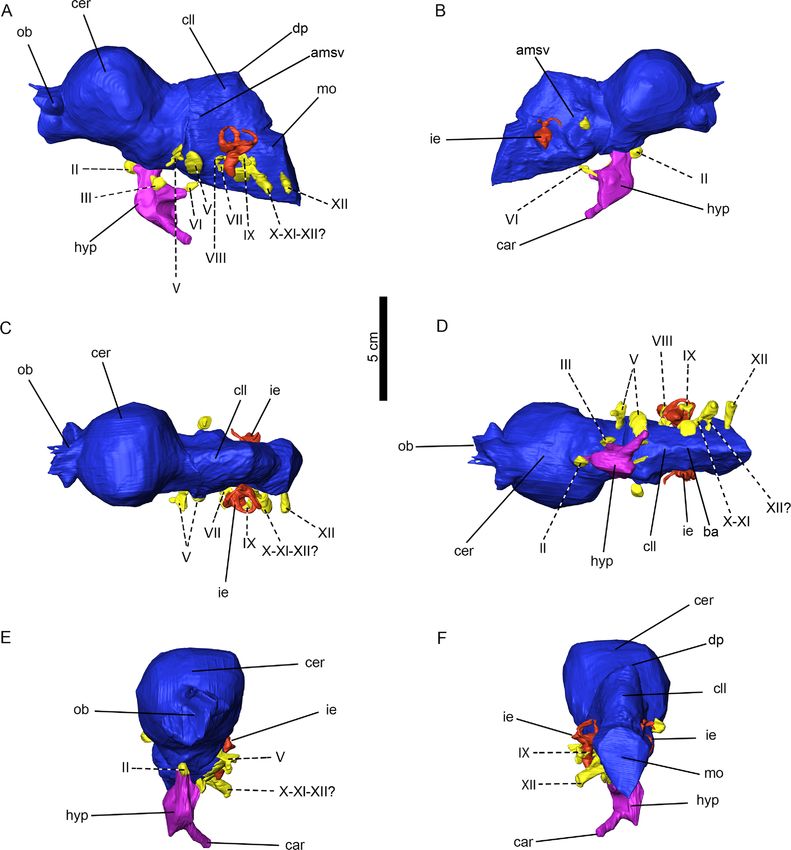
Figure 1 A 3D reconstruction of the braincase of Arenysaurus ardevoli. (A) Braincase opaque, (B)
                                      Semitransparent braincase with the brain cavity endocast opaque.

                                      The main goals of the present paper are (1) to describe the first 3D endocast of a
                                   European hadrosaurid, (2) compare the neuroanatomy of the European hadrosaurids
                                   with the other Laurasian ones, and (3) provide new insights into the paleobiology of
                                   the lambeosaurines, for which there has up to now been a scarcity of information in
                                   comparison with hadrosaurines (Evans, Ridgely & Witmer, 2009; Lauters et al., 2013).

                                   MATERIAL AND METHODS
                                   Studied material: MPZ2008/1 (Fig. 1), skull remains of the holotype of the taxon
                                   Arenysaurus (Pereda-Suberbiola et al., 2009b). The remains are from the Blasi 3 locality
                                   in the town of Arén (Huesca province, NE Spain). Postcranial remains of Arenysaurus have
                                   also been recovered (see Cruzado-Caballero et al., 2013).
                                   Computed tomography: The cranial material of Arenysaurus was CT scanned at the
                                   “Laboratorio de Evolución Humana” (LEH) of the Universidad de Burgos (Spain) using
                                   an industrial CT scanner, the Yxlon Compact (Yxlon Compact; YXLON International;
                                   Hamburg, Germany). The braincase is broken into two pieces (one including the frontal,
                                   parietal, left postorbital and left squamosal while the other includes the right postorbital
                                   and right squamosal), and these were scanned separately. In both cases, the material was
                                   scanned at 200 kV and 2.8 mA and an output of 1024 × 1024 pixels per slice, with an
                                   inter-slice space of 0.3 mm. In the part of the skull with the frontal, parietal, left postorbital
                                   and left squamosal, there were 543 slices, providing an in-plane pixel size of 0.24 mm, while
                                   in the other part including the right postorbital and right squamosal there were 582 slices,
                                   providing an in-plane pixel size of 0.2 mm. Due to the density of the bone and internal
                                   matrix, the CT images present several artifacts such as beam hardening, cupping artifacts
                                   and ring artifacts. These artifacts made automatic thresholding impossible, because the
                                   grey pixel value changes. For example, the beam hardening artifact makes the edge of the
                                   object brighter than the center, and ring artifacts produce bighting and dark concentric
                                   circles. Furthermore, the grey levels of regions of interest are very similar to those of matrix
                                   regions. Therefore, the endocast segmentation was done manually. The segmentation was
                                   done in the 3D Virtual Lab of the Institut Català de Paleontologia using Avizo 7.1 (VSG,

Cruzado-Caballero et al. (2015), PeerJ, DOI 10.7717/peerj.802                                                                       3/16
Germany), generating a 3D mesh of each CT scan. After the segmentation, the two 3D
surfaces were united using the same software and looking for contact points in the 3D
braincase surfaces. When these were perfectly fitted on the inside, the 3D endocast fitted
too. Then digital measurements, including the volume, were obtained using Rhinoceros 4.0
and ImageJ.
Repository of the ct-data sets: Figshare http://dx.doi.org/10.6084/m9.figshare.1287781,
http://dx.doi.org/10.6084/m9.figshare.1287779.
Cranial endocast
The braincase of Arenysaurus is almost complete, and the individual bones are heavily
co-ossified (Fig. 2, see Video S1). It has a slight lateral taphonomic deformation that
does not affect the validity of the three-dimensional digital model (see osteological
description in Pereda-Suberbiola et al., 2009b). By means of the CT scan, an almost
complete three-dimensional endocast has been reconstructed. The structures on the left
side of the endocast are well preserved and have been digitally rendered, while those on the
right side are poorly preserved and in some cases unable to be reconstructed. As a whole, it
is possible to observe the incomplete olfactory bulbs, the cerebral hemisphere, cerebellum,
beginning of the medulla oblongata, pituitary (hypophyseal) fossa, inner ear and the canal
for almost every nerve from II to XII (Fig. 2).
The Arenysaurus endocast, as is typical in hadrosaurids, is elongate anteroposteriorly
with an anteroposterior length of 116.5 mm from the base of the olfactory tract to
the caudal branch of the hypoglossal nerve. The maximum width across the cerebral
hemisphere is 48.4 mm, and the estimated volume of the endocast (including the olfactory
bulbs) is 126.2 cm3 . The total volume of the cerebral hemisphere is 65.4 cm3 , comprising
53.3% of the total endocranial volume (excluding the olfactory bulbs). This volume value is
close to the results obtained by Saveliev, Alifanov & Bolotsky (2012) for the adult specimen
of the lambeosaurine Amurosaurus AENM1/123 (see Table 1).
On the other hand, the Arenysaurus endocast is considerably constricted lateromedially
at the cerebellum level, with a maximum width of 31.3 mm in this region, and slightly
constricted at the medulla oblongata (26.3 mm). Unfortunately, the vallecula system,
described in the anterior part of the endocast of other hadrosaurids, cannot be observed in
Arenysaurus due to the hard matrix that covers this area.
The angle of the major axis of the cerebral hemisphere to the horizontal is close to 45◦ in
the endocast. According to Evans, Ridgely & Witmer (2009), this high angle corresponds to
a lambeosaurine shape as opposed to that of hadrosaurines and other ornithopods, where
the cerebral hemisphere is positioned more horizontally (Hopson, 1979).
The angle of flexure between the cerebellum and the cerebral hemispehere is very
small, close to 10◦ , revealing that in this respect the endocast is similar to previously
described adult Laurasian lambeosaurines (e.g., Hypacrosaurus altispinus ROM 702,
Amurosaurus riabinini IRSNB R 279, AENM nos. 1/232 and 1/240; Evans, Ridgely
& Witmer, 2009; Saveliev, Alifanov & Bolotsky, 2012; Lauters et al., 2013). According
to Giffin (1989), pontine flexures are virtually absent and the possession of a nearly
Cruzado-Caballero et al. (2015), PeerJ, DOI 10.7717/peerj.802 4/16Figure 2 Cranial endocast. (A) right lateral, (B) left lateral, (C) dorsal, (D) ventral, and (E) anterior
views. Abbreviations: car, cerebral carotid artery canal; cer, cerebral hemisphere; cll, cerebellum; dp, dural
peak; ie, inner ear; mo, medulla oblongata; ob, olfactory bulbs; pit; pituitary fossa; ts, transverse sinus;
vls, ventral longitudinal sinus. II–XII, nerves; II, optic nerve; III, oculomotor nerve; V, trigeminal nerve;
V1, ophthalmic branch of nerve V; g V, trigeminal ganglion of nerve V; VI, abducens nerve; VII, facial
nerve; VIII, vestibulocochlear nerve; IX, glossopharyngeal nerve; X, vagus nerve; XI, accessory nerve; XII,
hypoglossal nerve.
straight endocranial cavity is derived for “iguanodontids” and hadrosaurids. Further, in
lateral view the cerebral hemisphere is not very strongly arched, as is the case in adult
lambeosaurines and unlike young individuals (e.g., Parasaurolophus sp. RAM 14000).
These different angles are possibly a consequence of more strongly arched frontals in
young individuals (Farke et al., 2013). In Arenysaurus the angle of the dural peak is close to
114◦ (Lautenschlager & Hübner, 2013; Farke et al., 2013).
Cruzado-Caballero et al. (2015), PeerJ, DOI 10.7717/peerj.802 5/16Cruzado-Caballero et al. (2015), PeerJ, DOI 10.7717/peerj.802
Table 1 Measurements of length and volume for complete brain cavity and various brain regions. Measurements were obtained from Lambe (1920), Ostrom (1961),
Evans, Ridgely & Witmer (2009), Saveliev, Alifanov & Bolotsky (2012), Farke et al. (2013) and Lauters et al. (2013), and for Arenysaurus they were calculated from the
digital endocasts using digital segmentation in the Avizo 7.1 program.
Taxa Ontogenetic Specimen Total length Maximum width Volume total Cerebral hemi- % cerebral hemi- Olfactory bulbs
state number endocast of the cerebral of endocast spheres without spheres volumen volumen (cm3 )
without hemisphere without olfactory bulbs with respect total
olfactory (mm) olfactory (cm3 ) volume
bulbs (mm) bulbs (cm3 )
Lambeosaurus sp. Juvenile ROM 758 113.2 43 88.32 35.1 39.74 2.9
Corythosaurus sp. Juvenile ROM 759 110.1* 46.5 91.7 41.6 45.36 6.2*
Parasaurolophus sp. Juvenile RAM 14000 – 36* – – – –
Corythosaurus sp. Subadult CMN 34825 142 44.7 134.2 51.1 38.08 11.2*
Hypacrosaurus Adult ROM 702 204 63.2 275.9 117.5 42.59 14*
altispinus
Amurosaurus Adult AENM 1/123 230 72 370 210** 56.76** –
Amurosaurus Adult AENM 1/123 230 72 400 240** 60** –
Amurosaurus Adult IRSNB R 279 154 65 290 87 30 –
Arenysaurus Subadult-Adult MPZ2008/1 116.48 48.38 122.8 65.42 53.27 3.44*
Notes.
* Incomplete or stimate.
** Include the volume of the olfactory bubs.
–
No data.
6/16The olfactory bulbs are located anteroventral to the cerebral hemisphere; only the bases
of the bulbs are preserved. It has not been possible to reconstruct them completely, because
the skull is broken in the anterior part of the frontals. The left bulb is the more complete
one, while the right bulb only preserves its ventral part. In anterior view, the left olfactory
bulb has an inverted L-shaped morphology. In this view, it is also possible to observe that
the left olfactory bulb is almost half the height of the cerebral hemisphere, as also occurs in
the adult of Amurosaurus (IRSNB R 279, AENM nos. 1/232 and 1/240; Saveliev, Alifanov
& Bolotsky, 2012; Lauters et al., 2013) and the subadult of Corythosaurus sp. (CMN 34825;
Evans, Ridgely & Witmer, 2009). The olfactory bulbs are turned downward with an angle
on the dorsal side of 127.6◦ (measured between the anterodorsal surface of the cerebral
hemisphere and the dorsal surface of the olfactory bulb). The total volume of the partially
preserved olfactory bulbs is 3.4 cm3 .
Several authors have commented on the presence of vascular elements in endocasts
(Osmólska, 2004; Evans, 2005; Evans, Ridgely & Witmer, 2009; Lauters et al., 2013). In the
case of Arenysaurus, the transverse sinus can be seen on the lateral side of the cerebellum,
and on the ventral side of the cerebellum and in part of the medulla oblongata the ventral
longitudinal sinus can be discerned (Fig. 2).The Arenysaurus pituitary (or hypophyseal)
fossa is located posteroventrally to the optic nerve. It is deformed on its left side. It has a
length of 19.1 mm, a height of 32.8 mm, a width of 14.5 mm, and a volume of 3.6 cm3 . The
original volume of the pituitary fossa was probably bigger, but taphonomical deformation
has caused a volume artifact. The size of the pituitary body appears relatively large, as
in other hadrosaurids (Lauters et al., 2013). Posteroventrally, it is possible to observe the
joining of two big cerebral carotid arteries (Fig. 2).
Cranial nerves
The canals for almost all the cranial nerves, excluding nerve I and IV, can be seen to be pre-
served on the left side. Through these canals other structures also accompanied the nerves
(e.g., meninges, venous structures, arteries, etc.). The cranial nerves present the same
configuration as in other hadrosaurids (see Hopson, 1979; Evans, Ridgely & Witmer, 2009).
Nerve II, or the optic nerve (CN II), is the most anterior nerve preserved. It is very
small, tubular, and parallels the ventral side of the cerebral hemisphere (with a lateromedial
width of 4.8 mm, and a dorsoventral height of 5.5 mm). It is located under the cerebral
hemisphere and is joined to the pituitary anteriorly. This nerve is very small in comparison
with hadrosaurids; for example, Hypacrosaurus (Evans, Ridgely & Witmer, 2009) and
Amurosaurus (Lauters et al., 2013; Saveliev, Alifanov & Bolotsky, 2012). The optic chiasm
can only be seen in left view and is represented by a low, rounded protrusion dorsal to
the pituitary fossa. Nerve III, or the oculomotor nerve (CN III), is posterior to nerve II.
It is located in the middle of the juncture between the pituitary and the midbrain. It is
small and has a very short, tubular morphology (with a lateromedial width of 4.8 mm, a
dorsoventral height of 6.5 mm and an anteroposterior length of 5.9 mm).
The next nerve preserved towards the posterior portion is nerve V, or the trigeminal
nerve (CN V). From this nerve the ophthalmic branch (CN V1 ) and the base of the
Cruzado-Caballero et al. (2015), PeerJ, DOI 10.7717/peerj.802 7/16trigeminal ganglion are preserved. However, the maxillary and mandibular branches
(CN V2-3 ) are not observed. The ophthalmic branch is 7 mm in height dorsoventrally and
2.4 mm in length anteroposteriorly.
The ventral side of the endocast preserves nerve VI, or the abducens nerves (CN VI).
These are joined to the pituitary, and exits from it posteriorly to connect ventrally with the
cerebellum. The nerves are flattened lateromedially and are wider than high.
Nerve VII, or the facial nerve (CN VII), is positioned anterior to the cochlea and
near nerve VIII. This nerve is tube-like, very small and thin, with a slight widening
dorsomedially on its distal side. It is ventral to nerve VIII and runs lateroposteriorly with
respect to the anteroposterior axis of the endocast.
Nerve VIII, or the vestibulocochlear nerve (CN VIII), is dorsal to nerve VII. This nerve is
only partially preserved, showing a very small portion of the base dorsoventrally flattened.
Nerve IX, or the glossopharyngeal nerve (CN IX), is posterior to the cochlea and runs
laterally, touching the cochlea in its anteriormost part. This nerve is very slim in its
basal part and is tubular in shape. At its lateral extreme the nerve is extremely expanded
dorsomedially (3.1 mm) and lateromedially (3 mm).
Nerves X and XI, the vagus and accessory nerves respectively (CN X and XI), are joined
and these possibly also join with a branch of nerve XII to form a single nerve. These joined
nerves are very broad anteroposteriorly (6.8 mm) and are clearly lateroposteriorly directed.
Nerve XII, or the hypoglossal nerve (CN XII), is possibly formed by two branches. The
more anterior branch could be joined with nerves X and XI. The second branch, which
is more posterior presents an anteroposteriorly narrow base (2.2 mm) and a dorsoventral
height (3.9 mm) that is expanded distally (with an anteroposterior width of 4.7 mm and a
dorsoventral height of 5.58 mm). This nerve is only laterally directed.
Inner ear
The digital reconstruction of the inner ear is complete on the left side, whereas the right
side just conserves part of the cochlea and the anterior and posterior semicircular canals.
The general form of the inner ear is similar to that described in other hadrosaurids (Brown,
1914; Langston, 1960; Ostrom, 1961; Evans, Ridgely & Witmer, 2009; Farke et al., 2013),
and, as discussed in Evans, Ridgely & Witmer (2009), it resembles the condition in extant
crocodilians. The three semicircular canals are oriented in approximately the three planes
of space, where the anterior semicircular canal is slightly higher dorsoventrally and longer
(Fig. 3). This configuration is the most common one in vertebrates (Knoll et al., 2013). The
arch of the anterior and lateral semicircular canals is circular in shape while the posterior
semicircular canal is ellipsoidal. The anterior semicircular canal is slightly taller than
the posterior semicircular canal (when the lateral canal is oriented horizontally). This
difference between the dorsoventral heights of the canals is reflected in the ratio between
them, which is 0.98 in Arenysaurus. With regard to their ampullae, the lateral ampulla
is larger than the posterior ampulla and the anterior ampulla, as in Parasaurolophus sp.
RAM 14000 (Farke et al., 2013) and unlike in Hypacrosaurus altispinus ROM 702 and
Lambeosaurus sp. ROM 758 (Evans, Ridgely & Witmer, 2009), where the anterior ampulla
Cruzado-Caballero et al. (2015), PeerJ, DOI 10.7717/peerj.802 8/16Figure 3 Left inner ear. (A) lateral, (B) anterior, (C) posterior, and (D) dorsal views. Abbreviations: asc,
anterior semicircular canal; asca, ampulla of anterior semicircular canal; c, cochlear duct (= lagena); crc,
crus communis; fv, fenestra vestibuli (= oval window); lsc, lateral semicircular canal; lsca, ampulla of
lateral semicircular canal; psc, posterior semicircular canal; psca, ampulla of posterior semicircular canal;
ve, vestibule of inner ear.
is the largest, followed by the lateral ampulla. Moreover, in lateral view, the cochlea is
boomerang-like, convex laterally and concave medially. In anterior view, it presents an
S-shape with a sharp distal border and it has a length of 10.7 mm from the foramen
vestibulea (Table 3).
DISCUSSION
The endocranial morphology among hadrosaurid dinosaurs is similar and characteristic
of the family. Hadrosaurid endocranial possess a greatly inflated, smoothly rounded
cerebrum, do not have a pontine flexure and the orientation of the cranial cavity within
the skull is obliquely anterodorsal (Hopson, 1979). At a subfamily level (hadrosaurine-
lambeosaurine) there are characters that can help to distinguish them, such as a brain
cavity that is anteroposteriorly shorter or the angle of the major axis of the cerebral
hemisphere to the horizontal in lambeosaurines (Evans, Ridgely & Witmer, 2009).
Both characters are present in the endocast of Arenysaurus and are consistent with the
lambeosaurine affinity of this taxon.
A previous paper (Pereda-Suberbiola et al., 2009b) considered that this Arenysaurus
specimen belongs to a presumably adult individual on the basis of several osteological
characteristics. The paleoneuroanatomical evidence supports this ontogenetic assignment,
with the following features referred to adult hadrosaurid animals present in this specimen:
an angle of flexure between the cerebellum and cerebral hemisphere that is very small as
in lambeosaurine adults, as described by Evans, Ridgely & Witmer (2009), and the cranial
sutures that are difficult to discern in the CT scan, as is usual in adult specimens.
However, some paleoneuroanatomical features herein reported are indicative of a
subadult ontogenetic stage for this specimen: the total volume of the endocast without
olfactory bulbs; the volume of the cerebral hemispheres without olfactory bulbs; the
maximum width of the cerebral hemisphere (see Table 1). This puzzling mixture of
characters from adult and subadult stages may reflect a possible first case of a certain
degree of dwarfism evidenced by a hadrosaurid endocast. The hypothesis of a reduction
Cruzado-Caballero et al. (2015), PeerJ, DOI 10.7717/peerj.802 9/16Table 2 Measurement of the angle of the dural peak for several hadrosaurines and lambeosaurines
calculated from drawings and digital endocasts using ImageJ. Measurements were obtained from the
Arenysaurus endocast, Lambe (1920), Ostrom (1961), Evans, Ridgely & Witmer (2009), Saveliev, Alifanov
& Bolotsky (2012), Farke et al. (2013) and Lauters et al. (2013).
Taxa Angle of dural peak
Edmontosaurus regalis 110.66
(N.M.C. No. 2289)
Edmontosaurus 133.79
(A.M.N.H. No. 5236)
Kritosaurus notabilis 132.28
(A.M.N.H. No. 5350)
Corythosaurus sp. 130.4
(CMN 34825)
Hypacrosaurus altispinus 139.08
(ROM 702)
Lambeosaurus sp. 106.71
(ROM 758)
Amurosaurus 123.77
(AENM 1/123)
Amurosaurus 138.56
(IRSNB R 279)
Arenysaurus 117.08
(MPZ2008/1)
Parasaurolophus sp. 90
(RAM 14000)
in size due to insularism in European hadrosaurids has been proposed by several authors
in the last decade and is supported by bone as well as track records (Vila et al., 2013 and
references).
Moreover, Farke et al. (2013) have hypothesized that hadrosaurids such as the small
ornithopod Dysalotosaurus lettowvorbecki present a dural peak (the angulation of the
dorsal margin of the cerebellum, not its prominence) that is mostly unchanged through
the ontogenetic stages. These authors suggest that the phylogenetic differences between
the lambeosaurini and parasaurolophini tribes could be assessed in the light of the
angle of the dural peak. In these terms, the lambeosaurins presented a wider angle
(around 120◦ ) while parasaurolophins presented a more acute angle (approximately
90◦ ). We have observed hadrosaurins and lambeosaurins to display an angle of no less
than 100◦ . In the case of Arenysaurus, this angle is approximately 114◦ (see Table 2).
In sum, the angle of the dural peak may indeed be informative, suggesting that the
condition with a greater angle could be a basal character and anangle less than 100◦ may
be exclusive to the genus Parasaurolophus. Regarding the inner ear, although the general
form is similar to the other hadrosaurids, it is possible to observe small differences in
the semicircular canals with respect to the ornithopod clade (see Fig. 4). The anterior
semicircular canal is tallest at the base of the clade (Dysalotosaurus and Iguanodon; the
ratio of anterior/posterior semicircular canal height is 1.11 in Iguanodon), by contrast
Cruzado-Caballero et al. (2015), PeerJ, DOI 10.7717/peerj.802 10/16Table 3 The maximum length of the digital cochlea of Arenysaurus casts and of other lam-
beosaurines. The maximum length of the digital cochlea of Arenysaurus casts determined using the Avizo
7.1 program, and of other lambeosaurines from Evans, Ridgely & Witmer (2009).
Taxa Ontogenetic state Specimen no. Cochlea length (mm)
Lambeosaurus sp. Juvenile ROM 758 9.2
Corythosaurus sp. Juvenile ROM 759 11.9
Parasaurolophus sp. Juvenile RAM 1400 7.6*
Corythosaurus sp. Subadult CMN 34825 12.3
Hypacrosaurus altispinus Adult ROM 702 16.7
Arenysaurus Subadult-Adult? MPZ2008/1 10.72
Notes.
* Not complete.
Figure 4 Endosseous labyrinths of the inner ears. Endosseous labyrinths of the inner ears redrawn for:
Dysalotosaurus, Lautenschlager & Hübner, (2013; Fig. 2(h)); Iguanodon, Norman, Witmer & Weishampel
(2004; Fig. 19.9); Edmontosaurus, Ostrom; (1961; Fig. 59a); Lophorhothon, Langston (1960; Fig. 163a);
Parasaurolophus, Farke et al. (2013; Fig. 16d); Hypacrosaurus and Lambeosaurus, Evans, Ridgely & Witmer;
(2009; Fig. 8a,e) and Arenysaurus ardevoli, displayed on a cladogram redrawn from Horner, Weishampel &
Forster (2004), with additional data from McDonald (2012) and Cruzado-Caballero et al. (2013). Left inner
ear: Edmontosaurus, Arenysaurus, Hypacrosaurus and Lambeosaurus; right inner ear: Dysalotosaurus,
Iguanodon, Lophorhothon and Parasaurolophus.
with some hadrosaurines, where the posterior semicircular canal is slightly taller than the
others (Edmontosaurus; the ratio of anterior/posterior semicircular canal height is 0.92).
Later, in the Lambeosaurinae subfamily, Parasaurolophus and Arenysaurus present anterior
semicircular canals that are slightly taller (the ratio of anterior/posterior semicircular canal
height is 0.97 in Parasaurolophus and 0.98 in Arenysaurus), while in the lambeosaurini
tribe they are similar in proportions to those seen in Dysalotosaurus or Iguanodon (the
ratio of anterior/posterior semicircular canal height is 1.58 in Hypacrosaurus and 1.16 in
Lambeosaurus). In addition, Parasaurolophus and Arenysaurus share a lateral ampulla that
is larger than the posterior and the anterior ampullae.
The vestibular system is involved in the coordination of movement, gaze control and
balance, detecting head movement (sensing angular acceleration) in space and maintaining
visual and postural stability (Paulina Carabajal et al., 2013). The morphology and size
Cruzado-Caballero et al. (2015), PeerJ, DOI 10.7717/peerj.802 11/16of the semicircular canals are related to locomotor agility and neck mobility and a
decrease in the compensatory movements of eyes and head (see references in Knoll et
al., 2013 and Paulina Carabajal, Carballido & Currie, 2014). According to Witmer et al.
(2008), the reduction in the difference between the length of the anterior and posterior
semicircular canals, and perhaps also in the height of these canals, may reflect a decrease
in the compensatory movements of eyes and head in Edmontosaurus, Parasaurolophus and
Arenysaurus. If true, this could be related with behavioral patterns that require less agility
in the head movements (Sereno et al., 2007).
Likewise, we hypothesize that these differences in the vestibular system, i.e., the different
ratios between the height of the anterior and posterior semicircular canal and the size
of the ampullae, could be used as a phylogenetic signal to differentiate Edmontosaurus,
Parasaurolophus and Arenysaurus from the rest of the hadrosaurids. However, more
data are necessary to know the possible influences that these differences could have on
phylogenetic interpretations or on behavior.
CONCLUSION
We provide the first complete 3D reconstruction of the brain cavity and inner ear of
a European lambeosaurine, Arenysaurus. This cranial endocast presents the general
pattern known for hadrosaurids and add to the record of hadrosaurid brain cavities from
Laurasia. The osteological and paleoneuroanatomical data suggest that Arenysaurus was
an adult individual that probably presented a certain degree of dwarfism due to insularity.
Thus, Arenysaurus could be the first evidence of how dwarfism could affect hadrosaurid
paleoneuroanatomy. Moreover, it presents an optic nerve that is unusually small, indeed
very much smaller than that of other known hadrosaurid. Furthermore, the structure
of the inner ear shows differences from the ornithopod clade with respect to the height
of the semicircular canals. These differences can be explained principally in terms of a
probable decrease in the compensatory movements of eyes and head, which would affect
the paleobiology and behavior of these animals. We hypothesize that these differences in
the vestibular system could be used as a phylogenetic signal.
Institutional abbreviations
AEHM Amur Natural History Museum, of the Amur Complex Integrated Research
Institute of the Far Eastern Branch of the Russian Academy of Sciences,
Blagoveschensk, Russia (Amur KNII FEB RAS)
CMN Canadian Museum of Nature, Ottawa, Canada
IRSNB Institut Royal des Sciences Naturelles de Belgique, Brussels, Belgium
MPZ Museo de Ciencias Naturales de la Universidad de Zaragoza, Zaragoza, Spain
RAM Raymond M. Alf Museum of Paleontology, Claremont, California, USA
ROM Royal Ontario Museum, Toronto, Canada
Cruzado-Caballero et al. (2015), PeerJ, DOI 10.7717/peerj.802 12/16ACKNOWLEDGEMENTS
We acknowledge the academic editor Dr. Andrew A. Farke (Raymond M. Alf Museum of
Paleontology, Claremon, United States of America), Dr. Pascaline Lauters (Department of
Palaeontology, Royal Belgian Institute of Natural Sciences, Bruxelles, Belgium and Service
d’Anthropologie et Génétique Humaine, Université Libre de Bruxelles, Bruxelles, Belgium)
and an anonymous reviewer for their comments on the manuscript. The authors sincerely
thank Dr. Andrew A. Farke and Dr. Ariana Paulina Carabajal for valuable discussions, Dr.
Elena Santos for the CT scanning, as well as Rupert Glasgow, who revised the translation of
the text into English.
ADDITIONAL INFORMATION AND DECLARATIONS
Funding
This research has been supported by a grant from the Instituto de Estudios Altoaragoneses
(Huesca) to the first author in 2009 to perform the CT scans. This research has been
supported by the Government of Aragón (“Grupos Consolidados” H54, and “Dirección
General de Patrimonio Cultural”) and the University of Zaragoza (221-393). P Cruzado-
Caballero is supported by a postdoctoral grant from the Ministerio de Ciencia, Tecnologı́a
e Innovación Productiva Consejo Nacional de Investigaciones Cientı́ficas y Técnicas
(CONICET) of the Government of Argentina. The funders had no role in study design,
data collection and analysis, decision to publish, or preparation of the manuscript.
Grant Disclosures
The following grant information was disclosed by the authors:
Instituto de Estudios Altoaragoneses.
Government of Aragón.
University of Zaragoza: 221-393.
Ministerio de Ciencia, Tecnologı́a e Innovación Productiva Consejo Nacional de
Investigaciones Cientı́ficas y Técnicas (CONICET) of the Government of Argentina.
Competing Interests
The authors declare there are no competing interests.
Author Contributions
• P Cruzado-Caballero analyzed the data, wrote the paper, prepared figures and/or tables,
reviewed drafts of the paper.
• J Fortuny analyzed the data, contributed reagents/materials/analysis tools, wrote the
paper, prepared figures and/or tables, reviewed drafts of the paper.
• S Llacer contributed reagents/materials/analysis tools.
• JI Canudo analyzed the data, wrote the paper, reviewed drafts of the paper.
Cruzado-Caballero et al. (2015), PeerJ, DOI 10.7717/peerj.802 13/16Data Deposition
The following information was supplied regarding the deposition of related data:
Figshare
http://dx.doi.org/10.6084/m9.figshare.1287781
http://dx.doi.org/10.6084/m9.figshare.1287779.
Supplemental Information
Supplemental information for this article can be found online at http://dx.doi.org/
10.7717/peerj.802#supplemental-information.
REFERENCES
Brown B. 1914. Corythosaurus casuarius, a new crested dinosaur from the Belly River Cretaceous,
with provisional classification of the family Trachodontidae. Bulletin of the American Museum
of Natural History 33:559–565.
Casanovas ML, Pereda-Suberbiola X, Santafé JV, Weishampel DB. 1999. First lambeosaurine
hadrosaurid from Europe: palaeobiogeographical implications. Geological Magazine
136:205–211 DOI 10.1017/S0016756899002319.
Company J, Galobart À, Gaete R. 1998. First data on the hadrosaurid dinosaurs (Ornithischia,
Dinosauria) from the Upper Cretaceous of Valencia, Spain. Oryctos 1:121–126.
Cruzado-Caballero P. 2012. Restos directos de dinosaurios hadrosáuridos (Ornithopoda,
Hadrosauridae) del Maastrichtiense superior (Cretacico Superior) de Aren (Huesca). Doctoral
thesis, University of Zaragoza.
Cruzado-Caballero P, Pereda Suberbiola X, Ruiz-Omeñaca JI. 2010. Blasisaurus canudoi gen. et
sp. nov., a new lambeosaurine dinosaur (Hadrosauridae) from the latest Cretaceous of Arén
(Huesca, Spain). Canadian Journal of Earth Sciences 47(12):1507–1517 DOI 10.1139/E10-081.
Cruzado-Caballero P, Ruiz-Omeñaca JI, Canudo JI. 2010. Evidencias de la coexistencia de
hadrosaurinos y lambeosaurinos en el Maastrichtiano superior de la Penı́nsula Ibérica (Arén,
Huesca, España). Ameghiniana 47(2):153–164 DOI 10.5710/AMGH.v47i2.6.
Cruzado-Caballero P, Canudo JI, Moreno-Azanza M, Ruiz-Omeñaca JI. 2013. New material
and phylogenetical position of Arenysaurus ardevoli, a lambeosaurine dinosaur from the Late
Maastrichtian of Arén (Northern Spain). Journal of Vertebrate Paleontology 33(6):1376–1384
DOI 10.1080/02724634.2013.772061.
Dalla Vecchia FM. 2009. Tethyshadros insularis, a new hadrosauroid dinosaur (Ornithischa)
from the Upper Cretaceous of Italy. Journal of Vertebrate Paleontology 29(4):1100–1116
DOI 10.1671/039.029.0428.
Evans DC. 2005. New evidence on brain-endocranial cavity relationships in ornithischian
dinosaurs. Acta Palaeontologica Polonica 50(3):617–622.
Evans DC, Ridgely R, Witmer LM. 2009. Endocranial anatomy of lambeosaurine hadrosaurids
(Dinosauria: Ornithischia): a sensorineural perspective on cranial crest function. The
Anatomical Record 292:1315–1337 DOI 10.1002/ar.20984.
Farke AA, Chok DJ, Herrero A, Scolieri B, Werning S. 2013. Ontogeny in the tube-crested
dinosaur Parasaurolophus (Hadrosauridae) and heterochrony in hadrosaurids. PeerJ 1:e182
DOI 10.7717/peerj.182.
Cruzado-Caballero et al. (2015), PeerJ, DOI 10.7717/peerj.802 14/16Giffin EB. 1989. Pachycephalosaur paleoneurology (Archosauria: Ornithischia). Journal of
Vertebrate Paleontology 9(1):67–77 DOI 10.1080/02724634.1989.10011739.
Gilmore CW. 1924. On the genus Stephanosaurus, with a description of the type specimen of
Lambeosaurus lambei, parks. Canada Department of Mines. (Geological Survey of Canada)
Bulletin 38:29–48.
Godefroit P, Bolotsky YL, Lauters P. 2012. A new saurolophine dinosaur from the latest
Cretaceous of far Eastern Russia. PLoS ONE 7(5):e36849 DOI 10.1371/journal.pone.0036849.
Horner JR, Weishampel DB, Forster CA. 2004. Hadrosauridae. In: Weishampel DB, Dodson P,
Osmólska H, eds. The Dinosauria. Oakland: University of California Press, 438–463.
Hopson JA. 1979. Paleoneurology. In: Gans C, ed. Biology of the reptilian, vol. IX. New York:
Academic Press, 39–146.
Knoll F, Ridgely RC, Ortega F, Sanz JL, Witmer LM. 2013. Neurocranial osteology and
neuroanatomy of a Late Cretaceous titanosaurian sauropod from Spain (Ampelosaurus sp.).
PLoS ONE 8(1):e54991 DOI 10.1371/journal.pone.0054991.
Lambe LM. 1920. The hadrosaur Edmontosaurus from the Upper Cretaceous of Alberta. Canada
Department of Mines. (Geological Survey of Canada) Memoir 120:1–79.
Langston Jr W. 1960. The vertebrate fauna of the Selma Formation of Alabama. Pt. VI. The
Dinosaurs. Fieldiana Geology Memoirs 3:313–361.
Lautenschlager S, Hübner TH. 2013. Ontogenetic trajectories in the ornithischian endocranium.
Journal of Evolutionary Biology 26(9):2044–2050 DOI 10.1111/jeb.12181.
Lauters P, Vercauteren M, Bolotsky YL, Godefroit P. 2013. Cranial endocast of the lambeosaurine
hadrosaurid Amurosaurus riabinini from the Amur Region, Russia. PLoS ONE 8(11):e78899
DOI 10.1371/journal.pone.0078899.
Lull RS, Wright NE. 1942. Hadrosaurian dinosaurs of North America. Especial Paper - Geological
Society of America 40:1–242.
McDonald AT. 2012. Phylogeny of Basal Iguanodonts (Dinosauria: Ornithischia): an update. PLoS
ONE 7(5):e36745 DOI 10.1371/journal.pone.0036745.
Nopcsa F. 1900. Dinosaurierreste aus Siebenbergen: Schadel von Limnosaurus transsylvanicus
nov. gen. et specie. Denkschriften der Kaiserlichen Akademie der Wissenschaften in Wien,
Mathematisch-Naturwissenschaftliche Klasse 65:555–591.
Norman DB, Witmer LM, Weishampel DB. 2004. Basal Ornithischia. In: Weishampel DB, Dodson
P, Osmólska H, eds. The Dinosauria. Oakland: University of California Press, 325–334.
Paulina Carabajal A, Sterli J, Müller J, Hilger A. 2013. Neuroanatomy of the Marine Jurassic
Turtle Plesiochelys etalloni (Testudinata, Plesiochelyidae). PLoS ONE 8(7):e69264
DOI 10.1371/journal.pone.0069264.
Paulina Carabajal A, Carballido JL, Currie P. 2014. Braincase, neuroanatomy, and neck
posture of Amargasaurus cazaui (Sauropoda, Dicraeosauridae) and its implications for
understanding head posture in sauropods. Journal of Vertebrate Paleontology 34(4):870–882
DOI 10.1080/02724634.2014.838174.
Pereda-Suberbiola X, Canudo JI, Company J, Cruzado-Caballero P, Ruiz-Omeñaca JI. 2009a.
Hadrosaurids from the latest Cretaceous of the Iberian Peninsula: new interpretations. Journal
of Vertebrate Paleontology 29(3):946–951 DOI 10.1671/039.029.0317.
Pereda-Suberbiola X, Canudo JI, Cruzado-Caballero P, Barco JL, López-Martı́nez N, Oms O,
Ruiz-Omeñaca JI. 2009b. The last hadrosaurid dinosaurs of Europe: a new lambeosaurine
from the Uppermost Cretaceous of Aren (Huesca, Spain). Comptes Rendus Palevol 8:559–572
DOI 10.1016/j.crpv.2009.05.002.
Cruzado-Caballero et al. (2015), PeerJ, DOI 10.7717/peerj.802 15/16Prieto-Márquez A, Dalla Vecchia FM, Gaete R, Galobart À. 2013. Diversity, relationships,
and biogeography of the lambeosaurine dinosaurs from the European Archipielago,
with description of the new aralosurin Canardia garonnensis. PLoS ONE 8(7):e69835
DOI 10.1371/journal.pone.0069835.
Osmólska H. 2004. Evidence on relation of brain to endocranial cavity in oviraptorid dinosaurs.
Acta Palaeontologica Polonica 49(2):321–324.
Ostrom JH. 1961. Cranial morphology of the hadrosaurian dinosaurs of North America. Bulletin
of the American Museum of Natural History 122(2):37–186. Available at http://digitallibrary.
amnh.org/dspace/handle/2246/1260.
Saveliev SV, Alifanov VR, Bolotsky YL. 2012. Brain anatomy of Amurosaurus riabinini and some
neurobiological peculiarities of duck-billed dinosaurs. Paleontological Journal 46(1):79–91
DOI 10.1134/S003103011201011X.
Sereno P, Wilson JA, Witmer M, Whitlock JA, Maga A, Ide O, Rowe TA. 2007. Structural
extremes in a Cretaceous dinosaur. PLoS ONE 2:11 DOI 10.1371/journal.pone.0001230.
Serrano-Brañas CI, Hernández-Rivera R, Torres-Rodrı́guez E, Espinosa Chávez B. 2006. A
natural hadrosaurid endocast from the Cerro del Pueblo Formation (Upper Cretaceous) of
Coahuila, Mexico. In: Lucas SG, Sullivan RM, eds. Late cretaceous vertebrates from the Western
interior, New Mexico Museum of Natural History and Science Bulletin, 35. 317–321.
Vila B, Oms O, Fondevilla V, Gaete R, Galobart À, Riera V, Canudo JI. 2013. The Latest
Succession of Dinosaur Tracksites in Europe: Hadrosaur Ichnology, Track Production and
Palaeoenvironments. PLoS ONE 8(9):e72579 DOI 10.1371/journal.pone.0072579.
Witmer LM, Ridgely R, Dufeau DL, Semones MC. 2008. Using CT to peer into the past: 3D
visualization of the brain and inner ear regions of birds, crocodiles, and nonavain dinosaurs.
In: Endo H, Frey R, eds. Anatomical imaging: towards a new morphology. Tokyo: Springer-Verlag,
67–87.
Young CC. 1958. The dinosaurian remains of Liayang, Shantung. Palaeontol Sinica 142:1–139.
Cruzado-Caballero et al. (2015), PeerJ, DOI 10.7717/peerj.802 16/16You can also read