Molecular diagnostics on tissue samples obtained through EBUS- TBNA: review on practice guidelines - BJMO
←
→
Page content transcription
If your browser does not render page correctly, please read the page content below
Practice Guidelines

Molecular diagnostics on tissue
samples obtained through EBUS-
TBNA: review on practice guidelines
C. Dooms, MD, PhD1,4, B. Weynand, MD, PhD2,4, S. Vander Borght, PhD2,4, L. Vliegen, MSc3,4, E. Ver-
beken, MD, PhD2,4, J. Vansteenkiste, MD, PhD1,4, P. Vandenberghe, MD, PhD3,4

Endobronchial ultrasonography is a minimally invasive endoscopic technique that enables a real-time
transbronchial needle aspiration. Endobronchial ultrasound guided transbronchial needle aspiration
(EBUS-TBNA) specimens have a high diagnostic accuracy in the detection of intrathoracic lymph node
metastasis for a variety of malignancies. Predictive biomarker testing is gaining wide importance to tailor
the treatment with the largest benefit to the patient. Endobronchial ultrasound guided transbronchial
needle aspiration also results in an accurate analysis of molecular alterations (by ImmunoHistoChemistry,
Fluorescence In Situ Hybridisation, or gene sequencing) provided that the endoscopist takes sufficient
tumour samples and a dedicated cytopathologist is involved in the mastery of the specimens.
Endobronchial ultrasound guided transbronchial needle aspiration samples can be handled in different
ways. Liquid-based cytology and smears are mostly used. The choice of the testing method should be
based primarily on the nature of the sample to be tested, testing laboratory’s expertise, and available
equipment. ImmunoHistoChemistry, Fluorescence In Situ Hybridisation and targeted polymerase chain
reaction-based sequencing can be performed on > 80% of the endobronchial ultrasound guided
transbronchial needle aspiration specimens, as the latter is more sensitive in terms of limit of detection than
Sanger sequencing. The next step are the next-generation sequencing assays, with only 10-20 ng of DNA
sample input per gene mutation, which will minimise rejected samples due to insufficient sample quantity.
(Belg J Med Oncol 2016;10(1):15-20)

Introduction
Over the past two decades, endobronchial ultrasound                         pathology practice.
(EBUS) guided transbronchial needle aspiration                              Several groups have reported on EBUS-TBNA for the
(TBNA), or EBUS-TBNA, has emerged as a highly effec-                        diagnosis of intrathoracic lymph node metastases in
tive minimally invasive endoscopic technique to sam-                        patients with an extrathoracic malignancy.1-3 A recent
ple peribronchial hilar or mediastinal lymph nodes for                      meta-analysis calculated the diagnostic accuracy of
pathologic examination. Subtyping and genotyping on                         EBUS-TBNA in the detection of intrathoracic lymph
EBUS-TBNA specimens has long been considered lim-                           node metastases for a variety of extrathoracic malig-
ited by the lack of tissue architecture in these small                      nancies as 86%, with a negative likelihood ratio and
tissue samples, but their performance in tumour sub-                        diagnostic sensitivity of 16% and 85%, respectively.4
and genotyping has been proven accurate in modern                           Furthermore, the high degree of diagnostic accuracy

1
    Department of Respiratory Diseases and Respiratory Oncology Unit, 2Department of Pathology, 3Center for Human Genetics, 4Translational Re-
search Unit, University Hospitals KU Leuven, Leuven, Belgium.
Please send all correspondence to: C. Dooms, MD, PhD, University Hospitals KU Leuven, Department of Respiratory Diseases and Respiratory
Oncology Unit, Herestraat 49, 3000 Leuven, Belgium, tel: +32 1 634 68 01, email: christophe.dooms@uzleuven.be.
Conflict of interest: The authors have nothing to disclose and indicate no potential conflict of interest.
Keywords: carcinoma, EBUS-TBNA, FISH, gene sequencing, IHC.

Belgian Journal of Medical Oncology			                                                           Volume 10, Issue 1, February 2016

15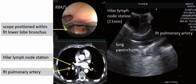
Table 1. Thoracic and extrathoracic malignancies referred for an EBUS-TBNA investigation of suspected hilar and/or mediastinal lymph node
 metastases.*
 Malignancy                   Diagnosis                         Predictive for Treatment
                              Morphology & IHC                  IHC                         FISH                    PCR/NGS
 NSCLC                        +                                 ALK                         ALK ; ROS1              EGFR
 Breast Ca                    +                                 ER ; PR ; HER2              HER2-Neu                -
 Melanoma                     +                                 -                           -                       BRAF ; (NRAS); KIT
 RCC                          +                                 -                           -                       -
 Colorectal Ca                +                                 MSI                         -                       RAS ; BRAF
 Prostate Ca                  +                                 -                           -                       -
 Head + Neck Ca               +                                 (p16)                       -                       -
 GEJ + gastric Ca             +                                 HER2                        HER2                    -

*Others (< 5% of all extrathoracic malignancies) referred for EBUS-TBNA: bladder, ovarian, cervix, pancreatic, thyroid, ampulloma, GIST, testis,
hepatocellular carcinoma.

for tissue specimens from EBUS-TBNA also implies                           EBUS-TBNA specimen collection and
that these specimens can provide adequate material for                     characteristics
predictive biomarker testing (either by IHC, FISH or                       A dedicated EBUS-scope has a diameter of around 6
mutation analysis). In our routine practice, cellblocks                    mm and can visualise mediastinal and/or hilar lymph
prepared from EBUS-TBNA derived material are used                          nodes in contact with the central airway as distal as the
for predictive biomarker testing in a variety of extratho-                 lower lobe bronchus (Figure 1). EBUS allows the explo-
racic malignancies (Table 1). Similarly, the use of EBUS-                  ration of the same paratracheal and subcarinal medias-
TBNA to successfully acquire adequate cellular mate-                       tinal lymph nodes as a cervical mediastinoscopy. In
rial for molecular subtyping in non-small cell lung                        addition, EBUS allows exploration of hilar lymph
cancer (NSCLC) has been demonstrated. Successful                           nodes. It must be stressed that EBUS cannot access the
testing of some targets (such as EGFR mutation and/or                      para-oesophageal and pulmonary ligament nodes in
ALK translocation) was observed in 72-98% of the                           the lower mediastinum. Several operators have there-
samples in several studies.5-8                                             fore extended the use of the EBUS scope into the

Figure 1. Case study of a patient with an enlarged hilar lymph node on spiral CT scan and previous history of malignant
melanoma in whom an EBUS-TBNA investigation was performed; the specimen characteristics are discussed in Figures
3 and 4.

Belgian Journal of Medical Oncology			                                                           Volume 10, Issue 1, February 2016

                                                                                                                                              16
                                                                                                                                                   1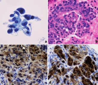
Practice Guidelines

                                                             evaluation and mastery of the EBUS-TBNA specimens
                                                             can be concluded. However, scant data exist on the
                                                             number of cases necessary to achieve and maintain
                                                             competency. In terms of safety, EBUS (within a recent
                                                             prospective registry) has a low complication or serious
                                                             adverse event rate of 1.4%.
                                                             Needle aspirations generate in general lower amounts
                                                             of DNA compared to bronchial biopsies, but they result
                                                             in an equally high success rate for mutation testing.10 In
                                                             one study, DNA extracted from formalin fixed paraffin
                                                             embedded (FFPE) small bronchial biopsies (10 un-
                                                             stained slides, 4 mm thick) yielded an average 1,690 ng
                                                             (range 250-3,600 ng) of DNA, while DNA extracted
                                                             from needle aspirations generated lower amounts of
Figure 2. Dedicated flexible bronchoscope with ultra-        DNA (average 230 ng; range 120-400 ng).10 In our ex-
sound transducer at its tip to perform real-time endosono-   perience, bronchoscopic forceps biopsy samples and
graphy and a fine needle aspiration.                         EBUS-TBNA cytology specimens exhibited a median
                                                             tumour cell proportion of 30 versus 20% and DNA
                                                             quantity of 1,610 versus 1,440 ng, respectively.7 This
oesophagus to perform an oesophageal ultrasound              observation is most likely related to the number of
(EUS) exploration with the EBUS-scope, which can             EBUS-TBNA needle passages performed by the endos-
reach the para-oesophageal and pulmonary ligament            copist, which was 5±1 needle aspirations per patient
nodes in the lower mediastinum, as well as the left          in our cohort.7
paratracheal and subcarinal mediastinal nodes similar
to the dedicated EUS scope that has a diameter of
around 12 mm. The inner diameter of the working
channel in the dedicated flexible bronchoscope is 2.0-
2.2 mm and a 21, 22 or 25 gauge needle can be used to
perform TBNA (Figure 2). Immediately after puncturing
a lymph node, the stylet is used to clear any bronchial
or cartilage debris and then the stylet is partially or
completely removed and suction can be connected in
the latter. At this time, the needle undergoes excur-
sions inside the lymph node. There is no general con-
sensus on the number of excursions or the exact loca-
tion within the lymph node that should be biopsied.
The number of needle passes needed to provide a sig-
nificant sample for molecular analysis remains un-
known, even though it has been reported in a recent
practice guideline for NSCLC that a total of four punc-
tures per lymph node provide diagnostic material in >
90% of patients.9 After each pass, the needle is with-       Figure 3. Representative image of the monolayer and
drawn, and a small amount of material can be applied         cellblock used for diagnosis and molecular diagnostics of
to a slide for preparation of smears. Alternatively, the     a malignant melanoma. (a) Papanicolaou stained mono-
aspirate can be collected directly into a preservative so-   layer showing a group of loosely cohesive neoplastic cells
lution (such as CytoLyt). The experience and skills of       with an abundant cytoplasm and polymorph nuclei with a
the bronchoscopist performing EBUS-TBNA directly             nucleolus. (b) corresponding HE stained cellblock. (c-d)
impacts the cytopathologist as they interpret the cyto-      immunohistochemical expression in tumour cells of res-
logic materials obtained from the TBNA. Furthermore,         pectively S100 protein and Melan A confirming the diagno-
a similar learning curve for the cytopathologist in the      sis of metastatic melanoma (magnification 400x).

Belgian Journal of Medical Oncology			                                        Volume 10, Issue 1, February 2016

17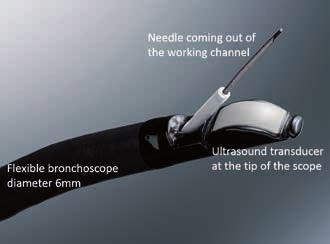
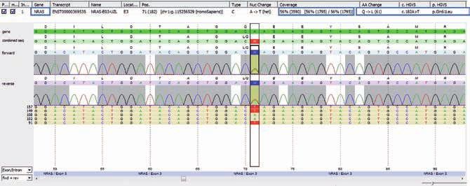
Figure 4. In the malignant melanoma (Figure 3: morphology) we found a NRAS p.Gln61Leu (p.Q61L), c.182A > T muta-
tion (NM_002524.4) in 56% of the reads (SeqNext JSI version 4.2.2) with next-generation-sequencing (Somatic 1 Multi-
plicom MASTRv2) on a Illumina platform.

Analytical phase cytopathology on                          routine use of ThinPrep-FISH is feasible and can reli-
smears or cellblocks                                       ably detect ALK gene rearrangements.8
Cytologic preparation, IHC and FISH (Figure 3)
Material obtained by EBUS-TBNA can be handled in           DNA extraction/quantitation
different ways depending on local preferences. Smears      The best results can be obtained if some technical is-
can immediately be prepared and stained if rapid on-       sues and procedures are optimised. The first issue re-
site evaluation (ROSE) is available. Several passes to     gards the use of a traditional cytologic smear or a cyto-
ensure adequate material for further cytological diag-     block. Although it has been shown that smears can be
nosis should always follow this. Liquid-based cytology     used for mutation assays, we preferred to use cellblocks
with subsequent cellblock preparation is a valid alter-    according to the guidelines from the College of Ameri-
native for diagnosis and molecular testing. In a retro-    can Pathologists.11 Cellblocks are recommended over
spective study on extrathoracic malignancies diag-         smears because of the ability to correlate with malig-
nosed by EBUS-TBNA including 117 patients, a               nant cell content, the possible retention of more mate-
diagnostic accuracy of more than 90% was shown.            rial for additional studies, and the proper fixation of
Cellblock material was available in 92% of the malig-      the material from cytological cell block preparations.
nant cases. Immunohistochemistry could be per-             Alcohol-based fixatives as the starting material for gene
formed in 80%, including hormonal receptor status          mutation testing are associated with better preserva-
and HER2 FISH in cases of metastatic breast carcino-       tion of DNA than formalin fixation.12 Molecular analy-
ma.2 For many years now, HER2 FISH has been per-           sis may be performed on samples fixed in alcohol, but
formed on paraffin-embedded tissue material for breast     the laboratory needs to extensively validate the tests to
tumours but FISH interpretation might be difficult be-     avoid false negative or false positive results.13 Success-
cause of signal loss by section artefacts, target DNA      ful sequencing is also reported on smears irrespective
integrity or incomplete penetration of probes. Several     of type of fixation or staining.14
advantages (e.g. assessment of the entire cell nucleus)    Depending on the amount of cells in the block, up to
can be envisioned for performing FISH directly on          ten consecutive 4 µm sections are prepared, of which
ThinPrep slides as compared to slides derived from         the first and last are stained with hematoxylin and eo-
FFPE cellblocks. A large NSCLC cohort with available       sin (H&E) and evaluated for the presence and amount
liquid-based cytology material (majority TBNA speci-       of tumour cells by an experienced pathologist. The pro-
men) for ALK status testing demonstrated that the          portion of tumour cells is estimated semi-quantitatively

Belgian Journal of Medical Oncology			                                      Volume 10, Issue 1, February 2016

                                                                                                                18
                                                                                                                        1
Practice Guidelines
Key messages for clinical practice
1. EBUS-TBNA is a minimally invasive endoscopic technique to sample peribronchial hilar/mediastinal
lymph nodes under local anaesthesia.
2. EBUS-TBNA specimens yield a high diagnostic accuracy in the detection of intrathoracic lymph
node metastases for a variety of malignancies.
3. EBUS-TBNA specimens can provide adequate material for predictive biomarker testing either by
IHC, FISH or mutation analysis.
4. EBUS-TBNA specimens can be handled in different ways depending on local preferences.
5. For IHC and FISH, either smears or liquid-based cytology with subsequent ThinPrep and cellblock
preparation can be used.
6. For mutation analysis, cellblocks are recommended over smears, in line with the guidelines from
the College of American Pathologists.
and the representative area is marked on the H&E slide. different between both sample types.14,16,18 However,
For EBUS-TBNA, macro-dissection is often impossible the amount of input DNA needed for performing NGS
due to spreading of the tumour cells. For most next- is quite variable, ranging from ten to > 250 ng, de-
generation sequencing (NGS) methods, a 10% malig- pending of the target capture and sequencing platform
nant cell fraction or a 5% mutant allele frequency is used. Irrespective of this feature, a lot of cytology sam-
warranted for a correct interpretation. NGS technolo- ples yielded suboptimal and insufficient DNA and
gies rely on high quality double stranded DNA that is could not be successful tested by NGS.14 False negative
suitable for library preparation followed by sequencing. and false positive events can be generated by preferen-
For low concentrations, any fluorophore-based method, tial amplification of non-tumour DNA or by amplifica-
for example Qubit 2.0, is well suited due to higher sen- tion of a homologous internal region of the desired am-
sitivity. Poor sample quality limits the amount of DNA plicon by multiplex PCR in tumour samples with low
that can be amplified. DNA of high molecular weight DNA amounts and/or low tumour content.19,20 A mini-
with little evidence of band shearing, containing no evi- mum read depth of 500x is required to reliably detect
dence of contamination from protein and RNA and a minor allele frequencies of 5-10%.14
260/280nm absorbance ratio of approximately 1.8-2.0 A great advantage of performing NGS on alcohol fixed
is therefore required.15 Also recommended, is to test the cellblocks or a smear is that no sequence artefacts as-
global DNA quality based on delta crossing point (ΔCp) sociated with formalin fixation are observed and this
values. Specifying the relation between sample quality does not hamper the variant calling. We believe that
and input requirement will help in selecting the correct all platforms will generate equal results but cost-effi-
range of input DNA. ciency, DNA input requirements, technical feasibility,
and turn-around time will be critical subjects in choos-
Sequencing (Figure 4) ing a good and reliable platform for the diagnostic ser-
NGS has emerged as a powerful tool for identifying ge- vice, especially for those samples with a low tumour
netic variants in a clinical laboratory setting. Defining content. NGS assays should have a reasonable turn-
the optimal workflow and use of the available cytologi- around time of ten working days from receipt of suit-
cal tumour material is a huge challenge for the patholo- able material in the testing laboratory to reporting the
gist. Recent, but still limited studies, stated that NGS results, consistent with the length of time previously
could also be performed on EBUS-TBNA specimens as accepted for a single gene testing. NGS testing is only
reliable and robust as on surgical specimens.14,16-18 The feasible in centres that have sufficient case through-
error rate, library complexity, enrichment performance put, appropriate equipment and technical/pathologi-
and depth of coverage does not seem to be significantly cal expertise.
Belgian Journal of Medical Oncology Volume 10, Issue 1, February 2016
19Conclusion lung carcinoma by FISH on ThinPrep slides with cytology material. J Thorac
EBUS-TBNA specimens have a high diagnostic accura- Oncol. 2014;9:464-8.
cy in the detection of intrathoracic lymph node metas- 9. van der Heijden EH, Casal RF, Trisolini R, et al. World Association for Bron-
tasis for a variety of malignancies, and can result in an chology and Interventional Pulmonology, Task Force on Specimen Guidelines.
accurate analysis of their molecular alterations (by IHC, Guideline for the acquisition and preparation of conventional and endobronchial
FISH, or gene sequencing) provided that the endosco- ultrasound-guided transbronchial needle aspiration specimens for the diagnosis
pist takes sufficient tumour samples and a dedicated and molecular testing of patients with known or suspected lung cancer. Respi-
cytopathologist is involved in the mastery of the speci- ration. 2014;88:500-17.
mens. The choice of testing method should be based 10. Arcila M, Oxnard G, Nafa K, et al. Rebiopsy of lung cancer patients with
primarily on the nature of the sample to be tested (cer- acquired resistance to EGFR inhibitors and enhanced detection of the T790M
tainly for cytology material), testing laboratory’s exper- mutation using a locked nucleic acid-based assay. Clin Cancer Res.
tise, and available equipment. Targeted methods based 2011;17:1169-80.
on real-time PCR can detect only specific mutations but 11. Lindeman N, Cagle P, Beasley M, et al. Molecular testing guideline for selec-
are more sensitive in terms of limit of detection than tion of lung cancer patients for EGFR and ALK tyrosine kinase inhibitors: guide-
Sanger sequencing. Probably a definitive answer will be line from the College of American Pathologists, International Association for the
given by NGS assays, with only 10-20 ng of DNA sam- study of Lung Cancer, and Association for Molecular Pathology. J Mol Diagn.
ple input per gene mutation, which will minimise re- 2013;15:415-53.
jected samples due to insufficient sample quantity. 12. Travis WD, Rekhtman N, Riley G, et al. Pathologic diagnosis of advanced
lung cancer based on small biopsies and cytology: a paradigm shift. J Thorac
References Oncol. 2010;5:411–4.
1. Tournoy K, Govaerts E, Malfait T, et al. Endobronchial ultrasound-guided 13. Jain D, Mathur S, Iyer V. Cell blocks in cytopathology: a review of preparative
transbronchial needle biopsy for M1 staging of extrathoracic malignancies. Ann methods, utility in diagnosis and role in ancillary studies. Cytopathology.
Oncol. 2011;22:127-31. 2014;35:356-71.
2. Sanz-Santos J, Cirauqui B, Sanchez E, et al. Endobronchial ultrasound- 14. Kanagal-Shamanna R, Portier B, Singh R, et al. Next-generation sequenc-
guided transbronchial needle aspiration in the diagnosis of intrathoracic lymph ing-based multi-gene mutation profiling of solid tumours using fine needle aspi-
node metastases from extrathoracic malignancies. Clin Exp Metastasis. ration samples: promises and challenges for routine clinical diagnostics. Mod
2013;30:521-8. Pathol. 2014;27:314-27.
3. Navani N, Nankivell M, Woolhouse I, et al. Endobronchial ultrasound-guided 15. Simbolo M, Gottardi M, Corbo V, et al. DNA Qualification Workflow for Next
transbronchial needle aspiration for the diagnosis of intrathoracic lymphade- Generation Sequencing of Histopathological Samples. PLoS One.
nopathy in patients with extrathoracic malignancy: A multicentre study. J Thorac 2013;8:e62692.
Oncol. 2011;6:1505-9. 16. Karnes HE, Duncavage EJ, Bernadt CT. Targeted next-generation sequenc-
4. Yang B, Li F, Shi W, et al. Endobronchial ultrasound-guided transbronchial ing using fine-needle aspirates from adenocarcinomas of the lung. Cancer Cy-
needle biopsy for the diagnosis of intrathoracic lymph node metastases from topathol. 2014;122:104-13.
extrathoracic malignancies: A meta-analysis and systematic review. Respirolo- 17. Qiu T, Guo H, Zhao H, et al. Next-generation sequencing for molecular diag-
gy. 2014;19:834-41. nosis of lung adenocarcinoma specimens obtained by fine needle aspiration
5. Garcia-Olive I, Monso E, Andreo F, et al. Endobronchial ultrasound-guided cytology. Sci Rep. 2015;5:11317.
transbronchial needle aspiration for identifying EGFR mutations. Eur Respir J. 18. Young G, Wang K, He J, et al. Clinical next-generation sequencing success-
2010;35:391-5. fully applied to fine-needle aspirations of pulmonary and pancreatic neoplasms.
6. Navani N, Brown JM, Nankivell M, et al. Suitability of endobronchial ultra- Cancer Cytopathol. 2013;121:688-94.
sound-guided transbronchial needle aspiration specimens for subtyping and 19. Vigliar E, Malapelle U, Belevicine C, et al. Outsourcing cytological samples
genotyping of non-small cell lung cancer: a multicentre study of 774 patients. to a referral laboratory for EGFR testing in non-small cell lung cancer: does
Am J Respir Crit Care Med. 2012;185:1316-22. theory meet practice? Cytopathology. 2015;26:312-7.
7. Dooms C, Vliegen L, Vander Borght S, et al. Suitability of small bronchoscopic 20. Vigliar E, Malapelle U, de Luca C, et al. Challenges and opportunities of
tumour specimens for lung cancer genotyping. Respiration. 2014;88:371-7. next-generation sequencing: a cytopathologist’s perspective. Cytopathology.
8. Minca E, Lanigan C, Reynolds J, et al. ALK status testing in non-small cell 2015;26:271-83.
Belgian Journal of Medical Oncology Volume 10, Issue 1, February 2016
20
1You can also read