Narrative review of hysteroscopy and endometriosis treatment
←
→
Page content transcription
If your browser does not render page correctly, please read the page content below
Review Article
                                                                                                                                           Page 1 of 10

Narrative review of hysteroscopy and endometriosis treatment
Ricardo Bassil Lasmar1, Bernardo Portugal Lasmar2,3,4
1
    Department of Surgery and Speciality of Federal Fluminense University (UFF), Brazil; 2Department of Maternal and Child, Federal Fluminense
University (UFF), Rio de Janeiro, Brazil; 3Department of Gynecological Endoscopy, Central Hospital Aristarcho Pessoa (HCAP-CBMERJ), Rio de
Janeiro, Brazil; 4Estacio da Sá University, Rio de Janeiro, Brazil
Contributions: (I) Conception and design: All authors; (II) Administrative support: All authors; (III) Provision of study materials or patients: All
authors; (IV) Collection and assembly of data: All authors; (V) Data analysis and interpretation: All authors; (VI) Manuscript writing: All authors; (VII)
Final approval of manuscript: All authors.
Correspondence to: Ricardo Bassil Lasmar. Rua Voluntários da Pátria 126/602, Botafogo, Rio de Janeiro, Brazil. Email: ricardo@lasmar.com.br.

                  Abstract: Endometriosis is defined as the presence of endometrial glands and stroma located outside
                  the uterine cavity. Adenomyosis is a benign uterine disease, characterized by the presence of glands and
                  endometrial stroma in the uterine musculature. Hysteroscopy is indicated in patients diagnosed with
                  endometriosis when there is also infertility, in the investigation of intrauterine causes of dysmenorrhea and
                  abnormal uterine bleeding. The investigation of the uterine cavity by hysteroscopy will only be indicated in
                  cases where the treatment of choice leads to uterine preservation, if there is an indication for hysterectomy,
                  this investigation will not be necessary, except when there is suspicion of cervical or endometrial cancer.
                  Literature review on endometriosis and uterine manifestations. Personal and college libraries searching
                  for texts on research methods and literature reviews. Hysteroscopy is indicated in patients diagnosed with
                  endometriosis when there is also infertility, in the investigation of intrauterine causes of dysmenorrhea
                  and abnormal uterine bleeding. Endometrial polyps, myomas and uterine malformations are related
                  to endometriosis. Those entities are related to infertility, pelvic pain and abnormal uterine bleeding.
                  Hysteroscopy is able to diagnose and treat the majority of uterine lesions associated to endometriosis.
                  Chronic endometritis, Endometrial Polyps, Myomas and uterine malformations should be investigated by
                  hysteroscopy in patients with endometriosis.

                  Keywords: Endometriosis; hysteroscopy; infertility; endometrial polyps; uterine malformations

                  Received: 11 September 2020; Accepted: 30 September 2020; Published: 25 March 2021.
                  doi: 10.21037/gpm-2020-es-01
                  View this article at: http://dx.doi.org/10.21037/gpm-2020-es-01

Introduction                                                                    female population at reproductive age, and may affect 25%
                                                                                of women with infertility. What worries most researchers is
Endometriosis is defined as the presence of endometrial
                                                                                that it takes 7 to 10 years to be diagnosed (1).
glands and stroma located outside the uterine cavity,
                                                                                   The basis of the investigation of a patient with
occurring most commonly in the cul-de-sac, ovaries, and                         endometriosis and pain involves anatomical and functional
pelvic visceral and parietal peritoneum. It represents a                        knowledge of the pelvic and abdominal organs and
chronic, inflammatory, eminently benign and estrogen                            characterization of the type of pain and its location. Colic-
dependent disease. This ectopic endometrial tissue can                          type pain may represent involvement of hollow muscle
present glands and/or stroma, usually found in the pelvic                       viscera. If it is central, retropubic, it may be related to
region, but can be present in any location. It is called                        uterine involvement, likely, adenomyosis, especially if
adenomyosis when tissue with endometrial characteristics is                     associated with increased menstrual flow. Dysmenorrhea
present in the myometrium.                                                      is now considered the greatest marker of endometriosis. If
   The incidence of endometriosis is 10% to 15% of the                          this colic pain, coinciding with menstruation, has associated

© Gynecology and Pelvic Medicine. All rights reserved.                       Gynecol Pelvic Med 2021;4:7 | http://dx.doi.org/10.21037/gpm-2020-es-01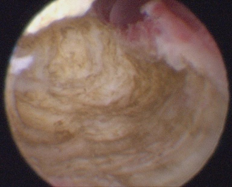
Page 2 of 10                                                                                 Gynecology and Pelvic Medicine, 2021

diarrhea, dyschezia, and sometimes hematochezia, we may           apoptosis, with greater expression of bcl-2. Several studies
have bowel endometriosis, possibly in the rectosigmoid.           have shown a higher incidence of endometrial polyps in
   Adenomyosis is a benign uterine disease, characterized         patients with endometriosis, especially when infertility
by the presence of glands and endometrial stroma in the           is associated (3). Kim et al. reported the prevalence of
uterine musculature. The presence of this tissue leads to         endometrial polyps in the endometriosis group and
hypertrophy and hyperplasia of the adjacent myometrium.           in the non-endometriosis group of 46.7% and 16.5%,
Until very recently, the diagnosis was made only by               respectively (4). Based on that information, it is important
anatomopathology, from the study of post-hysterectomy             for endometriosis patients to undergo hysteroscopy
uterine specimens. Thus, the prevalence estimate of the           to examine endometrial polyps, which can be resected
disease has always been underestimated, since the diagnosis       together, especially for infertile patients. There are some
was only made in patients undergoing hysterectomy.                data that suggest that there could be also links between
In addition, the various studies that address this theme          endometriosis and endometrial cancer. Cancer-related
applied different histological criteria for diagnosis, making     genetic changes such as loss of heterozygosis, and altered
it difficult to carry out a more comprehensive assessment,        methylation and expression patterns have been reported
leading to a huge variation in the prevalence of the disease      for endometriosis (5). Numerous endometrial cancer-
in each study, which ranged from 5% to 70% (2).                   associated genes, including PTEN and other genes in the
   Hysteroscopy is indicated in patients diagnosed                Ingenuity “endometrial cancer pathway,” have been shown
with endometriosis when there is also infertility, in the         to be dysregulated in endometriosis (6-8). This is still under
investigation of intrauterine causes of dysmenorrhea and          debate in the scientific community, due to the heterogeneity
abnormal uterine bleeding. The investigation of the uterine       of the research and low quality of evidence.
cavity by hysteroscopy will only be indicated in cases where         In addition to endometrial polyps, patients with
the treatment of choice leads to uterine preservation, if         endometriosis appear to have a higher incidence of uterine
there is an indication for hysterectomy this investigation        fibroids. Once again, the estrogen route appears to be
will not be necessary, except when there is suspicion of          the link between the two diseases. Nezhat et al., from the
cervical or endometrial cancer.                                   surgical evaluation of patients who underwent myomectomy
   In our evaluation, hysteroscopy is indicated in patients       or hysterectomy for symptomatic myoma, found the
diagnosed with endometriosis when there are complaints            presence of endometriosis, confirmed with biopsy, in 87.1%
of dysmenorrhea, abnormal uterine bleeding or findings of         of the patients (9). Nicolaus et al. found 57 patients (25.6%)
intrauterine changes in imaging tests or infertility.             with endometriosis among 223 who underwent laparoscopic
   We present the following article in accordance with the        myomectomy for symptomatic fibroids. They identified
Narrative Review reporting checklist (available at http://        that endometriosis was associated with infertility, nulliparity
dx.doi.org/10.21037/gpm-2020-es-01).                              and smaller fibroid size (10). Because of the significant
                                                                  overlap of symptoms, it is often difficult to discern which
                                                                  pathology is responsible for the patient’s complaints.
Discussion
                                                                  Thus, it is essential to properly investigate the patient with
Endometriosis is associated with a higher incidence               endometriosis, since the failure to identify a fibroid can
of uterine diseases, with hysteroscopy being the main             keep the patient symptomatic even after surgical resection
diagnostic and therapeutic tool in most cases. We can have        of the endometriosis. It is known that the submucosal
a greater association between endometriosis and:                  myoma is the main one associated with symptoms, AUB,
   (I) Uterine polyps and fibroids;                               and hysteroscopy is the method of choice for the diagnosis
   (II) Uterine malformation;                                     and treatment of these lesions.
   (III) Endometritis;                                               Hysteroscopy is an important tool in the evaluation
   (IV) Uterine factor infertility (adenomyosis).                 of uterine morphology. Endometriosis is associated with
   Like endometriosis, the pathophysiology of endometrial         obstructive anomalies and nonobstructive malformations,
polyps is still not well understood, but both are associated      especially those concerning the septated uterus. According
with an estrogenic environment. The two diseases share            to Sampson’s theory, a change in uterine morphology with
genetic changes with greater expression of estrogen and           obstruction of the outflow would lead to a higher incidence
aromatase receptors, in addition to dysregulation of              of endometriosis due to a significant tubal reflux. Song et al.

© Gynecology and Pelvic Medicine. All rights reserved.          Gynecol Pelvic Med 2021;4:7 | http://dx.doi.org/10.21037/gpm-2020-es-01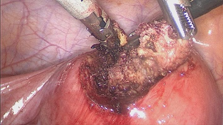
Gynecology and Pelvic Medicine, 2021                                                                                      Page 3 of 10

demonstrated that women with cervical atresia had an               undergoing hysterectomy, found chronic endometritis in
increased frequency of endometriosis (11). LaMonica et al.         52.94% of the endometriosis group and 27.02% of the non-
reported that the incidence of septated uterus in women            endometriosis group. The rate of chronic endometritis was
with infertility and/or pelvic pain ranges from 27% to 37%,        analyzed at each stage of endometriosis. Chronic endometritis
being significantly higher in women with endometriosis             was found in 40.0% of stage I endometriosis, 50.0% of stage
and mores so with stage IV disease (12). In an observational       II, 70.0% of stage III, and 46.7% of stage IV. The presence of
study of 52 patients with uterine malformation, Boujenah           ovarian endometrioma showed no difference in the prevalence
et al. identified a higher incidence of moderate and severe        of CE in this study (18).
endometriosis (endometrioma and DIE) in infertile women                Higher bacterial contamination of menstrual blood and
with uterine malformations (13). In this study, there was          an increased endotoxin level in menstrual and peritoneal
no difference in relation to peritoneal endometriosis or the       fluids have been found in women with endometriosis as
type of associated uterine malformation.                           compared with control women (19). The number of colony-
    It cannot be forgotten that the internal orifice               forming units (CFU/mL) of Gardnerella, a-Streptococcus,
stenosis, diagnosed and treated in hysteroscopy, causes            Enterococci, and E. coli was statistically significantly
dysmenorrhea.                                                      higher in endometrial samples obtained from women with
    Thus, in patients with endometriosis and associated            endometriosis than in controls.
infertility, hysteroscopy allows the evaluation of the uterine         CE can be focal or diffuse and the definitive diagnosis
cavity and the identification or exclusion of a morphological      is made by anatomopathological study of the endometrial
alteration, obstructive or not, concomitantly.                     material, with or without immunohistochemical study. This
    Chronic endometritis (CE) can be asymptomatic or have          biopsy can be done in several ways, being hysteroscopy
nonspecific symptoms such as intermenstrual bleeding,              the main method of choice, as it allows a direct view of the
pelvic pain, dyspareunia, leukorrhea or intermittent cystitis.     endometrial cavity, allowing the diagnosis of endometritis
It is characterized by infiltration of plasma cells in the         even if focal. The directed biopsy should be done, being the
endometrial stroma, where they are not normally present,           biopsy of the region with the greatest visual alteration.
except in the menstrual period, in addition to dissociation            The association of adenomyosis and endometriosis is
of cell maturation, increased stromal cellularity and edema.       very frequent. There is a constant debate in the literature as
It is a persistent inflammatory process, with a direct impact      to whether they are different entities or just different sites
on endometrial quality, and important repercussions on             of the same disease. Like endometriosis, the etiology of
the patient’s fertility. It has been identified in 12–46% of       adenomyosis is not known, and shares some of the theories
infertile patients, 30% of repeated implantation failures          of the former (20-22).
after in vitro fertilization-embryo transfer, 28% of                   The main theory is based in the rupture of the
unexplained infertility, and 12% of unexplained recurrent          endometrial-myometrial layer, allowing the invasion
miscarriages (14-16). Cicinelli et al., based on post              of glands and endometrial stroma in the myometrium.
hysterectomy endometrial analysis of patients with stage           This region is devoid of a submucosal layer, probably to
IV endometriosis and without endometriosis, found a 2.7            facilitate trophoblastic invasion in the gestational period
greater risk of endometritis in patients with endometriosis.       and, therefore, pro-inflammatory states or local trauma can
According to the authors, an etiopathogenetic link                 weaken this site, leading to a rupture. There is no transition
between CE and endometriosis may rely on altered uterine           tissue between the endometrium and the myometrium,
contractility. In women with CE, there is a decrease in the        therefore, uterine curettages, cesarean sections and multiple
anterograde subendometrial contractions present during             pregnancies can lead to a rupture of the endometrial-
menses, which contribute to the forward emptying of                myometrial interface, generating hyperplasia of the basal
menstrual blood, which thus facilitates retrograde reflux of       layer and infiltration in the injured myometrium (21-23).
menstrual bleeding through the fallopian tubes. This leads         Likewise, uterine hyperperistalsis or dysperistalsis can lead
us to speculate that, according to Sampson’s theory, CE            to local fissures, favoring the development of the disease.
may represent a facilitating factor for the development of             The immune system has also been described as causing
endometriosis (17).                                                adenomyosis, due to the action of activated T and B
    Takebayashi et al., using endometrial immunohistochemical      macrophages, which would produce antibodies and cytokines
study (CD138) in patients with and without endometriosis           capable of weakening the endomyometrial interface and

© Gynecology and Pelvic Medicine. All rights reserved.           Gynecol Pelvic Med 2021;4:7 | http://dx.doi.org/10.21037/gpm-2020-es-01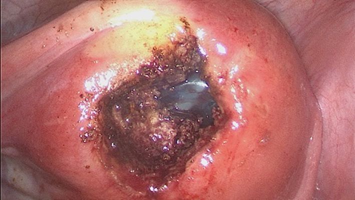
Page 4 of 10                                                                                   Gynecology and Pelvic Medicine, 2021

 Table 1 Adenomyosis classification
                                        Focal (adenomyoma)                                         Diffuse
 Type
                                             Superficial                        Intermediate                         Deep

 Invasion                                      80%

allowing the invagination of the basal layer (22).                 lymphatic spread of endometrial tissue, or through the
   There is evidence of a higher local estrogen                    transformation of stem cells into endometrial tissue.
concentration in patients with adenomyosis. The                    Endometrial regeneration in the menstrual cycle is
eutopic endometrium of patients with adenomyosis and               associated with the presence of stem cells, which could
endometriosis are sources of estrogen. Kitawaki and                lead to the development of endometrial tissue within the
collaborators (24) analyzed the concentrations of estradiol        myometrium (30,31).
in the topical endometrium (menstrual blood) and in
the peripheral circulation of patients with adenomyosis,
                                                                   Junction zone (ZJ)
endometriosis and in those with normal menstrual cycle.
The result found was a high concentration of estradiol             The transition layer from the endometrium to the
in the menstrual blood of patients with adenomyosis,               myometrium is called the subendometrial myometrium
followed by those with endometriosis, and a low                    or ZJ. This region shares greater similarities with the
concentration in patients without disease. Blood levels            endometrium than with the myometrium. Its origin,
showed no significant difference. These results, according         like that of the endometrium, is Mullerian, and changes
to the authors, prove the local production of estrogens,           in its constitution and thickness during the phases of
which is corroborated by the expression of aromatase P450          the menstrual cycle (1,21). Like the endometrium, its
in high concentrations in the endometrium of patients              thickness is maximum around the 8th to the 16th day of
with adenomyosis, and its absence in the endometrium of            the menstrual cycle. The use of hormonal contraceptives,
disease-free patients (1,22).                                      GnRH analogues and menopause reduce the thickness of
   Other hormonal changes have been described as inducing          this region. ZJ can be absent in up to 20% of women.
adenomyosis. In animal models, hyperprolactinemia led                  The normal thickness of the ZJ is up to 6–8 mm,
to the development of adenomyosis in rats. High rates of           being well defined on magnetic resonance or transvaginal
prolactin, together with the presence of steroid hormones,         ultrasonography. In the presence of adenomyosis, this
would be associated with degeneration of myometrial cells          region is thickened >12 mm, corresponding to myometrial
and, therefore, would favor the invasion of endometrial            hyperplasia, which may contain endometrial cysts (1).
tissue in the adjacent myometrium (22,25-28). Some                     Analyzes of uterine contractility, outside the pregnancy
classes of antidepressant drugs are related to the onset of        period, showed that these contractions originate exclusively
hyperprolactinemia, with the use of these substances being         in the ZJ. The amplitude and frequency of this peristalsis is
frequent in patients with adenomyosis, due to depression           related to the phase of the menstrual cycle, but the control
associated with chronic pelvic pain.                               is still not well understood (32). Another function of ZJ is to
   Adenomyosis shares with endometriosis the theory                allow trophoblastic invasion to occur properly, which may
of origin from embryonic remains (1,29). Mullerian                 be related to abortion, preeclampsia, restricted intrauterine
tissue present in the myometrium would differ from                 growth and premature birth, when there are changes in this
endometrial tissue. This theory corroborates the presence          region.
of adenomyotic tissue without continuity or proximity to
the uterine cavity, close to serosa, for example. In addition,
                                                                   Classification (Table 1)
comparative analyzes between these foci of adenomyosis and
the topical endometrium showed different compositions,             Adenomyosis can be divided into focal—adenomyoma—
suggesting different origins.                                      and diffuse. Adenomyoma is characterized by a myometrial
   Other theories, similar to that of endometriosis, associate     mass, with poorly defined contours, which differs from
the development of the disease to the intramiometrial              leiomyoma because it does not present a pseudocapsule or

© Gynecology and Pelvic Medicine. All rights reserved.           Gynecol Pelvic Med 2021;4:7 | http://dx.doi.org/10.21037/gpm-2020-es-01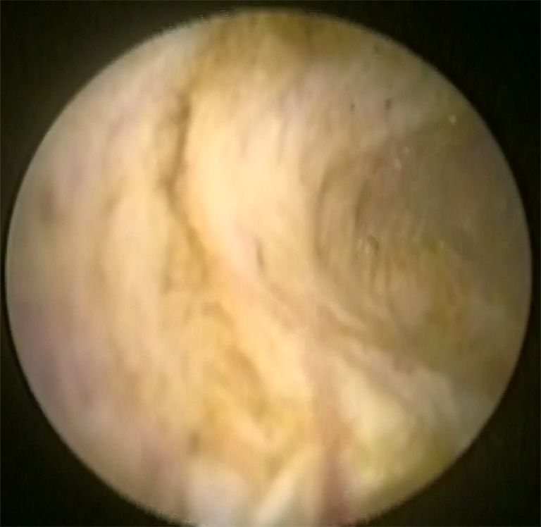
Gynecology and Pelvic Medicine, 2021 Page 5 of 10
a cleavage plane with the myometrium. There is also the embryo implantation, are at lower concentrations in patients
adenomyomatous polyp and cystic adenomyosis (32). The with adenomyosis when compared to patients free of the
first corresponds to an intracavitary polypoid formation disease. The HOXA 10 gene expression, associated with
with muscle cell composition and endometrial foci. Cystic endometrial receptivity and embryonic development, also
adenomyosis consists of cystic formations with hematic seems to be altered in these patients in relation to fertile
content surrounded by myometrium, being more frequent patients (37). The unfavorable intrauterine environment is
in adolescents or young adults. Cysts change in size associated with a higher risk of miscarriage in patients with
throughout the menstrual cycle, and there may be clinical adenomyosis.
resistance to the use of hormonal treatment. The identification of uterine adenomyosis in
There are several proposals to classify the disease. The hysteroscopy depends on the phase of the cycle in which
most widely used is the one that uses as a parameter the the exam is performed, should be performed in the recent
degree of invasion of ectopic endometrial tissue in the post-menstrual period and the lesion must be affecting the
myometrium. It is classified as superficial when the invasion uterine cavity. As adenomyosis is a myometrial disease,
is less than 40%, intermediate when it reaches 40 to 80% diagnosis by hysteroscopy will only be possible when it
and deep when it exceeds 80% penetration. is superficial or, when myometrial, it has a portion in the
Adenomyosis is associated with infertility for a few main uterine cavity. For this reason, the diagnosis of adenomyosis
reasons (33-37): in hysteroscopy makes the surgeon require imaging exams
Uterine peristalsis disorder and sperm transport. for myometrial evaluation seeking to assess the extent of
Abnormalities in the uterine cavity. adenomyosis, which may be magnetic resonance or 3D
Change in endometrial function and receptivity. ultrasound. In this way, it will be possible to decide the
The direction of uterine contractions has a fundamental access route for treatment, hysteroscopic or abdominal,
factor in the reproductive process, as it allows the proper knowing that the hysteroscopic is indicated only in
transport of sperm towards the egg. Two main moments superficial adenomyosis and the abdominal in others.
in the menstrual cycle mark uterine peristalsis: Menstrual Hysteroscopy to adenomyosis can present itself as a
period, where the sense of contractions is fundus-cervix, bluish-colored cystic formation, which, when sectioned, has
to facilitate the flow of endometrial desquamation; a chocolate content inside.
Periovulatory period, with cervical-fundus contractions,
to allow adequate transport of sperm, directed to the
Hysteroscopic treatment
tubal ostium ipsilateral to the dominant follicle. Uterine
peristalsis originates in the junctional zone, a region affected The presence of polyps, fibroids and uterine malformations,
by adenomyosis, generating hyperperistalsis and uterine as previously mentioned, is often associated with
dysperistalsis, in addition to increased intrauterine pressure. endometriosis. Uterine polyps should be removed by
In this way, sperm transport is compromised, leading to hysteroscopy, as well as fibroids with a submucosal
infertility (25). component, usually those most associated with AUB (38).
The presence of adenomyoma (focal adenomyosis) in Some uterine malformations can also be treated
the uterine cavity, as with submucosal fibroids, leads to hysteroscopically, such as uterine septa for example.
distortion in the architecture of this region, which can lead Hysteroscopic myomectomy should be indicated according
to infertility by obstructing the internal orifice and/or tubal to some hysteroscopic criteria. The Lasmar or STEP-W
ostia, making it difficult or impossible for sperm to pass (37). classification (39,40) is able to predict the complexity and
The endometrial response seems to be altered in viability of the procedure (Tables 2,3).
patients with adenomyosis. The high concentration of Hysteroscopic treatment of adenomyosis is possible when
P450 aromatase in the endometrium of these patients, as the disease is located close to the uterine cavity. Sometimes
previously discussed, is related to the worse response in in outpatient hysteroscopy, drainage of the adenomyotic
assisted reproduction methods. In addition, inflammatory cyst and partial resection of its capsule is possible, using
markers, such as interleukin 6 and 10 (IL-6, IL-10), and free biopsy-punch, with improvement of dysmenorrhea. Always
radicals are in high concentrations in this region, promoting depending on the patient’s pain tolerance (Figure 1).
a toxic effect on the embryo. Some adhesion molecules When adenomyosis is superficial and focal and the
(integrins, selectins and cadherins), which are essential for patient wants pregnancy, resection of the lesion with mono
© Gynecology and Pelvic Medicine. All rights reserved. Gynecol Pelvic Med 2021;4:7 | http://dx.doi.org/10.21037/gpm-2020-es-01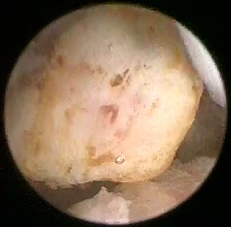
Page 6 of 10                                                                                        Gynecology and Pelvic Medicine, 2021

 Table 2 STEP-W classification
 Points                   Penetration              Size                Bases               Third             Lateral wall           Total

 0                            0                   ≤2 cm                 ≤1/3               Lower                 +1

 1                          ≤50%                1/3 to 2/3            Mild

 2                          5 cm                 >2/3               Upper

 Score                        +                     +                      +                 +                    =

 Table 3 STEP-W classification
 Score          Group               Suggested treatment

 0 to 4         I                   Low complexity hysteroscopic myomectomy

 5 to 6         II                  Complex hysteroscopic myomectomy, consider preparing with GnRH analog/or two-stage surgery

 7 to 9         III                 Recommend an alternative nonhysteroscopic technique

                      A                                                B

                      C                                                D

Figure 1 Ambulatory hysteroscopy with drainage of the adenomyotic cyst.

or bipolar resectoscope is possible. The procedure may                     of the uterus, the lesion is removed, with all its depth and
or may not be preceded by the GnRH analog, or it may                       extension (Figure 2). In those with no pregnancy desires,
be performed at the beginning of the first phase of the                    endometrial ablation may be an option as well as the
cycle, with a newly peeled endometrium. With the semi-                     association of ablation with the placement of levonorgestrel
circle handle, the most nodular area is “palpated”. With                   IUDs.
the movement of the energized loop towards the cervix                         Laparoscopic, robotic or vaginal hysterectomy assisted

© Gynecology and Pelvic Medicine. All rights reserved.                 Gynecol Pelvic Med 2021;4:7 | http://dx.doi.org/10.21037/gpm-2020-es-01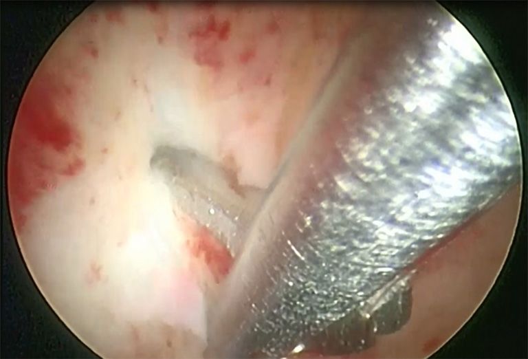
Gynecology and Pelvic Medicine, 2021                                                                                        Page 7 of 10

                   A                                               B

                   C                                               D

Figure 2 Adenomyosis resection.

by laparoscopy has become an excellent alternative to                  experience from the surgeon and an adequate analysis of
conventional laparotomy treatment, bringing with it all                the path chosen for the approach. Sometimes it is necessary
the benefits of endoscopic surgery in terms of length of               to monitor hysteroscopic resection of adenomyosis by
stay, morbidity and recovery of patients. Hysterectomy                 laparoscopy at the same time. We usually choose this
is an option for patients with constituted offspring. In               complementation when there is infiltration up to the
specific situations where it is necessary to preserve fertility,       proximity of the uterine serosa. Another option is to start
laparoscopy for resection of adenomyosis can be performed,             the procedure by performing a hysteroscopic resection
being a great challenge in cases of diffuse and extensive              to a certain myometrial depth and, in the same surgical
extension. In these cases, an attempt is made to resect as             procedure, to perform a laparoscopic resection of the rest of
much tissue as possible, always keeping in mind the need               the infiltrated area (Figure 3).
to preserve uterine function, and it is often impossible to               Pregnancies that occur after a deep surgical approach to
resect the entire disease (Figure 3).                                  adenomyosis have a higher incidence of miscarriage, uterine
   For adenomyoma the resection of the nodules is                      ruptures, which can be silent, and a higher incidence of
performed as in the case of myomectomy, however, in these              anomalous placentation. The latter are comparable to post-
cases, the cleavage plan is less defined, requiring more               cesarean section findings and myomectomy.

© Gynecology and Pelvic Medicine. All rights reserved.             Gynecol Pelvic Med 2021;4:7 | http://dx.doi.org/10.21037/gpm-2020-es-01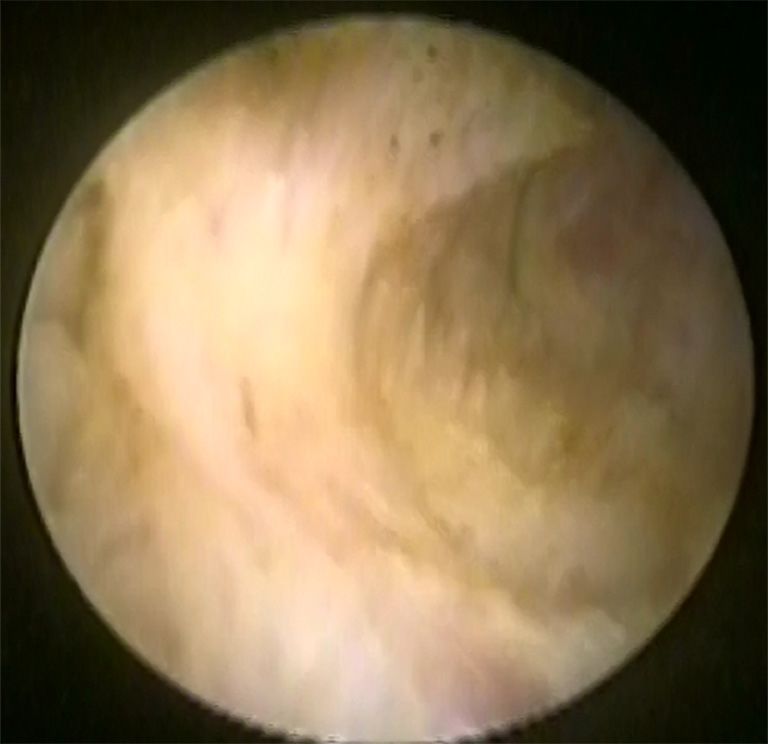
Page 8 of 10                                                                                     Gynecology and Pelvic Medicine, 2021

                   A                                                B

                   C                                                D

                   E                                     F                            G

Figure 3 Combining hysteroscopic and laparoscopic resection of adenomyosis. (A,B) MRI showing adenomyosis close to fundus serosa
(and a adenomyoma at anterior wall). (C,D) Hysteroscopic resection. (E,F,G) Trasnmural resection of adenomyosis by lapaparoscopy, after
hysteroscopic resection.

Final considerations                                                    Acknowledgments

Hysteroscopy brings important collaboration in the                      Funding: None.
investigation and treatment of intrauterine diseases in
patients with endometriosis who have no indication for
                                                                        Footnote
hysterectomy.
   The investigation of causes for infertility, dysmenorrhea            Provenance and Peer Review: This article was commissioned
and abnormal uterine bleeding may originate in the uterine              by the Guest Editor (Andrea Tinelli) for the series
cavity, as well as the identification of uterine malformations.         “Endometriosis Surgery” published in Gynecology and
   Our guidance is to have hysteroscopy performed                       Pelvic Medicine. The article has undergone external peer
before laparoscopic or robotic surgery in patients with                 review.
endometriosis. If office hysteroscopy is not possible, it
should be performed at the beginning of the same surgery                Reporting Checklist: The authors have completed the
to treat endometriosis. Further studies must be performed               Narrative Review reporting checklist. Available at http://
to evaluate the origin and better treatment of the disease.             dx.doi.org/10.21037/gpm-2020-es-01

© Gynecology and Pelvic Medicine. All rights reserved.              Gynecol Pelvic Med 2021;4:7 | http://dx.doi.org/10.21037/gpm-2020-es-01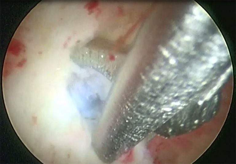
Gynecology and Pelvic Medicine, 2021                                                                                     Page 9 of 10

Conflicts of Interest: Both authors have completed the                  analyses. Cancer Med 2018;7:1978-87.
ICMJE uniform disclosure form (available at http://dx.doi.        9.    Nezhat C, Li A, Abed S, et al. Strong Association Between
org/10.21037/gpm-2020-es-01). The series “Endometriosis                 Endometriosis and Symptomatic Leiomyomas. JSLS
Surgery” was commissioned by the editorial office without               2016;20:e2016.00053.
any funding or sponsorship. The authors have no other             10.   Nicolaus K, Bräuer D, Sczesny R, Unexpected coexistent
conflicts of interest to declare.                                       endometriosis in women with symptomatic uterine
                                                                        leiomyomas is independently associated with infertility,
Ethical Statement: The authors are accountable for all                  nulliparity and minor myoma size. Arch Gynecol Obstet
aspects of the work in ensuring that questions related                  2019;300:103-8.
to the accuracy or integrity of any part of the work are          11.   Song X, Zhu L, Ding J, Clinical characteristics of
appropriately investigated and resolved.                                congenital cervical atresia and associated endometriosis
                                                                        among 96 patients. Int J Gynaecol Obstet 2016;134:252-5.
Open Access Statement: This is an Open Access article             12.   LaMonica R, Pinto J, Luciano D, Incidence of Septate
distributed in accordance with the Creative Commons                     Uterus in Reproductive-Aged Women With and Without
Attribution-NonCommercial-NoDerivs 4.0 International                    Endometriosis. J Minim Invasive Gynecol 2016;23:610-3.
License (CC BY-NC-ND 4.0), which permits the non-                 13.   Boujenah J, Salakos E, Pinto M, et al. Endometriosis and
commercial replication and distribution of the article with             uterine malformations: infertility may increase severity of
the strict proviso that no changes or edits are made and the            endometriosis. Acta Obstet Gynecol Scand 2017;96:702-6.
original work is properly cited (including links to both the      14.   Féghali J, Baker J, Mayenga JM, et al. Systematic
formal publication through the relevant DOI and the license).           hysteroscopy prior to in vitro fertilization. Gynecol Obstet
See: https://creativecommons.org/licenses/by-nc-nd/4.0/.                Fertil 2003;31:127-31.
                                                                  15.   Polisseni F, Bambirra EA, Camargos AF. Detection
                                                                        of chronic endometritis by diagnostic hysteroscopy in
References
                                                                        asymptomatic infertile patients. Gynecol Obstet Invest
1.   Abbott JA. Adenomyosis and Abnormal Uterine Bleeding               2003;55:205-10.
     (AUB-A)-Pathogenesis, diagnosis, and management. Best        16.   Cicinelli E, Resta L, Nicoletti R, et al. Detection of
     Pract Res Clin Obstet Gynaecol 2017;40:68-81.                      chronic endometritis at fluid hysteroscopy. J Minim
2.   Bird CC, McElin TW, Manalo-Estrella P. The elusive                 Invasive Gynecol 2005;12:514-8.
     adenomyosis of the uterus—revisited. Am J Obstet             17.   Cicinelli E, Trojano G, Mastromauro M, et al. Higher
     Gynecol 1972;112:583-93.                                           prevalence of chronic endometritis in women with
3.   Zhang YN, Zhang YS, Yu Q, Higher Prevalence                        endometriosis: a possible etiopathogenetic link. Fertil
     of Endometrial Polyps in Infertile Patients with                   Steril 2017;108:289-95.e1.
     Endometriosis. Gynecol Obstet Invest 2018;83:558-63.         18.   Takebayashi A, Kimura F, Kishi Y, et al. The association
4.   Kim MR, Kim YA, Jo MY, High frequency of endometrial               between endometriosis and chronic endometritis. PLoS
     polyps in endometriosis. J Am Assoc Gynecol Laparosc               One 2014;9:e88354.
     2003;10:46-8.                                                19.   Khan KN, Kitajima M, Hiraki K, et al. Escherichia coli
5.   Kokcu A. Relationship between endometriosis and                    contamination of menstrual blood and effect of bacterial
     cancer from current perspective. Arch. Gynecol. Obstet             endotoxin on endometriosis. Fertil Steril 2010;94:2860-
     2011;284:1473-9.                                                   3.e1.
6.   Aghajanova L, Giudice LC. Molecular evidence for             20.   Struble J, Reid S, Bedaiwy MA. Adenomyosis: A Clinical
     differences in endometrium in severe versus mild                   Review of a Challenging Gynecologic Condition. J Minim
     endometriosis. Reprod Sci 2011;18:229-51.                          Invasive Gynecol 2016;23:164-85.
7.   Dentillo DB, Meola J, Ferriani RA, Common dysregulated       21.   Benagiano G, Brosens I, Habiba M. Adenomyosis: a life-
     genes in endometriosis and malignancies. Rev Bras                  cycle approach. Reprod Biomed Online 2015;30:220-32.
     Ginecol Obstet 2016;38:253-62.                               22.   Benagiano G, Habiba M, Brosens I. The pathophysiology
8.   Painter JN, O’Mara TA, Morris AP, et al. Genetic overlap           of uterine adenomyosis: an update. Fertil Steril
     between endometriosis and endometrial cancer: evidence             2012;98:572-9.
     from cross-disease genetic correlation and GWAS meta-        23.   Taylor AH, Kalathy V, Habiba M. Estradiol and tamoxifen

© Gynecology and Pelvic Medicine. All rights reserved.          Gynecol Pelvic Med 2021;4:7 | http://dx.doi.org/10.21037/gpm-2020-es-01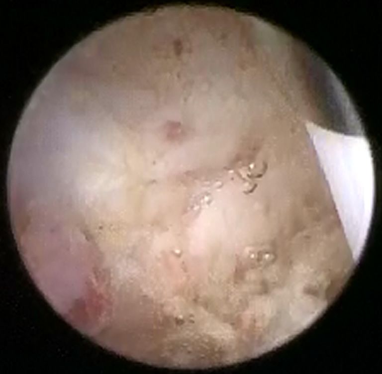
Page 10 of 10                                                                                    Gynecology and Pelvic Medicine, 2021

      enhance invasion of endometrial stromal cells in a three-             ‘endometrial-subendometrial myometrium unit disruption
      dimensional coculture model of adenomyosis. Fertil Steril             disease’ are two different entities. Reprod Biomed Online
      2014;101:288-93.                                                      2008;17:281-91.
24.   Kitawaki J, Noguchi T, Amatsu T, Expression of aromatase        33.   Koike H, Egawa H, Ohtsuka T, Correlation between
      cytochrome P450 protein and messenger ribonucleic acid                dysmenorrheic severity and prostaglandin production in
      in human endometriotic and adenomyotic tissues but not                women with endometriosis. Prostaglandins Leukot Essent
      in normal endometrium. Biol Reprod 1997;57:514e519.                   Fatty Acids 1992;46:133-7.
25.   Kissler S, Zangos S, Wiegratz I, Utero-tubal sperm              34.   Bazot M, Fiori O, Darai E. Adenomyosis in
      transport and its impairment in endometriosis and                     endometriosis—prevalence and impact on fertility.
      adenomyosis. Ann N Y Acad Sci 2007;1101:38-48.                        Evidence from magnetic resonance imaging. Hum Reprod
26.   Yamashita M, Matsuda M, Mori T. Increased expression of               2006;21:1101-2, author reply 1102-3.
      prolactin receptor mRNA in adenomyotic uterus in mice.          35.   Kunz G, Beil D, Huppert P, Adenomyosis in
      Life Sci 1997;60:1437-46.                                             endometriosis—prevalence and impact on fertility.
27.   Fiçicioğlu C, Tekin HI, Arioğlu PF, A murine model of                 Evidence from magnetic resonance imaging. Hum Reprod
      adenomyosis: the effects of hyperprolactinemia induced                2005;20:2309-16.
      by fluoxetine hydrochloride, a selective serotonin reuptake     36.   Kissler S, Hamscho N, Zangos S, Uterotubal transport
      inhibitor, on adenomyosis induction in Wistar albino rats.            disorder in adenomyosis and endometriosis - a cause for
      Acta Eur Fertil 1995;26:75-9.                                         infertility. BJOG 2006;113:902-8.
28.   Bedaiwy MA, Dahoud W, Skomorovska-Prokvolit Y,                  37.   Campo S, Campo V, Benagiano G. Adenomyosis and
      Expression of progesterone receptor isoforms-A and-B in               infertility. Reprod Biomed Online 2012;24:35-46.
      adenomyosis. Fertil Steril 2013;100:S387.                       38.   Lasmar RB, Lasmar BP. The role of leiomyomas in the
29.   Benagiano G, Brosens I, Habiba M. Structural                          genesis of abnormal uterine bleeding (AUB). Best Pract
      and molecular features of the endomyometrium in                       Res Clin Obstet Gynaecol 2017;40:82-8.
      endometriosis and adenomyosis. Hum Reprod Update                39.   Lasmar RB, Lasmar BP, Celeste RK, A new system to
      2014;20:386-402.                                                      classify submucous myomas: a Brazilian multicenter study.
30.   Bergeron C, Amant F, Ferenczy A. Pathology and                        J Minim Invasive Gynecol 2012;19:575-80.
      physiopathology of adenomyosis. Best Pract Res Clin             40.   Lasmar RB, Barrozo PR, Dias R, Submucous myomas: a
      Obstet Gynaecol 2006;20:511-21.                                       new presurgical classification to evaluate the viability of
31.   Sasson IE, Taylor HS. Stem cells and the pathogenesis of              hysteroscopic surgical treatment--preliminary report. J
      endometriosis. Ann N Y Acad Sci 2008;1127:106-15.                     Minim Invasive Gynecol 2005;12:308-11.
32.   Tocci A, Greco E, Ubaldi FM. Adenomyosis and

 doi: 10.21037/gpm-2020-es-01
 Cite this article as: Lasmar RB, Lasmar BP. Narrative review
 of hysteroscopy and endometriosis treatment. Gynecol Pelvic
 Med 2021;4:7.

© Gynecology and Pelvic Medicine. All rights reserved.              Gynecol Pelvic Med 2021;4:7 | http://dx.doi.org/10.21037/gpm-2020-es-01
You can also read