Neural Emotion Regulation Circuitry Underlying Anxiolytic Effects of Perceived Control over Pain
←
→
Page content transcription
If your browser does not render page correctly, please read the page content below

Neural Emotion Regulation Circuitry Underlying
           Anxiolytic Effects of Perceived Control over Pain

                        Tim V. Salomons1,2, Robin Nusslock3, Allison Detloff1,4,
                             Tom Johnstone1,2, and Richard J. Davidson1

Abstract
■ Anxiolytic effects of perceived control have been observed        trol. Controllable pain was associated with decreased state
across species. In humans, neuroimaging studies have sug-           anxiety, decreased activation in amygdala, and increased activa-
gested that perceived control and cognitive reappraisal reduce      tion in nucleus accumbens. In participants who perceived con-
negative affect through similar mechanisms. An important lim-       trol over the pain, reduced state anxiety was associated with
itation of extant neuroimaging studies of perceived control in      increased functional connectivity between each of these regions
terms of directly testing this hypothesis, however, is the use      and ventral lateral/ventral medial pFC. The location of pFC
of within-subject designs, which confound participantsʼ affec-      findings is consistent with regions found to be critical for the
tive response to controllable and uncontrollable stress. To com-    anxiolytic effects of perceived control in rodents. Furthermore,
pare neural and affective responses when participants were          interactions observed between pFC and both amygdala and
exposed to either uncontrollable or controllable stress, two        nucleus accumbens are remarkably similar to neural mechanisms
groups of participants received an identical series of stressors    of emotion regulation through reappraisal in humans. These
(thermal pain stimuli). One group (“controllable”) was led to       results suggest that perceived control reduces negative affect
believe they had behavioral control over the pain stimuli,          through a general mechanism involved in the cognitive regula-
whereas another (“uncontrollable”) believed they had no con-        tion of emotion. ■

INTRODUCTION
                                                                    led to advances in our understanding of the role of pFC
Perceived control has been defined as “the belief that one          in perceived behavioral control (Salomons, Johnstone,
has at oneʼs disposal a response that can influence the aver-       Backonja, Shackman, & Davidson, 2007; Wiech et al.,
siveness of an event” (Thompson, 1981). A broad scientific          2006; Salomons, 2004). Of particular note, the ventro-
literature has demonstrated the link between perceived              lateral pFC (vlPFC) and vmPFC appear to be critically
control and mental and physical health. Animals exposed             involved in modulating pain responses based on the per-
to uncontrollable stress experience deficits in learning and        ception of control (Salomons et al., 2007; Wiech et al.,
motivation as well as increased stress responses compared           2006). Although these studies have provided a prelimi-
with animals exposed to similar amounts of controllable             nary understanding of how perceived control alters the
stress (Weiss et al., 1994; Maier & Seligman, 1976). In hu-         neural response to pain, they were not optimized for
mans, perception of control over life stressors is associated       contrasting how a sustained level of perceived control
with reduced levels of depression and disease (Mineka,              alters neural and affective responses to repeated expo-
1985). Maier and Watkins (1998) have argued that behav-             sure to pain. These studies employed within-participant
ioral and neurochemical responses to uncontrollable stress          designs where participants received an equal amount of
are particularly relevant for understanding anxiety.                controllable and uncontrollable painful stress, such that
   The neural mechanisms by which perceived control                 the affective responses to controllable and uncontrollable
reduces negative emotional responses have been well                 stress were intermixed. Thus, participantsʼ affective state
delineated at the brainstem level in rodents (Maier &               reflected mixed success at controlling the painful stressor.
Watkins, 2005). Recent evidence suggests that, although             In contrast, previous studies in which participants were
brainstem regions are critical, their involvement is depen-         exposed to either only controllable or only uncontrollable
dent on the pFC and, in particular, the ventromedial pFC            stressors allowed for examination of how a sustained sense
(vmPFC; Amat et al., 2005). Functional neuroimaging has             of control might alter the affective state. These studies
                                                                    evoked a range of behavioral responses in both humans
                                                                    and animals including deficits in learning and motivation
1
 University of Wisconsin-Madison, 2University of Reading, 3North-   and, of particular interest to the study at hand, affective
western University, 4Duke University                                responses resembling anxiety (Maier & Watkins, 1998;

© 2014 Massachusetts Institute of Technology                                 Journal of Cognitive Neuroscience 27:2, pp. 222–233
                                                                                                       doi:10.1162/jocn_a_00702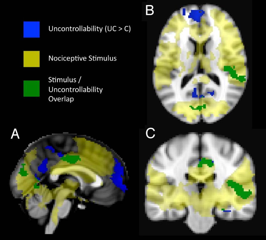
Weiss et al., 1994; Maier & Seligman, 1976). Although the       regions involved in generating affect (amygdala, NAcc)
neural mechanisms of these effects have been examined           and cortical regions involved in reappraisal and detection
in rodents, they have not been investigated in humans           of control (vlPFC, vmPFC). We predicted that increased
using in vivo neuroimaging techniques. The goal of this         functional connectivity between these cortical and sub-
study was to examine the neural mechanisms through              cortical regions would be associated with anxiety reduction
which sustained levels of perceived control over a stressor     by perceived control.
(in this case pain) alters the affective response. Accord-         An additional objective of this study was to further
ingly, we exposed two groups of healthy participants to a       investigate the effect of perceived control on the neural
matched set of painful stressors and provided differential      and perceptual response to pain. Previous neuroimaging
visual feedback such that one group believed they had           studies (Salomons et al., 2007; Wiech et al., 2006; Salomons,
behavioral control over the pain stimulus whereas the           2004) have converged on common regions involved in this
other group had the perception of a sustained lack of           response (e.g., vlPFC, ACC) but have diverged in the condi-
control over the pain stimulus.                                 tions that elicit these responses. Similarly controversial are
   On the basis of conceptual and anatomical overlap, it        the effects of perceived control on pain perception, with
has been suggested (Wiech et al., 2006) that perceived          some studies finding clear effects of perceived control on
control may alter the response to stressors through a           pain perception and others demonstrating null findings
mechanism similar to reappraisal (where the meaning             (Arntz & Schmidt, 1989; Thompson, 1981). Thus, although
of a stressful event is reinterpreted to alter the emotional    the primary focus of this report is the examination of the
response; Lazarus, 1999; Lazarus & Folkman, 1984). Neuro-       neural mechanisms underlying modulation of affective
imaging studies of reappraisal and other forms of voluntary     responses by perceived control, we also sought to clarify
regulation of negative affect have primarily focused on         these controversies.
the interplay between top–down cortical processing and
bottom–up responses in subcortical regions such as the
amygdala (Kim et al., 2011; Ochsner & Gross, 2005). The         METHODS
amygdala is differentially activated when individuals have
perceived control over stress (Salomons, 2004). The amyg-       Participants
dala has also been implicated in the generation of nega-        Participants were recruited using campus advertisements.
tive affective responses (Shin & Liberzon, 2010; Bishop,        Individuals were excluded if they were left-handed,
2007), making it an ROI for examining how perceived             pregnant, claustrophobic, or had a current psychiatric or
control alters anxiety. The interaction between amygdala        chronic pain disorder or a history of such disorders. They
and vmPFC has been implicated in the regulation of nega-        were screened for medical conditions that could affect
tive emotion (Kim et al., 2011). Additionally, extensive        pain sensitivity or regular use of drugs such as opioids or
evidence points to a role for the vlPFC in regulating activa-   NSAIDS that could alter pain perception. As the experimen-
tion in the amygdala when reappraisal is used to down-          tal manipulation involved deception and was dependent
regulate negative affect (Kalisch, 2009; Goldin, McRae,         on participants believing the instructions, psychology
Ramel, & Gross, 2008; Johnstone, van Reekum, Urry, Kalin,       majors were excluded on the grounds that they might have
& Davidson, 2007; Ochsner et al., 2004; Ochsner, Bunge,         familiarity with previous manipulations (e.g., learned help-
Gross, & Gabrieli, 2002; Schaefer et al., 2002). A recent       lessness experiments) in which participants were deceived
reappraisal study (Wager, Davidson, Hughes, Lindquist, &        about the amount of control they were able to exert. Par-
Ochsner, 2008) found that downregulation of negative af-        ticipants signed informed consent and were randomized to
fect was associated with interactions not only between vlPFC    the controllable and uncontrollable groups. Three par-
and amygdala but also between vlPFC and the nucleus             ticipants were excluded because the post experimental
accumbens (NAcc) suggesting an additional subcortical           questionnaire indicated that they had determined the
ROI. The potential involvement of the NAcc is consistent        intent of the experiment. Seven participants were excluded
with not only its role in reappraisal but also a proposed       from the controllable group because they failed to reliably
role of the striatum in processing the affectively beneficial   identify the response pattern to elicit positive visual feed-
effects of choice and perceived control (Leotti, Iyengar,       back (see Experimental Session section). This yielded a
& Ochsner, 2010). This proposed role is based on the            final sample of 52 participants with 23 in the controllable
demonstrated role of NAcc in reward (Haber & Knutson,           group (12 women; M[SD] = 20.8 [2.6] years) and 29 in
2010) as well as findings that perceived control is inher-      the uncontrollable group (14 women; M[SD] = 20.2 [2.1]).
ently rewarding (Leotti & Delgado, 2011).                       The Health Sciences institutional review board of the
   On the basis of the link between uncontrollable stress,      University of Wisconsin-Madison approved the protocol.
anxiety, and the neural mechanisms of perceived control
and reappraisal, the primary goals of this study were (1)
                                                                Familiarization Session
to examine how perceived control alters state anxiety in
response to sustained exposure to painful stimuli and           A separate familiarization session was used to determine the
(2) to understand the interaction between subcortical           level of thermal stimulation to be used in the subsequent

                                                                                                      Salomons et al.     223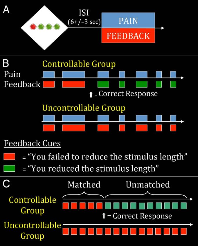
fMRI imaging session. Thermal stimulation was delivered
using a stimulator (TSA-II; www.medoc-web.com) con-
nected to a 30 × 30 mm, MRI-compatible Peltier device
affixed to the dorsal surface of the left forearm. Stimulation
began at 32°C and increased by 0.7°C/sec. Participants were
instructed to terminate stimulation when their pain reached
an 8 on an 11-point numeric rating scale anchored by 0
(no pain) and 10 (worst pain imaginable). This was re-
peated 10 times, with 30-sec breaks between presentations.
The mean temperature from the final five trials defined the
painfully hot stimulus. This strategy for determining a level
of thermal stimulation mirrors the one used in previous
studies of perceived control (Salomons, 2004). The maxi-
mum temperature used in the experiment was not allowed
to exceed 49°C. After titrating the thermal stimulation,
participants were familiarized with the MR environment
using a mock scanner and were given one 10-sec (“long”),
five 5-sec (“medium”) and four 2-sec (“short”) heat stimuli
to ensure that the experimental stimuli were painful but
tolerable.
   Thermal stimuli delivered during the experimental
session (see below) were delivered to the dorsal surface
of the left forearm with a ramp speed of 10°C/sec for all
participants.

                                                                    Figure 1. Study design. (A) One single trial. Participants were given a
Experimental Session                                                button box and told that they could shorten the painful stimulation if
                                                                    they found the correct pattern of presses on the buttons corresponding
On the day of the experimental session, participants were           to the green stars. (B) 2-, 5- and 10-sec stimuli were presented in the
given a four-button keypad and were instructed that they            same proportion and order in both groups. The groups differed only in
would receive a series of short (2 sec), medium (5 sec), and        the visual feedback received. Participants in the uncontrollable group
long (10 sec) pain stimuli in a random, preset order. Each          received consistent feedback indicating they had failed to exert control
                                                                    over the length of the heat (indicated in red). After figuring out the
trial began with a 6-sec visual cue 12 (±3) sec before the
                                                                    pattern, participants in the controllable group received feedback indicating
onset of pain. Following onset of the pain stimulus, there          that they had successfully controlled the length of the heat (indicated in
was a 5-sec gap, followed by a 7-sec rating screen and a            green). (C) The analytic focus was the medium (5 sec) stimuli. On the first
20- (±3) sec gap, resulting in a total ISI of 32 (±3) sec. During   five (“matched”) trials, participants received identical painful stimuli and
the ISI, participants rated pain intensity and unpleasantness       identical feedback. On the subsequent 20 (“unmatched”) trials, participants
                                                                    received identical painful stimuli but differed in feedback and therefore
on a 0–10 numeric rating scale (for intensity: 0 = No Pain,
                                                                    perception of control.
10 = Most Intense Pain Imaginable; for unpleasantness:
0 = Not Unpleasant, 10 = Extremely Unpleasant).
   The cue consisted of four stars (three green, one red),          not identify the correct pattern or who did not persist with
and participants were told that they could shorten the              the correct pattern following initial success feedback were
length of the subsequent painful stimuli by finding the             excluded (n = 7).
correct sequence of button presses on the keys cor-                    To prevent the responses from becoming stereotyped
responding to the green stars. They were told that if they          and to maintain a level of interest, the red star appeared
pressed the correct sequence on a trial in which they were          in a different position on each trial. The sequence of button
supposed to receive a 10-sec pain stimulus they would               presses, however, remained the same so that once partic-
receive a 5-sec stimulus and if they were supposed to re-           ipants identified the correct response sequence they could
ceive a 5-sec stimulus and made the correct response they           use it on all subsequent trials. For example, in Figure 1A,
would receive a 2-sec stimulus. They were told that 2-sec           if the correct sequence was “Left, Middle, Right,” they
stimuli could not be shortened. Participants were in-               would press Buttons 2, 3, and 4. If the red star moved to
structed that, once the correct sequence had been dis-              Position 2 on a subsequent trial, then they would press
covered (as indicated by visual feedback), they would be            Buttons 1, 3, and 4 to maintain the “Left, Middle, Right”
able to shorten the heat on every subsequent trial by re-           pattern.
peating that sequence. To ensure that all participants in              All participants irrespective of their perceived control
the controllable group received identical feedback (and             group status received an identical sequence of 50 thermal
thus a similar affective experience), participants who did          stimuli (1 long, 25 medium, 24 short). Controllability was

224     Journal of Cognitive Neuroscience                                                                           Volume 27, Number 2
manipulated as follows: Following their discovery of the the controllable group received feedback that they had
correct button sequence, the Controllable (C) group re- discovered the correct response pattern.
ceived visual feedback concurrent with the presentation The state anxiety portion of the State–Trait Anxiety
of the thermal stimuli indicating that they had success- Inventory (STAI; Spielberger, Gorsuch, & Lushene, 1970)
fully reduced the duration of the thermal stimuli when was administered immediately before and immediately
they pressed the correct response sequence (or in the after the scanning session. The variable created by residua-
case of 2-sec trials which could not be shortened, feed- lizing scores on the “after” questionnaire with respect to
back simply indicated that they had made the correct the “before” questionnaire will hereafter be referred to
response). By contrast, the Uncontrollable (UC) group as “state anxiety change.” Residualized scores were used
always received feedback indicating that they had failed for all difference scores (including RT and pain intensity)
to make the correct response sequence and thus failed instead of simple difference scores as previous research
to control the duration of the heat (see Figure 1B). Thus, (Williams & Zimmerman, 1982) suggests that residualized
the Controllable and Uncontrollable groups received an scores are more reliable when the ratio of standard devia-
identical set of thermal stimuli but differed in the feed- tions of early to late trials is greater than the correlation
back they received indicating whether or not they had between early and late trials, which was found to be the
controlled the duration of the thermal stimuli. case in this study for state anxiety. Results did not change
To control for potential group differences in pain re- substantively if simple difference scores were used. One
sponse and response to failure feedback, we ensured that participant in the controllable group did not provide state
all participants in the controllable group had the same anxiety data; thus, analyses of state-related changes in
number of initial failure trials. This was done as follows: un- anxiety are conducted on the 51 remaining participants.
beknownst to participants in the Controllable group, the Following testing, participants completed a ques-
“correct” response button sequence was determined by tionnaire that assessed their understanding of the task,
the first novel response following the 12th trial. This motivation, degree of engagement, perceived control, and
allowed an initial set of trials (which included five 5-sec attributions for success/failure using a series of structured
stimuli; see Figure 1C) on which participants in both and unstructured questions. Each item was administered
groups received identical thermal stimuli and identical on a 5-point Likert scale.
feedback indicating that they had failed to control the Analyses of all behavioral data and correlations with
length of the heat. These initial trials in which both groups extracted neural data (see below) were conducted in SPSS
received failure feedback are hereafter referred to as (Chicago, IL). Group × Time interactions in dependent
“matched trials.” Subsequent trials in which thermal stimuli measures (state anxiety, neural activation in ROIs) were
were identical but the feedback provided was different analyzed with group (UC vs. C) as a between-subject
(contingent on participants in the Controllable group factor and time as a repeated-measures factor (pre- vs.
making the correct response) are referred to as “un- postexperiment for state anxiety and Matched 5-sec trials
matched trials” (this set of trials included twenty 5-sec vs. Unmatched 5-sec trials for all variables). Between-
trials; see Figure 1C). Thus, the 50 total trials included both group comparisons (e.g., group differences in posttesting
an initial set of matched trials and a subsequent set of questionnaire data) were conducted using a one-way
unmatched trials. To maintain the illusion that repeating ANOVA with group as a factor. Within-group analyses (e.g.,
the correct sequence would always shorten the heat, no comparing state-related changes in self-reported anxiety)
10-sec heat bursts were given following the 12th trial. were run as repeated-measures ANOVAs. p < .05 (two-
There was minimal variation in the number of trials needed tailed) was used as the a priori significance level for all
to achieve a first novel and successful button response: all analyses.
participants in the controllable group made a novel re-
sponse between the fifth and sixth 5-sec stimuli (cor-
fMRI Image Acquisition
responding to the last 5-sec trial of the matched set and
the first 5-sec trial of the unmatched set). Images were acquired on a General Electric Signa 3.0-T
The analytic focus of the experiment for both ROI and high-speed imaging device with a quadrature head coil.
whole-brain analyses was the medium (5-sec) trials, as par- Functional images consisted of 30 × 4 mm sagittal EPI
ticipants in the Controllable group were led to believe that slices covering the whole brain (1 mm interslice gap;
they had successfully reduced a long stimulus, whereas 64 × 64 in-plane resolution; field of view = 240 mm;
participants in the Uncontrollable group believed they repetition time/echo time/flip = 2000 msec/30 msec/90;
had failed to reduce the medium stimuli to short ones. 225 image volumes per run). Immediately preceding acqui-
There were five medium-length stimuli during the matched sition of functional images, a whole-brain high-resolution
period and 20 medium-length stimuli during the un- T1-weighted anatomical scan (3-D T1-weighted inversion
matched period (see Figure 1C). All subsequent analyses recovery fast gradient-echo; 256 × 256 in-plane resolution;
and references to painful stimuli will refer to these 5-sec field of view = 240 mm; 124 × 1.2 mm axial slices) was
medium trials. We use the label “Time” for this variable acquired. Functional images were collected in five scan
to reflect the fact that all matched trials occurred before runs, 7 min and 30 sec per run.
Salomons et al. 225fMRI Image Analysis seed regions during pain (the output of the interaction
term in the first GLM) was significantly associated with state
Data preprocessing consisted of slice time correction and
anxiety change.
motion correction using AFNI (Cox, 1996). All other anal-
Contrasts at group and individual level are provided
yses were carried out using FEAT (Beckmann, Jenkinson,
as z scores. For all neuroimaging analyses, a cluster-wise
& Smith, 2003; fMRI Expert Analysis Tool), part of FSL
correction for multiple comparisons (z = 2.3, p < .05,
(FMRIBʼs Software Library, www.fmrib.ox.ac.uk/fsl). Data
Gaussian Random Field Theory) was used, unless other-
were smoothed with a 5-mm FWHM Gaussian blur and
wise noted.
high-pass filtered with a 100-sec cutoff. Five volumes
were dropped at the beginning of the experiment for
signal stabilization. Whole-brain Main Effect Analyses of Pain
Data were analyzed in two steps. In the first general and Controllability
linear model (GLM; at the individual participant level), a
The neural response to painful stimulation has been well
separate regressor for each experimental condition (the
delineated and has been the subject of both quantitative
cue, the 3-sec anticipatory period, the rating screen, and
(Farrell, Laird, & Egan, 2005) and qualitative (Peyron,
the short, medium, and long pain stimuli, with matched
Laurent, & Garcia-Larrea, 2000) meta-analyses. Several
and unmatched 5-sec medium pain stimuli modeled sep-
regions, including the anterior cingulate, insula, secondary
arately) was derived by convolving a stimulus-based binary
somatosensory cortex, and thalamus are consistently acti-
boxcar function (from onset to offset of the experimental
vated when participants are exposed to painful experimen-
condition) with an ideal hemodynamic response. Main
tal stimuli (Johnstone, Salomons, Backonja, & Davidson,
effect analyses of controllability were assessed from the
2012). As a measure of data quality, we conducted an
results of this individual participant level GLM.
analysis to ensure that our findings were concordant with
To examine psychophysical interactions (PPI; Friston
this literature. Pain-related activations are presented in
et al., 1997) with our ROIs, the fMRI time series was ex-
Table 1. Figure 5 displays regions that were significantly
tracted from the seed region for each participant, and this
activated in both the Controllable and Uncontrollable
time series was entered as a regressor along with all events
group on the twenty “Unmatched” 5-sec stimuli.
modeled in the experiment. A regressor representing the
For the purpose of comparison with previous studies
interaction of the seed time series with the unmatched
of the effects of perceived control on pain (Wiech et al.,
pain regressor was also run to examine which regions of
2006; Salomons, 2004), the results of whole-brain analyses
the brain differed in their connectivity with the seed re-
comparing “Unmatched” pain trials between the Uncon-
gion as a function of pain. Additionally, scan runs and six
trollable group to the Controllable group are also reported
motion covariates (I-S, L-R, A-P, pitch, yaw, and roll) were
(UC > C). Paralleling the analysis method used in our
included as nuisance variables. The time series data for
previous published work (Salomons, 2004), we report
each voxel were then modeled as the linear sum of all re-
the main effects of perceived control (UC > C) as well as
gressors. Data were registered to MNI space using FLIRT.
the stimulus/controllability overlap (regions activated in
both conditions, but more significantly activated in the
uncontrollable condition).
ROI Analysis
The primary goal of ROI analysis was to examine the role
of two a priori ROIs (amygdala and NAcc) in processing
RESULTS
the effects of perceived control on state anxiety change.
In line with this goal, we extracted values from anatomically Consistent with expectation, the Controllable group re-
defined amygdala and NAcc seeds. ROIs were generated ported greater perceived control than the Uncontrollable
by creating a mask of these regions from the Harvard group (M/SD = 3.3/1.3 for C, 1.7/1.2 for UC; F(1, 50) =
Oxford Subcortical Structural Atlas. Probability maps were 20.7, p < .05) on the posttesting questionnaire. The groups
thresholded such that every voxel within the mask had at did not differ in the degree to which they found the task
least an 80% chance of being within the structure. This boring (M/SD = 2.5/0.8 for C; 2.2/1.0 for UC) or their
mask was then used to extract values from the appropriate self-reported level of motivation following the experiment
contrast maps (see below). We first examined the main (M/SD = 4.7/0.6 for C, 4.3/0.9 for UC).
effects of perceived control on activation in these regions, A repeated-measures ANOVA indicated a significant
analyzing extracted values by group (UC vs. C) and at sin- Group × Time interaction in state anxiety, F(1, 49) =
gle time points (Matched vs. Unmatched trials), as well as 14.2, p < .05. Consistent with our hypothesis that expo-
Group × Time interactions in SPSS. We were also inter- sure to uncontrollable stress would elicit anxiety, the
ested in patterns of connectivity that underlie state anxiety uncontrollable group reported more anxiety following the
change when participants perceived control. We therefore experiment (pre M/SD 31.48/5.02, post 34.62/7.15; paired
conducted a voxelwise GLM within the Controllable group t(1, 28) = 2.76, p < .05), whereas the controllable group
to search for regions where altered connectivity with the reported less anxiety (pre M/SD 33.14/6.35, post 29.09/4.39;
226 Journal of Cognitive Neuroscience Volume 27, Number 2Table 1. Activation Results
A
Region xyz Z-score Max Voxel
Medial frontal gyrus (BA 9/BA 10) “mPFC” 0, 60, 8 5.15
Posterior cingulate gyrus (BA 23) 0, −54, 24 3.98
Superior/middle frontal gyrus (BA 8) “dlPFC” −22, 32, 32 3.66
Anterior cingulate gyrus (BA 24) “ACC” 0, −12, 36 4.37
Cuneus (BA 19) 20, −82, 38 3.27
Lingual gyrus 22, −62, −4 3.59
−12, −56, −2 3.87
Lingual/parahippocampal gyrus 28, −40, −8 3.02
Fusiform gyrus (BA 37) 42, −48, −16 3.71
Superior temporal gyrus 56, −22, 0 2.86
Thalamus −20, −32, 0 2.65
Hippocampus 28, −20, −18 5.15
B
Region xyz Mean Z-statistic
Anterior cingulate gyrus (BA 32)/SMA (BA 6) 2, 14, 38 7.08
Insular cortex (BA 13) 36, 8, 6 10.13
40, −14, 14 9.77
−36, 4, 6 8.31
Thalamus 8, −12, 0 6.85
−10, −12, 2 4.3
Inferior parietal lobe (BA 40) 62, −20, 20 8.66
Middle frontal gyrus (BA 9/BA 6) 44, 8, 36 5.76
Middle frontal gyrus (BA 46) 42, 40, 16 6.24
Inferior frontal gyrus (BA 10) 42, 44, 4 6.33
Fusiform gyrus 26, −78, −12 8.46
−18, −82, −16 8.90
Putamen 22, 10, −4 4.87
Lingual gyrus 4, −88, 0 8.5
(A) Significant activations in group contrast (UC–C). Regions surviving the cluster-based correction for multiple comparisons in the group contrast
(uncontrollable > controllable). Coordinates are in MNI space. (B) Activation in pain-related regions. To examine consistent responses to painful
stimuli, we broke the twenty-five 5-sec stimuli into five sets. The following regions were significant in all five sets and also during presentation of
10-sec stimuli following the experiment (which was presented without visual stimuli to mask out regions associated with viewing feedback stimuli).
Mean Z-statistics represent the mean of activation in all those conditions.
paired t(1, 21) = −2.53, p < .05). There was no significant pronounced increase in pain (mean/SD matched trials =
group difference on pre-experiment state anxiety, F(1, 49) = 4.93/1.15, unmatched trials 6.23/1.54, t = 6.28, p < .01)
1.08, p = .30, but the post-experiment difference was than the Uncontrollable group (mean/SD matched trials =
significant, F(1, 49) = 10.26, p < .05. 4.79/1.39, unmatched trials 5.49/1.41, t = 4.09, p < .01).
There was a significant Group × Time interaction in self- There was also a trend toward a significant difference be-
reported pain intensity ratings, F(1, 50) = 5.02, p = .03, tween groups in pain intensity ratings on unmatched trials
such that the Controllable group experienced a more (F = 3.19, p = .08). There was no relationship between
Salomons et al. 227pain intensity and state anxiety (r = −.09, p = .54). There
was no Group × Time interaction in pain unpleasantness
ratings, F(1, 50) = 0.67, p = .42.
There was a significant Group × Time interaction in
RT, F(1, 50) = 64.6, p < .01. Compared with the matched
trials, the Uncontrollable groupʼs RT on the unmatched
trials increased and the Controllable groupʼs RT de-
creased (Controllable group mean/SD in milliseconds
preexperiment = 2808.57/480.79, postexperiment
2023.54/314.33; Uncontrollable group mean/SD in milli-
seconds preexperiment 2603.94/728.53, postexperiment
2757.29/660.77). There was no significant group differ-
ence in RT on the matched trials, F(1, 50) = 1.35, p =
.25. RTs may be understood as an indirect proxy for task
engagement, as longer RTs in the Uncontrollable group
likely reflect ongoing uncertainty about the response
required to shorten the nociceptive stimulus in the wake
of negative feedback. RT change negatively correlated
with pain intensity ratings (r = −.34, p = .02) and self-
reported perceived control (r = −.42, p < .01). These
findings indicate that engagement in the task appeared
to result in reduction of perceived pain intensity.
Figure 2. Group × Time interaction for amygdala (A) and NAcc (B)
ROI Analyses: Neural Circuits Underlying activation. The Group × Time interaction was significant for anatomically
defined clusters in both amygdala, F(1, 50) = 4.02, p = .05, and NAcc,
Anxiolytic Effects of Controllability
F(1, 50) = 5.95, p < .05. Groups did not differ on matched trials for
A repeated-measures ANOVA revealed a significant either region.
Group × Time interaction for bilateral amygdala, F(1,
50) = 4.02, p = .05. Follow-up tests demonstrated sig- predicted state anxiety change in the Controllable group.
nificantly more activation in the uncontrollable group in Increased functional connectivity between bilateral NAcc
the amygdala on unmatched trials, F(1, 50) = 5.88, p < and several prefrontal regions was significantly associated
.05 (see Figure 2A). There was no difference in amygdala with reduced state anxiety. These regions included the
activation between the groups on matched trials, F(1, 50) = ventral medial pFC (BA 10/BA 32) and bilateral ventral
0.59, p = .45. Within the Controllable group, amygdala lateral prefrontal/orbitofrontal cortex (BA 11/BA 44/BA 45/
activation was significantly correlated with state anxiety BA 47).
such that reduction in amygdala activation (unmatched Increased functional connectivity between bilateral
trials residualized with respect to matched trials) was asso- amygdala and right ventral lateral prefrontal/orbitofrontal
ciated with reduced state anxiety (posttesting STAI resid- cortex (BA 11/BA 44/BA 45/BA 47; peak 48, 34, −10; see
ualized with respect to pretesting STAI; r = .56, p < .05; Figure 3A) was significantly associated with reduced state
see Figure 4). anxiety. This ventral lateral prefrontal/orbitofrontal cortex
A repeated-measures ANOVA revealed a significant region (hereby “vlPFC“) largely overlapped with the cor-
Group × Time interaction for the bilateral NAcc, F(1, 50) = responding pFC cluster in the NAcc map. The correlation
5.95, p < .05 (see Figure 2B). There was significantly more between state anxiety reduction and connectivity between
NAcc activation in the controllable group on unmatched this overlapping vlPFC cluster and the amygdala (r = −.69)
trials, F(1, 50) = 5.95, p < .05. There was no difference was significantly stronger than the corresponding correla-
between the groups on matched trials, F(1, 50) = −0.85, tion in the Uncontrollable group (UC r = −.33, p = ns;
p = .36. NAcc activation was correlated with state anxiety z for difference between correlations = −1.67, p < .05).
change within the Controllable group (r = .6, p < .05) such Similarly, the association between state anxiety change
that higher NAcc activation was associated with higher and NAcc–vlPFC connectivity (r = −.74) was significantly
state anxiety. This correlation was not significant after stronger than in the Uncontrollable group (UC r = .03,
accounting for a single outlier >2SD from the mean in p = ns; z for difference between correlations = −3.25,
state anxiety change and will therefore not be discussed p < .05). Although these findings suggest that the relation-
further. Accounting for this outlier (as well as one similar ship between anxiety and pFC to amygdala and anxiety and
outlier in the UC group) did not affect the significance or pFC to NAcc connectivity might be unique to controllable
direction of any of the other results in this report. stress, they should be interpreted with caution. Specifically,
A PPI analysis was conducted to look for regions where the connected regions were derived from a voxel-wise
altered connectivity with amygdala and NAcc during pain search within the controllable group and thus may result
228 Journal of Cognitive Neuroscience Volume 27, Number 2in a bias toward that group. There was no group difference
in the mean level of connectivity between the amygdala
and vlPFC, F(1, 50) = 0.03, p = .87, or between NAcc
and vlPFC, F(1, 50) = 0.01, p = .94.
Although connectivity between amygdala and vmPFC
did not meet our a priori threshold for significance
(z = 2.3, p < .05 corrected), given the demonstrated
involvement of vmPFC in mediating the beneficial effects
of perceived control and, more generally, in interacting
with amygdala to regulate negative affect (Urry et al.,
2006), we were interested in investigating the role of this
region. We reran the analysis at a reduced voxelwise
Figure 4. Amygdala activation versus state anxiety change in the
controllable group. Postscan state anxiety is residualized with respect to
prescan anxiety. Amygdala activation on unmatched trials is residualized
with respect to activation on matched trials.
threshold while still applying correction for multiple
comparisons (z = 1.96, p < .05, corrected). At this level,
connectivity between amygdala and both vmPFC and left
vlPFC was significantly associated with state anxiety
change. This map largely overlapped the NAcc connec-
tivity mask (see Figure 3B for overlap). Increased connec-
tivity between vmPFC and amygdala was correlated with
state anxiety reduction in the Controllable group (r = −.7).
This relationship was nonsignificant in the Uncontrollable
group (r = −.14, p = .49) and significantly weaker than
the Controllable group (z = −2.41, p < .05). Similarly,
increased vmPFC–NAcc connectivity was associated with
state anxiety reduction in the Controllable (r = −.7) but
not Uncontrollable group (r = .17, p = .37). This difference
was significant (z = −3.44, p < .05). Although the focus of
this report is the right vlPFC region that met our a priori
threshold in both maps, these results nevertheless indicate
that functional connectivity between the two seed regions
and ventral lateral pFC is bilateral. Furthermore, they
confirm the role of vmPFC in the anxiolytic effects of
perceived control.
Whole-brain Analyses: Effects of Perceived Control
Figure 3. (A) Functional connectivity of vlPFC with amygdala and
NAcc predicts state anxiety change. Anatomically defined amygdala The Uncontrollable group displayed significantly more
and NAcc clusters were used as seeds in PPI analysis. Maps of regions activation during the pain stimuli on the unmatched trials,
whose connectivity with the ROI was associated with reduced anxiety
compared with the Controllable group, in a number of
were generated. The region of vlPFC pictured represents the overlap of
the amygdala and NAcc map, such that increased functional connectivity
regions (Table 1). These included regions such as the thala-
of vlPFC with both regions (indicated by the green arrow) significantly mus, insula, and anterior cingulate that are commonly acti-
predicted reduced anxiety (r = −.67 for amygdala, r = −.74 for NAcc). vated in pain, reinforcing our previous finding (Salomons,
z = 2.3 ( p < .05, corrected). (B) Extended map of amygdala and 2004) that perceived control reduces activation within re-
NAcc connectivity overlap. To investigate an a priori hypothesis about
gions commonly associated with pain when control is per-
involvement of vmPFC, we examined regions where increased connectivity
of both amygdala and NAcc predicted reduced state anxiety change at a ceived. We also observed activation differences (UC > C)
lower threshold of z = 1.96 ( p < .05, corrected). Images are shown at in posterior cingulate cortex as well as a region of parietal
the peak voxel (z = 4.0) for vmPFC. cortex (BA7) that has been linked with the integration
Salomons et al. 229relevance to the present findings are data linking dysregu-
lated amygdalar activation with anxiety disorders (Shin &
Liberzon, 2010; Bishop, 2007) and behavioral inhibition
(Oler et al., 2010). Animal and human work has focused
on the role of prefrontal regions in regulating amygdala re-
sponses and the lack of a regulatory relationship between
pFC and amygdala has been observed in major depressive
disorder ( Johnstone et al., 2007). Consistent with this
literature, studies in which participants are asked to re-
appraise aversive stimuli consistently observe a regulatory
relationship between prefrontal regions and the amygdala
(Ochsner & Gross, 2005), with the vlPFC most frequently
implicated (Goldin et al., 2008; Johnstone et al., 2007;
Ochsner et al., 2002; Schaefer et al., 2002). Consistent with
the role of amygdala in negative affect, we observed re-
duced activation in amygdala when individuals perceived
control and a positive relationship between amygdala
activation and state anxiety change. The amygdala has also
been demonstrated to underlie reappraisal of both pain
Figure 5. Activation associated with nociceptive stimulation, and other elicitors of negative affect within individuals
controllability, and their overlap. Activations in yellow are regions that (Lapate et al., 2012). Our finding that increased connec-
were activated by the 5-sec nociceptive stimuli in both Uncontrollable tivity of amygdala and vlPFC was associated with reduced
and Controllable groups. Activations in blue are regions where anxiety when participants perceived control is consistent
uncontrollable pain elicited significantly more activation in the
with previous observations linking interactions between
Uncontrollable group. Regions in green were significantly activated in
both conditions, but significantly more active in the Uncontrollable these regions in regulating negative affect.
condition. In addition to the previously observed role of vlPFC–
amygdala interactions, it has been hypothesized that stria-
tal regions play a role in processing affective responses
of visual and somatosensory input in threat assessment to choice and perceived control (Leotti et al., 2010). Fur-
(Dong, Chudler, Sugiyama, Roberts, & Hayashi, 1994; thermore, Wager et al. (2008) suggested a role for one
Robinson & Burton, 1980). There were no activation dif- particular region of the striatum, the NAcc, in cognitive
ferences (C > UC) that survived correction for multiple regulation of negative affect, with vlPFC up-regulating the
comparisons at the whole-brain level (see Figure 5). NAcc during reappraisal of negative affect. This region is
frequently associated with reward processing and reward-
Correlations between Activation and related affect (Haber & Knutson, 2010), leading to the
Behavioral Measures hypothesis that its role in volitional control of negative
emotion is increasing positive affect in parallel with
Increased activation in mPFC was also associated with amygdala-related reduction of negative affect ( Wager
longer RT (r = .34, p = .01). Within the uncontrollable et al., 2008). Our finding that perceived control was associ-
group, higher activation in mPFC was associated with ated with increased activation of NAcc and that connectivity
increased anxiety (r = .4, p = .04). between NAcc and right vlPFC was associated with de-
creased state anxiety in participants who perceived control
is consistent with evidence that perceived control is in-
DISCUSSION
herently rewarding and motivational (Leotti & Delgado,
These data provide evidence for the anxiolytic effects of 2011). These rewarding properties may therefore con-
perceived control over a stressor. Participants who per- tribute to the anxiolytic effects of control. Although per-
ceived control over pain experienced a significant reduc- ceiving a sense of control over oneʼs environment might
tion in state anxiety compared with participants who did be inherently rewarding, it should be noted that the cur-
not. Participants who perceived control also had reduced rent study design does not allow the effects of perceived
activation in the amygdala and increased activation in NAcc. control and reward to be disentangled, as perceived con-
In participants who perceived control, these anxiolytic effects trol was delivered in the form of success feedback, which
were associated with increased functional connectivity of was likely perceived as rewarding.
amygdala and NAcc with both vlPFC and vmPFC. There was a high degree of overlap in regions of the pFC
The amygdalaʼs involvement in the encoding of affec- showing anxiety-related functional connectivity changes
tive significance and in emotional learning and expression with the amygdala and NAcc. In particular, increased
is well documented (Morrison & Salzman, 2010; Phelps connectivity between the amygdala and the NAcc and
& LeDoux, 2005; Davis & Whalen, 2001). Of particular both the vlPFC and vmPFC was associated with reduced
230 Journal of Cognitive Neuroscience Volume 27, Number 2anxiety. This is consistent with a large body of literature A more likely explanation for this finding is that, on
documenting the role of these prefrontal regions in pro- the unmatched trials (following the Controllable group
cessing the effects of perceived control and in emotion finding the correct pattern), the Uncontrollable group
regulation more generally. The vmPFC has been demon- was more engaged in the cognitive task of identifying
strated to play a role in distinguishing between uncontrol- the correct pattern of button presses and therefore more
lable and controllable stress and mediating the anxiolytic distracted from the pain stimulus than the Controllable
effects of the latter. Furthermore, covariation between group. Higher engagement throughout the task in the
vmPFC and amygdala has been associated with extinction Uncontrollable group is supported by significantly longer
of fear (Delgado, Nearing, Ledoux, & Phelps, 2008; Quirk & RTs on the unmatched trials in that group. Longer RTs
Beer, 2006; Urry et al., 2006), although rodent studies indicate a combination of continued effort and uncer-
strongly suggest that the role of vmPFC in the anxiolytic tainty, as participants who had either figured out the cor-
effects of perceived control is an expression of fear rather rect pattern or given up serious efforts to figure out the
than altered learning (Baratta, Lucero, Amat, Watkins, & pattern would be expected to respond more quickly.
Maier, 2008). Furthermore, similar effects are observed if Given that feedback about the correctness of the response
vmPFC is activated during the expression of fear even in was coincident with the pain stimuli, it is likely that par-
the absence of perceived control. Collectively, these find- ticipants who remained engaged in the task would be
ings suggest that the anxiolytic effects of perceived control actively evaluating their previous responses and perhaps
are not mediated by a dedicated neural circuit but utilize a formulating future responses during the pain stimuli, a
more general regulatory mechanism. process that likely distracted from the coincident sensory
Consistent with this assertion, a striking finding of this input. The possibility that this process might have dis-
study is how similar the neural mechanisms of the anxi- tracted participants from the sensory aspects of the pain
olytic effects of perceived control are to those observed stimuli is consistent with the observed correlation be-
in previously published reports of reappraisal of nega- tween RT and pain intensity ratings, such that slower RTs
tive emotion. More specifically, we found that functional (indicative of greater task engagement) were associated
connectivity of both amygdala and NAcc with vlPFC, a with decreased pain ratings.
circuit implicated in the reduction of negative affect by An attentional interpretation of this finding is also consis-
reappraisal (Wager et al., 2008) and compassion training tent with the fact that differences were observed between
( Weng et al., 2013), was associated with reductions in the groups in intensity, rather than unpleasantness. Two
state anxiety when participants had perceived behavioral studies examining the differential impact of emotion and
control over the duration of the pain stimuli, but no such attention on pain (Villemure & Bushnell, 2002, 2009) report
relationship existed for participants who did not perceive that emotional modulation of pain differentially affects pain
control. Averill (1973) has distinguished between behav- unpleasantness whereas attentional modulation affects
ioral control (where individuals perceive the availability pain intensity.
of a behavioral response, which will remove or modify This interpretation casts new light on previous findings
a stressor) and cognitive control (where individuals alter regarding the effects of perceived control on pain percep-
their evaluation of a stressor to reduce their stress re- tion and neural activation. A previous within-participants
sponse). Optimal coping with stress is thought to involve study found widespread increases throughout the so-called
both forms of control, with cognitive control hypothe- “pain matrix” when pain was perceived as uncontrollable,
sized to be an adaptive response when no behavioral although these activation increases were not associated
options for controlling stress are available (Carver, Scheier, with increased pain perception (Salomons, 2004). This
& Weintraub, 1989). These data put this assertion in new 2004 study diverged from a subsequent study by Wiech
light by suggesting that optimal regulation of negative et al. (2006), which found increased activation in regions
affect depends on the ability to find a situationally appro- such as ACC in the controllable (rather than uncontrol-
priate means of activating a common regulatory circuit. lable) condition, as well as reduced pain perception in
the controllable condition. These divergent findings
can be reconciled using more recent work, suggesting
Pain Perception and the Effect of Controllability
that activation within the “pain matrix” is largely unspecific
Anxiety has commonly been found to exacerbate the ex- for pain and rather has to do with the salience of the
perience of pain (Keefe et al., 2004). The finding that the stimulus—the degree to which the stimulus captures atten-
Uncontrollable group reported a significantly smaller tion and/or compels action.
increase in pain, despite increased anxiety is therefore In the experiment by Wiech and colleagues (Iannetti,
somewhat counterintuitive. In instances of extreme anxi- Salomons, Moayedi, Mouraux, & Davis, 2013), participants
ety, stress-induced analgesia has been observed (Amit & were asked to stop the stimuli in the controllable con-
Galina, 1986), but given the relatively low levels of anxiety dition when they could no longer tolerate the pain, likely
elicited and the fact that anxiety and pain intensity were drawing their attention toward the pain in that condition
uncorrelated, it would seem unlikely that the observed and increasing pain perception. In this case, behavioral
result is because of stress-induced analgesia. demands of the task likely drew attention to the pain
Salomons et al. 231stimulus in the controllable condition. In the 2004 study by Acknowledgments
Salomons et al., behavioral demands of the task were We would like to thank J. J. Curtin, J. B. Nitschke, J. P. Newman,
matched, but a cue preceding the pain drew participant M. M. Backonja, and L. Y. Abramson for helpful comments. This
attention toward the fact that the subsequent stimulus study was supported by National Institute of Mental Health
would be uncontrollable, likely increasing the salience of grants R01-MH43454 and P50-MH069315 to R. J. D. and a gift
from William Heckrodt and a Clinician-Scientist Award from
the stimulus in that condition. The current study partially the University of Toronto Centre for the Study of Pain to T. V. S.
replicates this latter finding, showing increased activation
in “pain matrix” regions (anterior cingulate and insula), Reprint requests should be sent to Tim V. Salomons, Centre for
Integrative Neuroscience and Neurodynamics, School of Psy-
but these activations are notably less widespread than in chology and Clinical Language Sciences, University of Reading,
the 2004 study. Within the context of the work on salience Early Gate, Whiteknights, RG6 6AL, United Kingdom, or via
discussed above, a likely explanation is that, although e-mail: t.v.salomons@reading.ac.uk.
overall the uncontrollable condition might have been more
behaviorally demanding (as evidenced by longer RTs) and
therefore more salient overall, these behavioral demands REFERENCES
competed with sensory input for attention, reducing acti- Amat, J., Baratta, M. V., Paul, E., Bland, S. T., Watkins, L. R.,
vation in this “salience” network and resulting in the rela- & Maier, S. F. (2005). Medial prefrontal cortex determines
tively small net increase in salience regions. This draws how stressor controllability affects behavior and dorsal
fresh attention to an often-overlooked aspect of the per- raphe nucleus. Nature Neuroscience, 8, 365–371.
ceived controllability literature, namely the demands that Amit, Z., & Galina, Z. H. (1986). Stress-induced analgesia: Adaptive
pain suppression. Physiological Reviews, 66, 1091–1120.
are placed on an individual seeking to regain control. These Arntz, A., & Schmidt, A. J. M. (1989). Perceived control and the
data suggest that it is not only the cognitive state of having experience of pain. In A. Steptoe & A. Appels (Eds.), Stress,
control that is relevant to pain perception and correspond- personal control and health (p. 131). Brussels: Wiley.
ing brain activation but also the behavioral demands of Averill, J. R. (1973). Personal control over aversive stimuli and its
regaining control that may be of relevance. Future work relationship to stress. Psychological Bulletin, 80, 286–303.
Baratta, M. V., Lucero, T. R., Amat, J., Watkins, L. R., & Maier,
should focus on examining how the interaction of per- S. F. (2008). Role of the ventral medial prefrontal cortex
ceived control and task difficulty (or the degree to which in mediating behavioral control-induced reduction of
the task draws attention away from sensory input) affects later conditioned fear. Learning & Memory (Cold Spring
pain perception and associated neural activation. Harbor, N.Y.), 15, 84–87.
Beckmann, C. F., Jenkinson, M., & Smith, S. M. (2003).
General multilevel linear modeling for group analysis in
fMRI. Neuroimage, 20, 1052–1063.
Conclusion Bishop, S. J. (2007). Neurocognitive mechanisms of anxiety: An
integrative account. Trends in Cognitive Sciences, 11, 307–316.
In summary, we found that when participants perceived Carver, C. S., Scheier, M. F., & Weintraub, J. K. (1989). Assessing
that they had successfully exerted control over a series coping strategies: A theoretically based approach. Journal
of Personality and Social Psychology, 56, 267–283.
of painful stimuli, they experienced reductions in state
Cox, R. W. (1996). AFNI: Software for analysis and visualization
anxiety and amygdala activation and increased activation of functional magnetic resonance neuroimages. Computers
in the NAcc. In participants who perceived control over and Biomedical Research, an International Journal, 29,
the painful stimuli, increased functional connectivity be- 162–173.
tween each of these subcortical regions and both ven- Davis, M., & Whalen, P. J. (2001). The amygdala: Vigilance
and emotion. Molecular Psychiatry, 6, 13–34.
tral lateral and ventral medial pFC was associated with
Delgado, M. R., Nearing, K. I., Ledoux, J. E., & Phelps, E. A. (2008).
decreased state anxiety. On the basis of the observed sim- Neural circuitry underlying the regulation of conditioned fear
ilarity between this anxiolytic circuitry and findings of and its relation to extinction. Neuron, 59, 829–838.
previous studies of negative affect reduction through Dong, W. K., Chudler, E. H., Sugiyama, K., Roberts, V. J., &
reappraisal, it is suggested that perceived control reduces Hayashi, T. (1994). Somatosensory, multisensory, and task-
related neurons in cortical area 7b (PF) of unanesthetized
anxiety by recruiting a general emotion regulation circuit.
monkeys. Journal of Neurophysiology, 72, 542–564.
Additionally, our finding partially replicate previous Farrell, M. J., Laird, A. R., & Egan, G. F. (2005). Brain activity
findings of increased activation in salience regions (e.g., associated with painfully hot stimuli applied to the upper
ACC, insula) when pain is uncontrollable. Although both limb: A meta-analysis. Human Brain Mapping, 25, 129–139.
groups experienced an increase in pain intensity as the ex- Friston, K. J., Buechel, C., Fink, G. R., Morris, J., Rolls, E., &
Dolan, R. J. (1997). Psychophysiological and modulatory
periment continued, this increase was significantly smaller
interactions in neuroimaging. Neuroimage, 6, 218–229.
in the Uncontrollable group. As pain intensity was unre- Goldin, P. R., McRae, K., Ramel, W., & Gross, J. J. (2008).
lated to anxiety, it is likely that this difference was the re- The neural bases of emotion regulation: Reappraisal and
sult of distraction because of higher task engagement in suppression of negative emotion. Biological Psychiatry,
the Uncontrollable group, calling attention to the impor- 63, 577–586.
Haber, S. N., & Knutson, B. (2010). The reward circuit: Linking primate
tance of examining task demands in perceived control
anatomy and human imaging. Neuropsychopharmacology:
experiments before attributing activation solely to agency Official Publication of the American College of
beliefs. Neuropsychopharmacology, 35, 4–26.
232 Journal of Cognitive Neuroscience Volume 27, Number 2You can also read