Original Article Microstructural changes of cartilage and subchondral bone in a guinea pig model of early- and middle-stage patellofemoral arthritis
←
→
Page content transcription
If your browser does not render page correctly, please read the page content below
Am J Transl Res 2023;15(2):847-857
www.ajtr.org /ISSN:1943-8141/AJTR0147265

Original Article
Microstructural changes of cartilage
and subchondral bone in a guinea pig model
of early- and middle-stage patellofemoral arthritis
Xuefeng Li1*, Shihui Zhang1*, Longlong Du2, Fan Ping3, Qimeng Gao4, Yafei Liu1
1
  Department of Orthopaedics, Honghui Hospital, Xi’an Jiaotong University, Xi’an 710054, Shaanxi, China;
2
  Traditional Chinese Medicine, Honghui Hospital, Xi’an Jiaotong University, Xi’an 710054, Shaanxi, China;
3
  Shaanxi University of Traditional Chinese Medicine School of Pharmacy, Xianyang 712046, Shaanxi, China;
4
  The First Clinical Medical College of Shaanxi University of Traditional Chinese Medicine, Xianyang 712046,
Shaanxi, China. *Equal contributors and co-first authors.
Received October 26, 2022; Accepted November 27, 2022; Epub February 15, 2023; Published February 28, 2023

Abstract: Objective: Patellofemoral arthritis is a common type of knee osteoarthritis and a prime cause of anterior
knee pain and disability. Most of the existing research on knee osteoarthritis focuses on tibial-femoral arthritis,
while studies on patellofemoral arthritis are relatively rare. This study aims to observe changes in osteochondral
and subchondral bone structure over time in the patella and femoral trochlea in an animal model of spontaneous
patellofemoral arthritis. Methods: A total of 24 1-, 3- or 5-month-old healthy female Hartley guinea pigs were used
for experiments. No intervention was applied, and the mechanical pain threshold was assessed prior to euthanasia.
Bilateral knee joints were collected in the animals at the different ages, and the patellofemoral joints were taken
to evaluate the bone microstructure of patellofemoral articular cartilage and subchondral bone by macroscopy, his-
topathology and micro-computed tomography (micro-CT). Results: There was a significant difference in the severity
of femoral trochlea injury assessed by the Macro score between 5- and 1-month-old groups (P
Microstructure of cartilage and subchondral bone in a guinea pig model of arthritis

vated when the knee is bent during weight-         (350.0±8.3) g, (535.0±15.5) g, and (648.0±
bearing activities. Pathological changes inclu-    18.3) g, respectively, were used for experi-
de cartilage degeneration, subchondral bone        ments, with 8 pigs in each age group. All ani-
remodeling and hyperosteogeny, etc. According      mals are provided by Xixian New area Jiadong
to the 2016 patellofemoral pain consensus          New City Experimental Animal Farm (license
statement [10], PFOA may be a precursor of         number: SCXK (Shaanxi) 2018-001). The
degenerative joint changes that eventuate in       Experimental Animal Ethics Committee at
KOA. It is speculated that the main cause of       Shaanxi University of Traditional Chinese Me-
PFOA is cartilage and subchondral bone-relat-      dicine ratified the experimental scheme.
ed lesions [11]. With the development of intel-    Animals were kept in a hygienic, well-ventilated
ligent science and technology, the imaging         environment and a 12-hour light/12-hour dark
research on KOA is becoming increasingly ma-       regime, with the temperature and relative
ture [12]. Micro-computed tomography (micro-       humidity controlled at 22±2°C and 60%±20%,
CT), with the advantages of high-resolution        respectively, and were fed with special feed
images and two-dimensional (2D) and three-         and vegetables. All animals were sacrificed
dimensional (3D) reconstruction of bone struc-     within the corresponding months of age.
ture, has become the main means to detect the
pathological morphology of bone tissue [13].       Experimental specimen collection and macro-
While micro-CT greatly assists in studying the     scopic observations
underlying mechanism of KOA, most of the
studies only demonstrated changes in bone          Animals were anesthetized and maintained
micro-structure in TFOA but not the changes of     with a mixture of 1.5 to 3% isoflurane and oxy-
cartilage and subchondral bone micro-struc-        gen. The anesthetized animals were then
ture in PFOA [14].                                 immediately transferred to a carbon dioxide
                                                   chamber for euthanasia. The knee joints of
At present, the establishment of a KOA animal      both hind limbs were stripped and washed with
model has become relatively mature [15]. An        0.9% sodium chloride solution (3B20070201,
animal model of PFOA is mainly constructed by      Qidu Pharmaceutical Industry), and the speci-
inducing joint injury, but there are few sponta-   mens were recorded by digital camera (Olym-
neous PFOA animal models that are character-       pus, Japan). The patella and femoral trochlea
ized by slow disease progression, long study       were scored and summarized based on the
duration and variable outcomes [16-18]. Har-       Guingamp [20] naked eye lesion grading. The
tley guinea pigs, with pathological features       specific grades are as follows: 0: normal ap-
similar to the degenerative changes of human       pearance; 1: yellowish discoloration of the car-
osteoarthritis, are ideal animals to model the     tilage surface; 2: erosion into the surface or the
natural degeneration of human joints. The          middle area; 3: downward erosion to the sub-
Osteoarthritis Research Society International      chondral bone area; 4: large erosion, large
(OARSI) has developed guidelines for histolo-      area of subchondral bone exposure.
gical examination of this species [18, 19].
Accordingly, this research aims to investigate     Mechanical pain threshold evaluation
the early and mid-term changes in patellofemo-
                                                   The mechanical withdrawal threshold (MWT) of
ral cartilage and subchondral bone in female
                                                   the guinea pigs was measured using a preci-
Hartley guinea pigs from different age groups.
                                                   sion sensory evaluation instrument (VonFrey
The novelty is to provide a basis for PFOA ani-
                                                   filament pain meter, Hong Kong Shengchang
mal experiments and references for clinical
                                                   Co., Ltd.) [21]. To keep the environment quiet,
treatment of PFOA by analyzing behavior chang-
                                                   the guinea pigs were placed in 30 cm × 20 cm
es, macromorphology and histopathology as
                                                   × 25 cm glass compartments (with foot mats)
well as micro-CT image of Hartley guinea pigs.
                                                   to adapt to the environment for 30 min.
Material and methods                               According to the up & down method, VonFrey
                                                   filaments (0.08, 0.2, 0.4, 0.6, 1.4, 2.0, 4.0, 6.0,
Experimental animals and groups                    8.0, 15.0, 26.0 g, 60.0 g, 100.0, 180.0 g) were
                                                   stimulated from 2.0 g (4.31 MN) to stimulate
Twenty-four 1-, 3- or 5-month-old healthy fe-      the center of both feet of guinea pigs vertically,
male Hartley guinea pigs with a body weight of     and the fibers were bent to “C” or “S” for 4-10

848                                                          Am J Transl Res 2023;15(2):847-857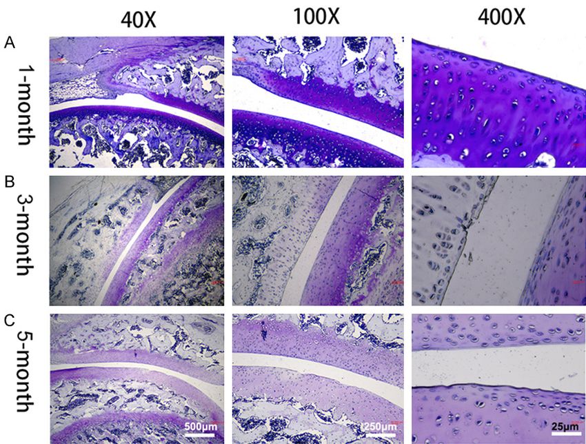
Microstructure of cartilage and subchondral bone in a guinea pig model of arthritis

seconds. Claw withdrawal or foot licking was        in 1% phosphotungstic acid solution for 1-3
considered a positive response. The mean            days. Microtomography system (Skyscan1276,
MWT values were obtained after 3 repeated           Bruker, Belgium) was used to image the patel-
experiments at 0, 4, 8, 12, 16, 20 and 24 days,     lofemoral joint. The parameters were 75 kV and
respectively.                                       200 μA, and the resolution was 18 μm. The
                                                    regions of interest (ROIs) of patellar cartilage
Observation of cartilage pathological morphol-      and femoral trochlear cartilage were deter-
ogy                                                 mined to be located in the medial center of the
                                                    patella and the trochlear, with an area of a rect-
The left knee joint was fixed in 4% parafor-        angle (1.2 × 2 mm), while the two ROIs of the
maldehyde solution (AR1068, Wuhan Bosher            subchondral bone were located in the cross
Biological Technology Co., LTD., China) for ab-     section under the subchondral plate of the
out 48 h, followed by decalcification in 10%        patella and the femur. After the scan, the
Ethylene Diamine Tetraacetic Acid (EDTA,            parameters were analyzed and calculated with
XK-011-00008, Tianjin Hedong District Hong-         the matching software. In addition, cartilage
yan Reagent Factory, China) for about 6 weeks.      analysis (bone volume, BV; bone surface, BS;
The decalcified solution was replaced every         trabecular thickness, Tb.Th) and subchondral
other week when about half of the decalcifica-      bone analysis (bone volume/trabecular vol-
tion process was done. The joint was cut open       ume, BV/TV; trabecular number, Tb.N; trabecu-
with a sharp blade (guided by the trochlear         lar thickness, Tb.Th; structure model index,
groove), and then the two halves of the joint       SMI) were performed to study patellofemoral
were put back into the decalcifying solution.       structure damage.
After that, the tissue was taken out and rinsed
properly with running water for 5 min, followed     Statistical processing
by three PBS rinses that lasted for 5 min each
time. After fine cutting of the tissue around the   The experimental results were statistically pro-
knee joint, the knee joint was resected in the      cessed by GraphpadPrism 8.0 (GraphPad, Inc.,
sagittal plane, dehydrated with gradient con-       San Diego, CA, USA). Data (denoted by x ± s)
centrations of ethanol, transparentized with        were analyzed by one-way analysis of variance
xylene and embedded in paraffin. The speci-         (ANOVA) plus Bonferroni post-hoc test and
mens were cut into 3-μm slices, with at least       t-test to conduct multi-group and between-
6 sections taken from each embedded wax             group comparisons, respectively. Data at differ-
block. After hematoxylin and eosin (H&E,            ent time points were compared by repeated
Solabio biotechnology co., Ltd., China) staining,   measures ANOVA, followed by the Bonferroni
fuchsin fast green staining and toluidine blue      post-hoc test. P0.05), which was significantly increased
70% ethanol for 2 hours, followed by immersion      when compared with the 1-month-old group

849                                                           Am J Transl Res 2023;15(2):847-857
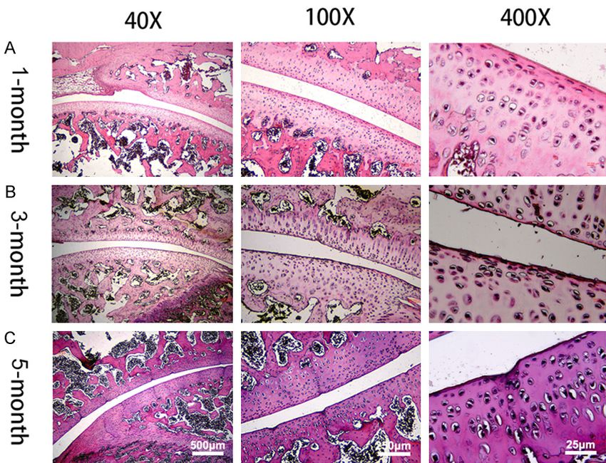
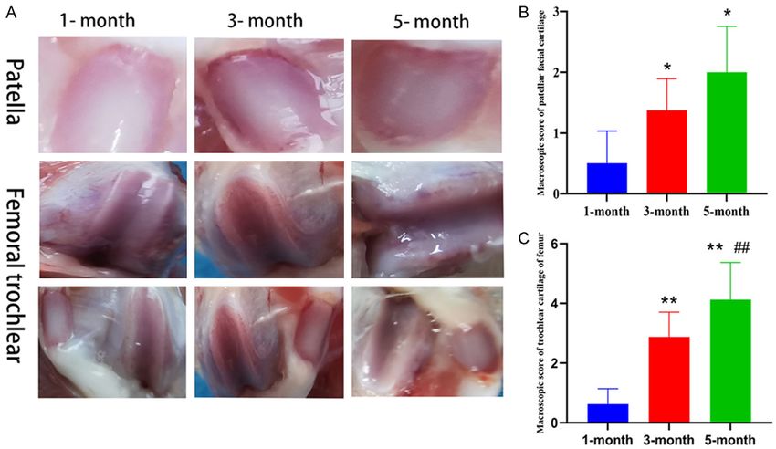
Microstructure of cartilage and subchondral bone in a guinea pig model of arthritis

Figure 1. Macroscopic observation and macroscopic scores of the patellofemoral joint of guinea pigs in each group.
A: Macroscopic observation; B: Macroscopic score of patellar facial cartilage; C: Macroscopic score of trochlear
cartilage of the femur. Compared to the 1-month-old group, *P
Microstructure of cartilage and subchondral bone in a guinea pig model of arthritis
face of femur was punctate or
sunken, with partial missing
of the matrix staining, partial
interruption of the subchon-
dral bone trabeculae, and ver-
tical cracks extending to the
middle layer (Figures 3C, 4C,
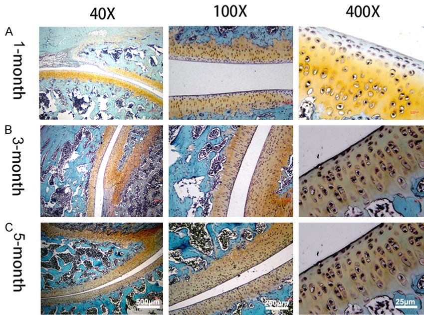
5C). The OARSI scores with
toluidine blue staining are
shown in Figure 6. It can be
found that the OARSI articu-
lar cartilage histopathological
score of patellar cartilage
and trochlear cartilage of
the femur increased gradually
over time (Figure 6). The
OARSI of trochlear cartilage
of femur was significantly in-
creased in the 5-month-old
Figure 3. Hematoxylin-eosin staining of patellofemoral cartilage tissue from group as compared with that
Hartley guinea pigs. A: 1-month-old; B: 3-month-old; C: 5-month-old. in the 1- and 3-month-old
groups (P0.05), but
cytes on the surface of patellar cartilage, there were significant differences in trochlear
uneven surface of cartilage on the trochlear cartilage of femur BV and Tb. (P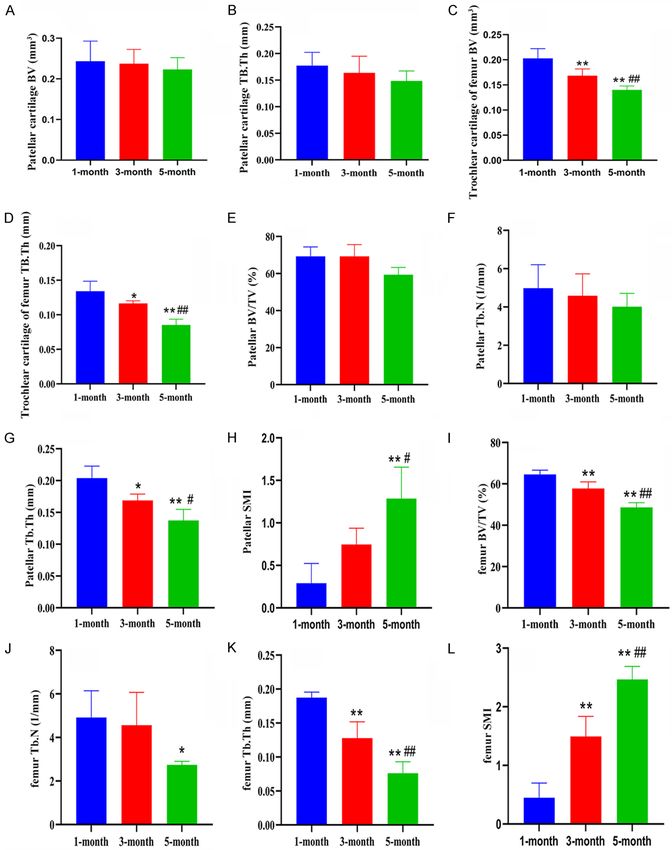
Microstructure of cartilage and subchondral bone in a guinea pig model of arthritis

                                                                                  the main research directions
                                                                                  of pathological changes in
                                                                                  PFOA [12]. In this study, we
                                                                                  successfully developed an ex-
                                                                                  perimental model of early-
                                                                                  and mid-stage PFOA that is
                                                                                  characterized by cartilage in-
                                                                                  jury deterioration and sub-
                                                                                  chondral bone loss with age.
                                                                                  There are a variety of meth-
                                                                                  ods to evaluate the success
                                                                                  of PFOA experimental models:
                                                                                  first, the statistical results of
                                                                                  Guingamp macroscopic score
                                                                                  and mechanical pain thresh-
                                                                                  old support the successful
                                                                                  establishment of the model;
                                                                                  second, histology shows rou-
                                                                                  gh and damaged surfaces of
Figure 5. Toluidine blue staining of patellofemoral cartilage tissue from Hart-   the patella and femoral troch-
ley guinea pig. A: 1-month-old; B: 3-month-old; C: 5-month-old.                   lear cartilage, obviously une-
                                                                                  ven staining associated with
                                                                                  hypertrophy and disordered
                                                                                  chondrocytes, and increased
                                                                                  pathological scores of OARSI
                                                                                  cartilage with age, consistent
                                                                                  with the pathological manifes-
                                                                                  tations of early- and middle-
                                                                                  stage OA; third, micro-CT con-
                                                                                  firms changes in cartilage
                                                                                  thickness and volume and
                                                                                  deterioration of subchondral
                                                                                  trabecular microstructure. In
                                                                                  addition, this is the first study
Figure 6. OARSI articular cartilage histopathological scores of guinea pig pa-    to investigate the relationship
tella and femoral trochlea in each group. A: Patella OARSI score; B: Femoral      between spontaneous PFOA
trochlea OARSI score. Compared with the 1-month-old group, **P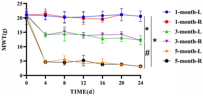
Microstructure of cartilage and subchondral bone in a guinea pig model of arthritis

Figure 7. Micro-CT measurements of the region of interest. A: Patellar cartilage; B: Femoral cartilage (the yellow
circled area is the patellar cartilage, and the blue circled area is the trochlear cartilage of the femur); C: Patellar
subchondral bone; D: Femoral subchondral bone (the green delineated area is the subchondral bone of the patella,
and the red delineated area is the subchondral bone of the femur); E: Overall image of the region of interest.

                                                                                    the natural development of
                                                                                    human PFOA.

                                                                            The degeneration of articular
                                                                            cartilage is one of the main
                                                                            characteristics of PFOA aff-
                                                                            ecting joints [28]. At present,
                                                                            cartilage degeneration in OA
                                                                            animal models is mainly bas-
                                                                            ed on macroscopic evaluation
                                                                            of bone tissue and histopath-
                                                                            ological sections, which has
                                                                            some limitations. Combined
                                                                            with micro-CT joint 3D recon-
                                                                            struction, specimens can be
                                                                            evaluated longitudinally, sub-
Figure 8. Micro-CT 3D reconstruction image. A: 1-month-old patellar sub-    tle bone structural lesions
chondral bone and femoral subchondral bone; B: 3-month-old femoral sub-     can be easily found in the
chondral bone and femoral subchondral bone; C: 5-month-old femoral sub-
chondral bone and femoral subchondral bone.
                                                                            early stage, and OA disease
                                                                            progression can be detected
                                                                            more sensitively [29]. In this
tion, Bei et al. [27] used patellar ligament short-       study, the ROI cartilage bone volume and bone
ening to establish a PFOA model, and found                thickness were measured by micro-CT. It was
that patellar cartilage and femoral trochlear             found that the index value of the 5-month-old
load-bearing area cartilage were significantly            group was markedly lower compared with the
damaged in the model group. In this study,                1-month-old group, and the trochlear thickness
female Hartley guinea pigs with primary OA                of the femur decreased significantly with age,
were used as animal models, without related               but there was no difference in the bone area in
intervention. Most importantly, this model will           the three-month-old group compared with the
not cause damage to the articular cavity and its          1-month-old group, which may be affected by
surrounding tissues, which is better than previ-          multiple separate cross-sectional sets of 2D
ous PFOA models and can approximately reflect             analysis. The histopathological scores of OARSI

853                                                                        Am J Transl Res 2023;15(2):847-857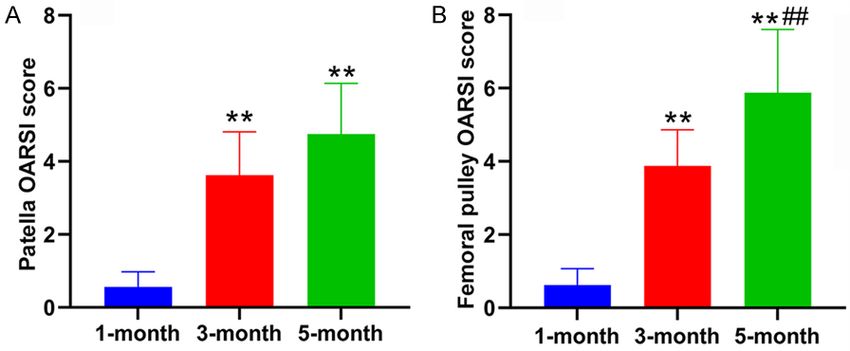
Microstructure of cartilage and subchondral bone in a guinea pig model of arthritis

Figure 9. Micro-CT subchondral bone analysis. A: Patellar cartilage bone volume (BV); B: Patellar trochlear tra-
becular thickness (Tb.Th); C: Femoral trochlear cartilage BV; D: Femoral cartilage Tb.Th; E: Patellar bone volume/
trabecular volume (BV/TV); F: Patellar trabecular number (Tb.N); G: Patellar Tb.Th; H: Patellar structure model index
(SMI); I: Femoral BV/TV; J: Femoral Tb.N; K: Femoral Tb.Th; L: Femoral SMI. Compared with the 1-month-old-group,
*
  P
Microstructure of cartilage and subchondral bone in a guinea pig model of arthritis

which was similar to that in guinea pigs with        Conclusion
spontaneous OA in different age groups [30].
Among them, there was a large area of carti-         In conclusion, this study confirmed that
lage erosion and cell loss in the femoral troch-     3-5-month-old female Hartley guinea pigs
lear weight-bearing area in the 5-month-old          could be used for an animal model of early- and
guinea pigs, but no significant destruction was      mid-stage spontaneous PFOA, and that both
found in patellar cartilage, which is supported      their cartilage and subchondral bone deterio-
by the clinical study on PFOA [31]. Some schol-      rated gradually with the passage of time, espe-
ars have reported more serious cartilage de-         cially the femur.
struction in 7-month-old guinea pigs than in
3-month-old guinea pigs [32]. However, the           Disclosure of conflict of interest
patellofemoral articular cartilage was only stud-
ied in the sagittal plane, and a comprehensive       None.
histopathological analysis of articular cartilage
on other horizontal planes is needed in future       Address correspondence to: Yafei Liu, Department
studies.                                             of Orthopaedics, Honghui Hospital, Xi’an Jiaotong
                                                     University, Xi’an 710054, Shaanxi, China. Tel: +86-
In addition to cartilage degeneration, PFOA is       029-33341362; E-mail: Liuyafei9521@126.com
characterized by abnormal bone remodeling of
the subchondral bone that is featured by a high      References
bone turnover rate and bone mass loss; high
bone turnover will damage the microstructure         [1]   Bijlsma JW, Berenbaum F and Lafeber FP. Os-
of subchondral bone, which plays a vital part in           teoarthritis: an update with relevance for clini-
OA progression [33]. The changes of BV/TV of               cal practice. Lancet 2011; 377: 2115-2126.
subchondral bone in 5-month-old guinea pigs          [2]   Zhang Z, Huang C, Jiang Q, Zheng Y, Liu Y, Liu
from another study [34] is consistent with the             S, Chen Y, Mei Y, Ding C and Chen M. Guide-
results of this study, and the change difference           lines for the diagnosis and treatment of osteo-
                                                           arthritis in China (2019 edition). Ann Transl
may be due to the different anatomic location
                                                           Med 2020; 8: 1213.
of ROIs. Some scholars have detected the OA
                                                     [3]   Lin W, Kang H, Dai Y, Niu Y, Yang G, Niu J, Li M
of female Hartley guinea pigs at the age of 1, 3,
                                                           and Wang F. Early patellofemoral articular car-
6 and 9 months, and found that the BV/TV of                tilage degeneration in a rat model of patellar
the tibia decreased gradually over time, which             instability is associated with activation of the
supports our findings [35]. This indicates that            NF-κB signaling pathway. BMC Musculoskelet
the patellar articular subchondral bone has                Disord 2021; 22: 90.
abnormal bone turnover and that the micro-           [4]   Zeng C, Wang H, Wu Z, Wang Y, Hu Y and
structure of subchondral bone has been                     Lei G. Interpretation of Chinese clinical prac-
destroyed in the early and middle stages of                tice guideline for patellofemoral osteoarthritis
PFOA, which further confirms the important                 (2020 edition). Chin J Orthop 2021; 129-132.
role of the subchondral bone in the onset of         [5]   Xu J, Zhou W and Luo X. Visual analysis of
PFOA.                                                      patellofemoral pain syndrome research hot-
                                                           spots and content. Chin J Tissue Eng 2022;
However, this study still has room for improve-            26: 1877.
ment. First, the establishment of a guinea pig       [6]   van Middelkoop M, Bennell KL, Callaghan MJ,
model is not easily comparable to PFOA in                  Collins NJ, Conaghan PG, Crossley KM, Eijken-
humans, warranting clinical exploration. Se-               boom JJFA, van der Heijden RA, Hinman RS,
cond, this experiment only studied alterations             Hunter DJ, Meuffels DE, Mills K, Oei EHG, Run-
in cartilage and subchondral bone in early- and            haar J, Schiphof D, Stefanik JJ and Bierma-Ze-
mid-stage PFOA, while spontaneous OA is a                  instra SMA. International patellofemoral osteo-
                                                           arthritis consortium: consensus statement on
relatively long-term process. In the future, a
                                                           the diagnosis, burden, outcome measures,
complete PFOA model should be constructed to
                                                           prognosis, risk factors and treatment. Semin
expand the age groups of the experimental ani-             Arthritis Rheum 2018; 47: 666-675.
mals. Third, abnormal joint load will also cause     [7]   Crossley K and Hinman R. The patellofemoral
changes in OA, so we should further compare                joint: the forgotten joint in knee osteoarthritis.
the pathological alterations in patellar articular         Osteoarthritis Cartilage 2011; 19: 765-767.
cartilage and subchondral bone, and explore          [8]   Li Z, Liu Q, Zhao C, Gao X, Han W, Stefanik JJ,
the influencing mechanism of cartilage and                 Jin Q, Lin J and Zhang Y. High prevalence of
subchondral bone on PFOA.                                  patellofemoral osteoarthritis in China: a multi-

855                                                             Am J Transl Res 2023;15(2):847-857
Microstructure of cartilage and subchondral bone in a guinea pig model of arthritis
center population-based osteoarthritis study. response study of loss of mobility, morphology,
Clin Rheumatol 2020; 39: 3615-3623. and biochemistry. Arthritis Rheum 1997; 40:
[9] Gaitonde DY, Ericksen A and Robbins RC. 1670-1679.
Patellofemoral pain syndrome. Am Fam Physi- [21] Chaplan SR, Bach FW, Pogrel J, Chung J and
cian 2019; 99: 88-94. Yaksh T. Quantitative assessment of tactile al-
[10] Schiphof D, van Middelkoop M, de Klerk BM, lodynia in the rat paw. J Neurosci Methods
Oei E, Hofman A, Koes BW, Weinans H and 1994; 53: 55-63.
Bierma-Zeinstra SM. Crepitus is a first indica- [22] Pritzker KP, Gay S, Jimenez SA, Ostergaard K,
tion of patellofemoral osteoarthritis (and not of Pelletier JP, Revell PA, Salter D and Van den
tibiofemoral osteoarthritis). Osteoarthritis Car- Berg WB. Osteoarthritis cartilage histopathol-
tilage 2014; 22: 631-638. ogy: grading and staging. Osteoarthritis Carti-
[11] Eijkenboom J, Waarsing J, Oei E, Bierma-Zein- lage 2006; 14: 13-29.
stra S and van Middelkoop M. Is patellofemo- [23] Kaymaz B, Atay OA, Ergen FB, Mermerkaya MU,
ral pain a precursor to osteoarthritis? Patello- Olgun ZD, Atesok K and Doral MN. Develop-
femoral osteoarthritis and patellofemoral pain ment of the femoral trochlear groove in rabbits
patients share aberrant patellar shape com- with patellar malposition. Knee Surg Sports
pared with healthy controls. Bone Joint Res Traumatol Arthrosc 2013; 21: 1841-1848.
2018; 7: 541-547. [24] Takahashi I, Matsuzaki T, Kuroki H and Hoso
[12] Bayramoglu N, Nieminen MT and Saarakkala M. Induction of osteoarthritis by injecting
S. Machine learning based texture analysis of monosodium iodoacetate into the patellofem-
patella from X-rays for detecting patellofemoral oral joint of an experimental rat model. PLoS
osteoarthritis. Int J Med Inform 2022; 157: One 2018; 13: e0196625.
104627. [25] Clark A, Leonard T, Barclay L, Matyas J and
[13] Du Longlong YP, Yang W, Li X and Gao Q. Ad- Herzog W. Opposing cartilages in the patello-
vantages of micro CT in three-dimensional re- femoral joint adapt differently to long-term cru-
construction of specimens and its application ciate deficiency: chondrocyte deformation and
in animal models of osteoarthritis. Chin J Tis- reorientation with compression. Osteoarthritis
sue Eng Res 2022; 26: 1931. Cartilage 2005; 13: 1100-1114.
[14] Li J, Su Y and Bai D. Morphological characteris- [26] Chang NJ, Shie MY, Lee KW, Chou PH, Lin CC
tics of subchondral bone in a mouse model of and Chu CJ. Can early rehabilitation prevent
early osteoarthritis. Chin J Tissue Eng Res posttraumatic osteoarthritis in the patellofem-
2022; 26: 1692. oral joint after anterior cruciate ligament rup-
[15] Kim JE, Song DH, Kim SH, Jung Y and Kim SJ. ture? Understanding the pathological features.
Development and characterization of various Int J Mol Sci 2017; 18: 829.
osteoarthritis models for tissue engineering. [27] Bei MJ, Tian FM, Xiao YP, Cao XH, Liu N, Zheng
PLoS One 2018; 13: e0194288. ZY, Dai MW, Wang WY, Song HP and Zhang L.
[16] Naruse K, Urabe K, Jiang SX, Uchida K, Kozai Y, Raloxifene retards cartilage degradation and
Minehara H, Mikuni-Takagaki Y, Kashima I and improves subchondral bone micro-architecture
Itoman M. Osteoarthritic changes of the patel- in ovariectomized guinea pigs with patella ba-
lofemoral joint in STR/OrtCrlj mice are the ear- ja-induced-patellofemoral joint osteoarthritis.
liest detectable changes and may be caused Osteoarthritis Cartilage 2020; 28: 344-355.
by internal tibial torsion. Connect Tissue Res [28] Bei M, Tian F, Liu N, Zheng Z, Cao X, Zhang H,
2009; 50: 243-255. Wang Y, Xiao Y, Dai M and Zhang L. A novel rat
[17] Salo PT, Seeratten RA, Erwin WM and Bray RC. model of patellofemoral osteoarthritis due to
Evidence for a neuropathic contribution to the patella baja, or low-lying patella. Med Sci Monit
development of spontaneous knee osteoar- 2019; 25: 2702.
throsis in a mouse model. Acta Orthop Scand [29] Zamli Z, Robson Brown K, Tarlton JF, Adams
2002; 73: 77-84. MA, Torlot GE, Cartwright C, Cook WA, Vassi-
[18] McCoy A. Animal models of osteoarthritis: levskaja K and Sharif M. Subchondral bone
comparisons and key considerations. Vet plate thickening precedes chondrocyte apop-
Pathol 2015; 52: 803-818. tosis and cartilage degradation in spontane-
[19] Cook JL, Kuroki K, Visco D, Pelletier JP, Schulz ous animal models of osteoarthritis. Biomed
L and Lafeber FP. The OARSI histopathology Res Int 2014; 2014: 606870.
initiative-recommendations for histological as- [30] Wang T, Wen CY, Yan CH, Lu WW and Chiu KY.
sessments of osteoarthritis in the dog. Osteo- Spatial and temporal changes of subchondral
arthritis Cartilage 2010; 18 Suppl 3: S66-S79. bone proceed to microscopic articular carti-
[20] Guingamp C, Gegout-Pottie P, Philippe L, Ter- lage degeneration in guinea pigs with sponta-
lain B, Netter P and Gillet P. Mono-iodoacetate- neous osteoarthritis. Osteoarthritis Cartilage
induced experimental osteoarthritis. A dose- 2013; 21: 574-581.
856 Am J Transl Res 2023;15(2):847-857Microstructure of cartilage and subchondral bone in a guinea pig model of arthritis
[31] Ryu J, Saito S and Yamamoto K. Changes in [34] Radakovich LB, Marolf AJ, Shannon JP, Pan-
articular cartilage in experimentally induced none SC, Sherk VD and Santangelo KS. Devel-
patellar subluxation. Ann Rheum Dis 1997; 56: opment of a microcomputed tomography scor-
677-681. ing system to characterize disease progression
[32] Yan JY, Tian FM, Wang WY, Cheng Y, Xu HF, in the Hartley guinea pig model of spontane-
Song HP, Zhang YZ and Zhang L. Age depen- ous osteoarthritis. Connect Tissue Res 2018;
dent changes in cartilage matrix, subchondral 59: 523-533.
bone mass, and estradiol levels in blood se- [35] Yan JY, Tian FM, Wang WY, Cheng Y, Song HP,
rum, in naturally occurring osteoarthritis in Zhang YZ and Zhang L. Parathyroid hormone
guinea pigs. Int J Mol Sci 2014; 15: 13578- (1-34) prevents cartilage degradation and pre-
13595. serves subchondral bone micro-architecture in
[33] Goldring MB and Goldring SR. Articular carti- guinea pigs with spontaneous osteoarthritis.
lage and subchondral bone in the pathogene- Osteoarthritis Cartilage 2014; 22: 1869-1877.
sis of osteoarthritis. Ann N Y Acad Sci 2010;
1192: 230-237.
857 Am J Transl Res 2023;15(2):847-857You can also read