Perioperative rifaximin is not associated with enhanced functional and volumetric recovery after major liver resection
←
→
Page content transcription
If your browser does not render page correctly, please read the page content below
www.nature.com/scientificreports

                OPEN              Perioperative rifaximin
                                  is not associated with enhanced
                                  functional and volumetric recovery
                                  after major liver resection
                                  Jan Bednarsch1, Zoltan Czigany1, Sven H. Loosen2,3, Lara Heij1,4, Lorenz Ruckgaber1,
                                  Henning Maes1, Jan‑Pit Krause1, Matthias Reen1, Beata Toteva1, Theresa Vosdellen1,
                                  Philipp Bruners5, Sven Arke Lang1, Tom Florian Ulmer1, Christoph Roderburg2,3,
                                  Tom Luedde2,3,7 & Ulf Peter Neumann1,6,7*

                                  The objective of this randomized controlled trial (RCT) was to assess the impact of rifaximin on the
                                  course of liver function, liver regeneration and volumetric recovery in patients undergoing major
                                  hepatectomy. The ARROW trial was an investigator initiated, single-center, open-label, phase 3 RCT
                                  with two parallel treatment groups, conducted at our hepatobiliary center from 03/2016 to 07/2020.
                                  Patients undergoing major hepatectomy were eligible and randomly assigned 1:1 to receive oral
                                  rifaximin (550 mg twice daily for 7–10 or 14–21 days in case of portal vein embolization preoperatively
                                  and 7 days postoperatively) versus no intervention. Primary endpoint was the relative increase in
                                  postoperative liver function measured by LiMAx from postoperative day (POD) 4 to 7. Secondary
                                  endpoint were the course of liver function and liver volume during the study period as well as
                                  postoperative morbidity and mortality. Between 2016 and 2020, 45 patients were randomized and
                                  35 patients (16 individuals in the rifaximin and 19 individuals in the control group) were eligible for
                                  per-protocol analysis. The study was prematurely terminated following interim analysis, due to the
                                  unlikelihood of reaching a significant primary endpoint. The median relative increase in liver function
                                  from POD 4 to POD 7 was 27% in the rifaximin group and 41% in the control group (p = 0.399). Further,
                                  no significant difference was found in terms of any other endpoints of functional liver- and volume
                                  regeneration or perioperative surgical complications following the application of rifaximin versus
                                  no intervention. Perioperative application of rifaximin has no effect on functional or volumetric
                                  regeneration after major hepatectomy (NCT02555293; EudraCT 2013-004644-28).

                                  Abbreviations
                                  AE	Adverse event
                                  ALPPS	Associating liver partition and portal vein ligation for staged hepatectomy
                                  ALT	Alanine aminotransferase
                                  AP	Alkaline phosphatase
                                  ASA	American society of anesthesiologists
                                  AST	Aspartate aminotransferase
                                  BMI	Body mass index
                                  CONSORT	Consolidated standards of reporting trials
                                  CRLM	Colorectal liver metastases
                                  CT	Computed tomography
                                  CVP	Central venous pressure

                                  1
                                   Department of Surgery and Transplantation, University Hospital RWTH Aachen, Pauwelsstrasse 30, 52074 Aachen,
                                  Germany. 2Department of Medicine III, University Hospital RWTH Aachen, Aachen, Germany. 3Department of
                                  Gastroenterology, Hepatology and Infectious Diseases, University Hospital Heinrich Heine University, Duesseldorf,
                                  Germany. 4Institute of Pathology, University Hospital RWTH Aachen, Aachen, Germany. 5Department of
                                  Radiology, University Hospital RWTH Aachen, Aachen, Germany. 6Department of Surgery, Maastricht University
                                  Medical Centre (MUMC), Maastricht, The Netherlands. 7These authors contributed equally: Tom Luedde and Ulf
                                  Peter Neumann. *email: uneumann@ukaachen.de

Scientific Reports |   (2021) 11:17936               | https://doi.org/10.1038/s41598-021-97442-w                                                1

                                                                                                                                            Vol.:(0123456789)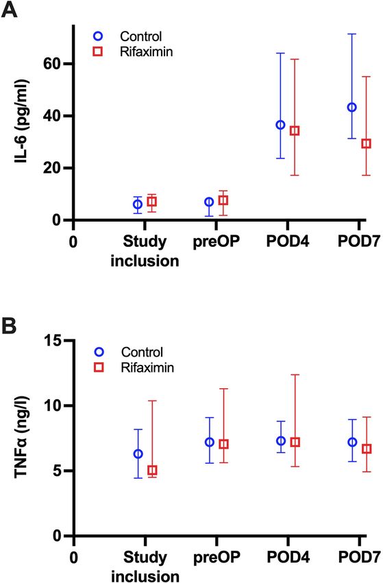
www.nature.com/scientificreports/
DAMP Damage-associated molecular patterns
FFP Fresh frozen plasma
FLR Future liver remnant
FLRF Future liver remnant function
FLRV Future liver remnant volume
GGT Gamma glutamyl transferase
IIT Investigator initiated trial
IL Interleukin
LiMAx Maximum liver function capacity
MRI Magnetic resonance imaging
NAFLD Nonalcoholic fatty liver disease
NASH Nonalcoholic steatohepatitis
pCCA Perihilar cholangiocarcinoma
POD Postoperative day
prePVE Prior to PVE
preOP Prior to surgery
PAMP Pathogen‐associated molecular patterns
PVE Portal vein embolization
RCT Randomized controlled trial
SAE Serious adverse event
TLR Toll-like receptor
TNF Tumor necrosis factor
RWTH Rheinisch-Westfälische Technische Hochschule
Liver resection (LR) is a major cornerstone in the therapy of primary and secondary liver tumors, displaying
compelling long-term oncological outcomes in comparison to interventional or medical treatment in various
hepatobiliary and oncological diseases1–4. Despite its broad acceptance, LR remains a highly invasive procedure
with reported mortality rates up to 15% depending on patient selection, indication and the particular technical
procedure5,6. Especially major LR—defined by the surgical removal of more than 2 liver segments—is associ-
ated with significant postoperative morbidity and mortality due to postoperative liver failure (POLF)7,8. POLF
is considered to be an acquired deterioration in the ability of the liver to maintain its synthetic, excretory and
detoxifying function after LR9. POLF is further reported to be the main driver of postoperative morbidity and
mortality in these patients and occurs in up to 10% of patients undergoing major LR. Subsequently, improv-
ing perioperative liver function and enhancing liver regeneration after LR has been a research focus of the last
decades10.
Liver regeneration is regulated by a complex interaction of hepatocytes and non-parenchymal cells directed by
cytokines, growth hormones and metabolic f actors11. Over the last years, the bidirectional relationship between
the liver and the intestine, the so-called gut-liver axis, and its role in liver regeneration and disease are gaining
more and more attention. LR is known to affect the integrity of the gut epithelial barrier, facilitate the transloca-
tion of bacteria and bacterial products to the liver were these products trigger an inflammatory r esponse12. As
liver regeneration is strongly inhibited by hepatic inflammation, any medical interventions to reduce inflamma-
tion in the early postoperative course appear reasonable13,14.
Rifaximin is a rifamycin derivative with a broad therapeutic range and approved for the treatment of gastroin-
testinal infections15. Further, rifaximin has shown efficiency to maintain remission from hepatic encephalopathy
and reduce the risk of hospitalization involving hepatic e ncephalopathy16. Thus, considering the aforementioned
direct link between gut microbiota translocation to the liver as a pathophysiological event following major LR
and its potential adverse role in substantial liver dysfunction and POLF, we hypothesized that perioperative anti-
biotic treatment with rifaximin may improve postoperative liver function and reduce morbidity after major LR.
Due to the low bioavailability of rifaximin with less than 0.5% of the oral dose being intestinally absorbed,
there is a low risk of systemic toxicity, allowing a safe use in the perioperative s etting15. In this RCT, we sys-
tematically assessed the impact of rifaximin on the course of liver function, liver regeneration and volumetric
recovery in patients undergoing major LR. The RCT is presented in accordance with the Consolidated Standards
of Reporting Trials (CONSORT) guidelines.
Material and methods
Study design. ARROW (Administration of Rifaximin to improve Liver Regeneration and Outcome follow-
ing Major Liver Resection) is a randomized, controlled, single-center, open-label superiority phase 3 trial with 2
parallel treatment groups. The study is an investigator-initiated trial conducted according to the requirements of
the German Medicinal Products Act (Arzneimittelgesetz-AMG). The study protocol was approved by the Ger-
man Federal Institute for Drugs and Medical Devices and was registered with ClinicalTrials.gov and EudraCT
(NCT02555293, first registration 21/09/2015; EudraCT 2013-004644-28, first registration 18/03/2014). The
RWTH Aachen University acted as the responsible sponsor for the trial. The ARROW trial was approved by
the institutional review board of the RWTH Aachen (EK 13-129) and all necessary regulatory approvals were
obtained. There were no major protocol amendments during the study period impacting trial design or trial
objectives. Informed consent was obtained from every patient and the trial has been conducted in accordance
with the current version of the Declaration of Helsinki, and the good clinical practice guidelines (ICH-GCP).
The ARROW trial was conducted and reported according to the CONSORT guidelines.
Scientific Reports | (2021) 11:17936 | https://doi.org/10.1038/s41598-021-97442-w 2
Vol:.(1234567890)
www.nature.com/scientificreports/
Study participation. Patients aged between 18 and 80 years who were scheduled for major LR were eligible
if they additionally presented with an BMI between 18 and 40 and were assessed with a performance status I to
III according to the American society of anesthesiologist (ASA) classification. Patients with the requirement of
concomitant extrahepatic surgical procedures, hyperthermic intraperitoneal chemotherapy (HIPEC) or associ-
ating liver partition and portal vein ligation for staged hepatectomy (ALPPS) were ineligible for trial participa-
tion.
Randomization and masking. Patients were randomly assigned 1:1 to the rifaximin group or the control
group. Treatment was not masked and no placebo was used in the control group. Random allocation was car-
ried out by a computer algorithm (Study Management Tool, RWTH Aachen University, Germany) that stratified
participants by the preoperative requirement of portal vein embolization.
Procedures. All patients were screened and recruited in the local outpatient department. Liver function
according to LiMAx (maximum liver capacity) was determined and computed-tomography (CT) volume-
try of the future liver remnant (FLR) was carried out. In need of preoperative hypertrophy induction, portal
vein embolization (PVE) was scheduled. In these particular patients, LiMAx and volumetric assessment were
repeated one day prior to actual LR. In the treatment group, rifaximin (550 mg) was given twice a day for 14
to 21 days in case of PVE or 7 to 10 days in cases without preoperative PVE prior to LR, respectively. After LR,
rifaximin was continued until the postoperative day (POD) 7 and then discontinued. Patients participating in
the trial were regularly visited until discharge and underwent LiMAx on the POD 4 and 7 as well as a magnetic
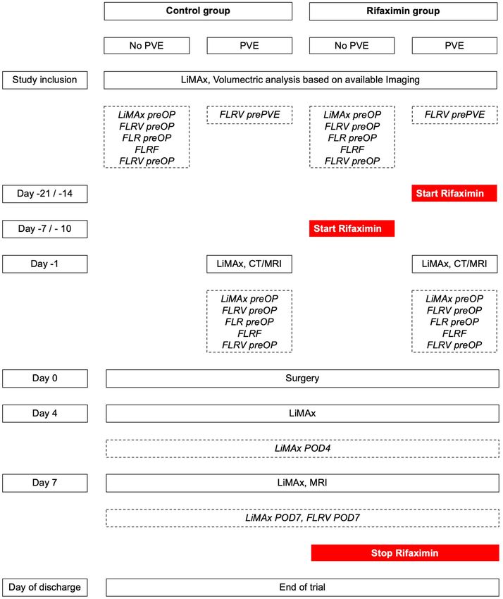
resonance imaging (MRI) on POD 7. A reduced trial overview including all study visits is shown in Fig. 1.
Methods. Liver function was determined by the LiMAx which represents a dynamic C13-breath test reflect-
ing enzymatic liver function capacity. During the test, a bodyweight-adjusted intravenous 13C-labeled methace-
tin bolus injection and continuous measurement of the 13CO2/12CO2 concentration ratio using a special device
(FLIP, Humedics GmbH, Berlin, Germany) is performed as previously d escribed17. LiMAx values > 315 μg/kg/h
are considered normal18.
CT- or MRI-based volumetry was carried out using a dedicated software (IntelliSpace Portal 8.0 software,
Philips healthcare, Amsterdam, The Netherlands). After manual delineation of margins in every slide, total
liver volume (TLV), tumor volume (TV) and future liver remnant volume (FLRV) were subsequently computed
automatically. TV was considered as non-functional liver parenchyma for all functional calculations. Finally,
the FLR was computed by the following formula:
FLRV
FLR[%] = × 100
TLV − TV
Direct postoperative liver function (future liver remnant function, FLRF) is estimated on the basis of preop-
erative LiMAx values and the results of the volumetric liver analysis using the following formula as previously
described8:
FLRF µg/kg/h = FLR × LiMAx
escribed19. Briefly,
PVE was carried out using a percutaneous transhepatic ipsilateral approach as previously d
a catheter was inserted into the right portal vein by transhepatic CT-guided puncture of the right portal branch.
Embolization of the right portal branches was carried out with a mixture of n-butyl-cyanoacrylate (Braun, Tut-
tlingen, Germany) and lipiodol (Guerbet, Roissy, France) in a ratio of 1:2 to 1:3. Successful embolization was
confirmed through repeated portography.
LR was carried in accordance to clinical standards as previously d escribed20.
An intraoperative ultrasound was performed to visualize the local tumor spread and other suspicious lesions.
Parenchymal transection was carried out using the Cavitron Ultrasonic Surgical Aspirator (CUSA, Integra LifeS-
ciences, Plainsboro NJ, USA) with low central venous pressure (CVP) and intermittent Pringle maneuvers if
necessary. In laparoscopic hepatectomy, parenchymal transection was commonly performed by Thunderbeat
(Olympus K.K., Tokyo, Japan), Harmonic Ace (Ethicon Inc. Somerville, NJ, USA) or laparoscopic CUSA (Inte-
gra life sciences, New Jersey, USA) in combination with vascular staplers (Echelon, Ethicon, Somerville, New
Jersey, USA) or polymer clips (Teleflex Inc., Pennsylvania, USA). The anesthesiologic management was based
on a restrictive fluid intervention strategy ensuring a low central venous pressure (CVP) during parenchymal
dissection.
Outcomes. The primary outcome of the trial was functional recovery after major LR, defined as the percen-
tal increase of LiMAx measured on POD 7 in relation to LiMAx measured on POD 4. Secondary outcomes were
volumetric recovery, defined as percental increase of FLRV measured on POD 7 in relation to preoperatively
determined FLRV, the hypertrophy of the FLRV after PVE and course of liver function over time determined by
LiMAx as well as postoperative morbidity and mortality.
Statistical analysis. An a priori sample size calculation of the trial was based on an estimated change of
30% of LiMAx values in the treatment group compared to the control and a dropout rate of 10% based on the
findings of Rayes et al. investigating liver regeneration after right hepatectomy determined by LiMAx in the
context of perioperative administration of p robiotics21. As such, 96 patients were required to detect a statistically
significant difference between the groups with a two-sided significance level of 5% and 0.90 power. Extensive
Scientific Reports | (2021) 11:17936 | https://doi.org/10.1038/s41598-021-97442-w 3
Vol.:(0123456789)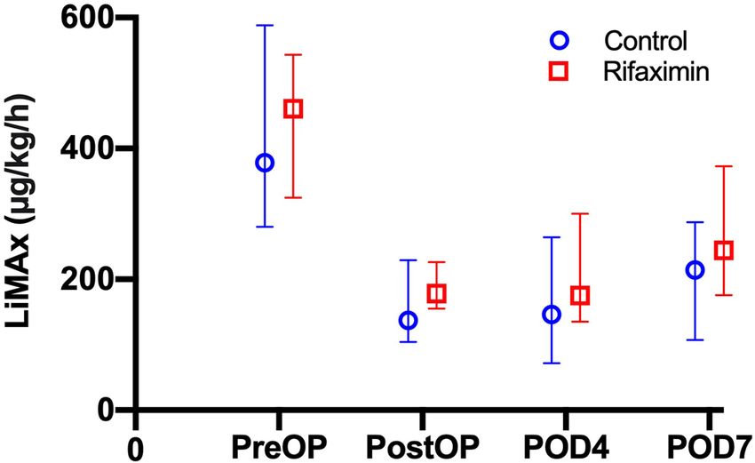
www.nature.com/scientificreports/

                                            Figure 1.  Trial overview. A reduced overview of the trial and all included study visits. Study events and tests
                                            are depicted in continues rectangles and measured variables with importance for statistical analysis in dashed
                                            rectangles. FLRF, future liver remnant function; FLRV, future liver remnant volume; LiMAx, maximum liver
                                            function capacity; MRI, Magnetic resonance imaging; POD, postoperative day; prePVE, prior to PVE; preOP,
                                            prior to surgery; PVE, portal vein embolization.

          Scientific Reports |   (2021) 11:17936 |               https://doi.org/10.1038/s41598-021-97442-w                                                    4

Vol:.(1234567890)
www.nature.com/scientificreports/
Figure 2. Trial profile. ALPPS, Associating liver partition with portal vein ligation for staged hepatectomy;
PVE, portal vein embolization.
statistical group comparisons were conducted between the rifaximin and control group. Categorical data are
presented as numbers and percentages and are statistically analyzed using the chi-squared test, fisher’s exact test
or linear-by-linear association in accordance to scale and number of cases. Continuous variables are presented as
median and interquartile range and compared by the Mann–Whitney-U-test. Perioperative complications were
classified according to the Clavien-Dindo scale22. The level of significance was set to p < 0.05 and p-values are
given for two-sided testing. Analyses were carried out using SPSS Statistics 24 (IBM Corp., Armonk, NY, USA).
Early termination. In 08/2020, an interims analysis was conducted to ensure trial safety. Here, no relevant
difference was found regarding the primary outcome of the trial (LiMAx increase from POD 4 to POD 7) by the
medical advisor board. Subsequently, the trial was prematurely stopped and the incomplete dataset was analyzed.
Role of the funding source. ARROW was an investigator-initiated trial predominantly using internal
departmental funds. However, limited external funding covering the study medication was provided by Norgine
GmbH (Wettenberg, Germany). Norgine GmbH had no role in running of the study, data collection, analysis
and interpretation or writing of the publication. Upon completion of all trial data, JB, TL and UPN had full access
to all the data and the corresponding authors had final responsibility for the decision to submit for publication.
Results
Between 03/2016 and 07/2020, at total of 45 patients were enrolled in the trial and randomly assigned to the
control (n = 24) and rifaximin group (n = 21). Of all randomized patients, a set of 10 individuals were excluded
from the trial as they were finally not treated by major LR (n = 3), underwent ALPPS (n = 3), were intraoperatively
assessed as technically not resectable (n = 2) or showed tumor progression after PVE which precluded further
surgical therapy (n = 2). No further withdrawal from trial treatment or consent were recorded during the study
period. All patients underwent the trial as scheduled and study medication was completed in all individuals of
the rifaximin group. As such, 19 patients in the control and 16 patients in the rifaximin group were eligible for
a per-protocol analysis. A detailed trial profile is shown in Fig. 2.
Patients’ characteristics. The groups were well balanced regarding clinical characteristics with no
observed difference in gender (p = 0.830), age (p = 0.481), BMI (p = 0.301) and ASA categorization (p = 0.501).
While there was a higher rate of perihilar cholangiocarcinoma (pCCA) and a lower rate of colorectal liver metas-
tases in the control group (CRLM; 7/19 vs. 2/16 and 4/19 vs. 9/16 respectively), this tendency did not gain
statistical significance (p = 0.131). Also, no difference was observed regarding laboratory liver function and clini-
cal chemistry (Table 1). Further, the applied surgical procedures (p = 0.613) as well as operative morbidity and
mortality (p = 0.731) were comparable between the groups. The total number of AEs and SAEs were 33 and 16
in the control and 29 and 14 in the rifaximin group (p = 0.688; p = 0.284). A detailed overview of patients’ char-
acteristics is given in Table 1.
Course of liver function. The median preoperative LiMAx was 378 µg/kg/h in the control and 461 µg/kg/h
in the Rifaximin group (p = 0.567). The median estimated FLRF based on preoperative LiMAx and volumetry of
the FLR was calculated to be 137 µg/kg/h in the control and 178 µg/kg/h in the rifaximin group (p = 0.125). In
the postoperative setting, median LiMAx was 146 µg/kg/h on POD 4 and 214 µg/kg/h on POD 7 in the control
as well as 175 µg/kg/h and 244 µg/kg/h in the rifaximin group (p = 0.142; p = 0.483). In the control group the
median increase from FLRF to POD 4 was -6% and 24% to POD 7, while in the increase from FLRF to POD 4
was 13% and 54% to POD 7 in the rifaximin group (p = 0.331; p = 0.815). The relative increase of liver function
Scientific Reports | (2021) 11:17936 | https://doi.org/10.1038/s41598-021-97442-w 5
Vol.:(0123456789)www.nature.com/scientificreports/
Control group vs. rifaximin group analysis
Variables Control group (n = 19) Rifaximin group (n = 16) p
Demographics
Sex, m/f (%) 10 (52.6)/9 (47.4) 9 (56.3)/7 (43.8) .830
Age (years) 60 (55–68) 62 (56–74) .481
BMI (kg/m2) 27 (24–31) 25 (24–28) .301
ASA, n (%) .515
I 1 (5.3) 0
II 10 (52.6) 8 (50.0)
III 8 (42.1) 8 (50.0)
Diagnosis, n (%) .131
Perihilar Cholangiocarcinoma 7 (36.8) 2 (12.5)
Intrahepatic Cholangiocarcinoma 1 (5.3) 3 (18.8)
Hepatocellular Carcinoma 3 (15.8) 1 (6.3)
Colorectal Liver Metastases 4 (21.1) 9 (56.3)
Adenoma 1 (5.3) 1 (6.3)
Hemangioma 3 (15.8) 0
Preoperative chemotherapy, n (%) 3 (15.8) 4 (25.0) .497
Preoperative PVE, n (%) 8 (42.1) 8 (50.0) .640
Laboratory liver function and clinical chemistry
AST (U/l) 35 (26–48) 29 (26–44) .443
ALT (U/l) 45 (23–63) 29 (18–46) .271
GGT (U/l) 201 (77–331) 111 (46–504) .317
Total bilirubin (mg/dl) 0.49 (0.34–0.92) 0.41 (0.35–0.59) .441
Platelet count (/nl) 310 (246–391) 288 (211–327) .151
Alkaline Phosphatase (U/l) 161 (95–262) 140 (94–321) .683
Prothrombin time (%) 99 (86–107) 97 (91–108) .935
Hemoglobin (g/dl) 13.1 (11.7–14.6) 12.7 (12.1–13.9) .502
Creatinine (mg/dl) 0.8 (0.7–1.0) 0.8 (0.7–1.0) .883
Operative characteristics
Operative procedure, n (%) .613
Right hepatectomy 9 (47.4) 7 (43.8)
Left hepatectomy 3 (15.8) 1 (6.3)
Extended right hepatectomy 4 (21.1) 5 (31.3)
Extended left hepatectomy 0 1 (6.3)
Right trisectionectomy 2 (10.5) 1 (6.3)
Left trisectionectomy 1 (5.3) 0
Central resection 0 1 (6.3)
Operative time (minutes) 375 (283–453) 386 (279–437) .935
Intraoperative blood transfusion, n (%) 8 (42.1) 4 (25.0) .288
Intraoperative FFP, n (%) 10 (52.6) 5 (31.3) .203
Postoperative complications, n (%) .731
No complications 7 (36.8) 6 (37.5)
I 1 (5.3) 4 (25.0)
II 5 (26.3) 2 (12.5)
IIIa 2 (10.5) 1 (6.3)
IIIb 1 (5.3) 1 (6.3)
IVa 0 0
IVb 1 (5.3) 1 (6.3)
V 2 (10.5) 1 (6.3)
ISGLS liver failure .561
None 14 (73.7) 12 (75.0)
Grade A 4 (21.1) 3 (18.8)
Grade B 1 (5.4) 0
Grade C 0 1 (6.3)
Number of AE, total per trial arm 35 30 .875
Number of SAE, total per trial arm 17 15 .241
Continued
Scientific Reports | (2021) 11:17936 | https://doi.org/10.1038/s41598-021-97442-w 6
Vol:.(1234567890)www.nature.com/scientificreports/
Control group vs. rifaximin group analysis
Variables Control group (n = 19) Rifaximin group (n = 16) p
Dynamic liver function analysis
LiMAx preOP (µg/kg/h) 378 (280–588) 461 (325–543) .567
FLRF (µg/kg/h) 137 (104–229) 178 (155–226) .125
LiMAx POD4 (µg/kg/h) 146 (72–264) 175 (135–300) .142
LiMAx POD7 (µg/kg/h) 214 (107–287) 244 (176–373) .483
Increase FLRF to POD4 (%) − 6 (− 5–40) 13 (0–56) .331
Increase FLRF to POD7 (%) 24 (9–94) 54 (3–89) .815
Increase POD4 to POD7 (%) 41 (3–99) 27 (− 3–63) .399
Volumetric analysis
FLRV prePVE (ml)* 543 (403–782) 398 (381–566) .234
FLRV preOP (ml)* 662 (532–769) 558 (474–735) .279
Increase prePVE to preOP (%)* 32 (5–36) 33 (18–44) .574
FLRV preOP (ml) 637 (503–798) 676 (509–939) .987
FLR preOP (%) 39 (33–44) 41 (34–54) .333
FLRV POD7 (ml) 962 (831–1352) 827 (711–982) .140
Increase FLRV preOP to FLRV POD7 52 (27–72) 45 (17–50) .180
Cytokine data
TNFa (ng/l)
Visit 1 (study inclusion) 6.3 (4.5–8.2) 5.1 (4.5–10.4) .825
Visit 3 (preOP) 7.2 (5.6–9.1) 7.1 (5.6–11.3) .798
Visit 4 (POD4) 7.3 (6.4–8.8) 7.2 (5.3–12.4) .953
Visit 5 (POD7) 7.2 (5.7–9.0) 6.7 (4.9–9.1) .597
IL-6 (pg/ml)
Visit 1 (study inclusion) 6.1 (2.6–9.2) 7.1 (3.4–9.7) .746
Visit 3 (preOP) 7.0 (1.5–7.9) 7.6 (1.9–11.0) .635
Visit 4 (POD4) 36.6 (23.9–63.3) 34.3 (18.1–59.5) .468
Visit 5 (POD7) 43.3 (30.0–84.3) 29.4 (17.8–50.7) .144
Table 1. Comparative analysis of the trial cohort. Data presented as median and interquartile range if not
noted otherwise. Categorical data were compared using the chi-squared test, fisher’s exact test or linear-
by-linear association according to scale and number of cases. Data derived from continuous variables of
different groups were compared by Mann–Whitney-U-Test. *Data only shown for patients who underwent
PVE. AE, adverse event; ALT, alanine aminotransferase; AP, alkaline phosphatase ASA, American society
of anesthesiologists classification; AST, aspartate aminotransferase; BMI, body mass index; CRP, c-reactive
protein; FFP, fresh frozen plasma; FLR, future liver remant; FLRF, future liver remnant function; FLRV, future
liver remant volume; GGT, gamma glutamyltransferase; IL, Interleukin; ISGLS, International Study Group for
Liver Surgery; LiMAx, Liver function capacity; POD, postoperative day; preOP, preoperative day 1; PVE, portal
vein embolization; SAE, serious adverse event; TNF, tumor necrosis factor.
from POD 4 to POD 7 was 41% in the control and 27% in the rifaximin group (p = 0.399). More details are pre-
sented in Table 1 and Fig. 3.
Course of liver volume. The median estimated FLRV was 637 ml in the control and 676 ml in the rifaxi-
min group (p = 987). On POD 7, the median measured FRLV was 962 ml in the control as well as 827 ml in the
rifaximin group (p = 0.140) which translates to a median postoperative increase from FLRV to POD 7 of 52%
in the control and 45% in the rifaximin group (p = 0.180). A similar sub-group analysis was carried out for PVE
patients (n = 16, 8/8) exclusively investigating hypertrophy after PVE. Here, the median volume increase of the
FRLV was 32% in the control and 33% in the rifaximin group (p = 0.574). More details are presented in Table 1
and Fig. 4.
Cytokine release. To explore to underlying effects of rifaximin, tumor necrosis factor alpha (TNFα) and
interleukin 6 (IL-6) were assessed at pre-defined time points over the course of the trial. Here, no significant
between-group differences were observed for IL-6 and TNFα at any time point. A detailed overview of the course
of serum cytokine release is provided in Table 1 and Fig. 5.
Scientific Reports | (2021) 11:17936 | https://doi.org/10.1038/s41598-021-97442-w 7
Vol.:(0123456789)www.nature.com/scientificreports/
Figure 3. Course of liver function with respect to trial group. Liver function assessed with LiMAx during the
trial period is presented as median and interquartile range. Liver function was significantly reduced due to liver
resection and did subsequently recover in the postoperative period. However, no time point showed statistical
significance between the Rifaximin and control group (preOP: p = 0.567; postOP: p = 0.125; POD 4: p = 0.142;
POD 7: p = 0.483). LiMAx, maximum liver function capacity; POD, postoperative day; postOP, after surgery;
preOP, prior to surgery.
Figure 4. Course of future liver remnant liver volume with respect to trial group. FRLV was assessed on
different time points during the trial period is presented as median and interquartile range (A) Volumetric
growth after PVE. FRLV subsequently increased after PVE in both groups with no statistical in any time point
(prePVE: p = 0.234; preOP: p = 0.279; only patients undergoing PVE were analyzed). (B) Volumetric growth
after liver resection. FRLV increased after surgery in both groups with no statistical in any time point (postOP:
p = 0.987; POD 7: p = 0.140). FLRV, future liver remnant volume; POD, postoperative day; prePVE, prior to PVE;
preOP, prior to surgery.
Discussion
In the ARROW trial, we investigated the impact of the perioperative application of rifaximin on functional and
volumetric recovery after LR. As no effect of the study medication on the primary readout of the trial—liver
function increase from POD 4 to POD 7 measured by LiMAx—was observed in the interim analysis, the trial
was prematurely discontinued and completely analyzed using the available data. Based on the results of the trial,
we were not able to demonstrate a significant benefit in functional regeneration or volumetric increase as well
as in perioperative morbidity and mortality in this dataset.
The gut-liver axis, which refers to the bidirectional relationship between the intestinal microbiome, the
gut and the liver, has been in the focus of gastrointestinal research in the last decade12. The microbiome is the
first interface between environment and the gut barrier and has been shown to be influenced by dietary habits,
ethanol and certain drugs (i.e. antibiotics among others)23–25. Treatment with antibiotics can significantly alter
the intestinal microbiome and can therefore play a role in liver damage following surgical resection and liver
transplantation. For example, administration of polymyxin B is associated with the reduction of total parenteral
nutrition induced steatosis in both rats and humans26. The pathophysiological background of this observation
might be explained by bacterial translocation which is the passage of bacteria or bacterial products to the liver
via the portal circulation27. Here, bacterial components can increase the expression of specific receptors, e.g. Toll-
Like Receptors (TLRs). TLRs can bind pathogen‐associated molecular patterns (PAMPs) and damage-associated
molecular patterns (DAMPs) leading to increased gene expression of pro-inflammatory mediators such as TNFα,
IL-1β, and interferons28. These effects are further facilitated by an increased gut permeability which is a well-
known pathophysiological mechanism in a broad spectrum of liver diseases29.
Scientific Reports | (2021) 11:17936 | https://doi.org/10.1038/s41598-021-97442-w 8
Vol:.(1234567890)www.nature.com/scientificreports/
Figure 5. Course of cytokines with respect to trial group. (A) IL-6. The course of serum IL-6 levels is presented
as median and interquartile range. There was no significant difference in IL-6 levels between the Rifaximin and
control group at any of the time points (study inclusion: p = 0.825; preOP: p = 0.798; POD 4: p = 0.953; POD 7:
p = 0.597). (B) TNFα. The course of TNFα is presented as median and interquartile range. Also, no time point
showed statistically significant difference between the Rifaximin and control group (study inclusion: p = 0.746;
preOP: p = 0.635; POD 4: p = 0.468; POD 7: p = 0.144). IL, Interleukin; POD, postoperative day; preOP, prior to
surgery. TNF, tumor necrosis factor.
The non-absorbable rifaximin is a potent treatment in various gastrointestinal diseases and has beneficial
clinical effects in irritable bowel syndrome, treatment and prevention of traveler’s diarrhea, small intestinal bac-
terial overgrowth, hepatic encephalopathy and diverticular d isease30. The underlying physiological mechanisms
are still a matter of debate. Rifaximin appears to have a minimal negative impact on overall gut microbiota, only
leading to a transient change in the concentrations of GI bacteria during therapy31. Further, studies on rifaximin
in cell lines indicate an ability to alter cytokine expression profiles (e.g. IL-8, etc.) suggesting an anti-inflammatory
activity32. Also reduced pathogen adherence in cell line studies investigating epithelial cell physiology has been
attributed to rifaximin exposure33. Additionally, rifaximin prevented the elevation of IL-17, IL-6, TNFα and the
stress-induced increase in mucosal inflammation and gut p ermeability31,34. While TNFα and IL-6 themself can
increase gut permeability by affecting tight junctions allowing bacterial translocation, both cytokines also play
a major role in liver homeostasis35. In the context of acute liver failure, high levels of circulating IL-6 and TNFα
are associated with impaired outcome in some studies, while in the hepatic compartment itself IL-6 seems to
be protective as it downregulates TNFα-induced hepatic apoptosis36,37. Also, both TNFα and IL-6 are known as
pivotal regulators during the early phase of liver regeneration as they modulate the interaction between non-
parenchymal cells and hepatocytes inducing the priming process, i.e., the G0/G1 transition of hepatocytes and
production of hepatic growth factor (HGF)38,39. As no differences in the release characteristics of TNFα and IL-6
were observed between treatment and control group in our study, we were not able to further elucidate the effects
of rifaximin on cytokines in the scenario of our clinical RCT. While the physiological basis of its effect remains to
be fully unraveled, rifaximin appears to interfere with the gut-liver axis by modifying the microbiome, enhancing
Scientific Reports | (2021) 11:17936 | https://doi.org/10.1038/s41598-021-97442-w 9
Vol.:(0123456789)www.nature.com/scientificreports/
the epithelial gut barrier and modulating inflammatory r esponse34. This observation is further supported by its
various clinical a pplications30.
However, within the setting of this RCT, investigating rifaximin in the context of major LR, we were not able
to detect any notable effect on liver function or volumetric recovery. Reasons for this negative observation might
be attributed to the trial design and the clinical context of major LR. As liver dysfunction or POLF usually arises
after significant loss of the liver parenchyma, we decided to restrict the trial to individuals undergoing major
LR40. Despite careful preoperative patient selection, major LR remains an invasive procedure with significant
perioperative morbidity as illustrated in our cohort by the rate of major morbidity (defined as any complica-
tions rated > Clavien Dindo II) in 32% (6/19) in the control and 25% (4/16) in the rifaximin group. Periopera-
tive complications are known to have a strong effect on liver function and regeneration41. LiMAx is a precise
diagnostic tool to measure liver function and has proven its diagnostic and predictive abilities in various clinical
scenarios8,17,42–44. This is also shown in our trial with both groups displaying a significant drop in liver function
due to LR and a continuous recovery in the postoperative course (Fig. 2). LiMAx is further capable to measure
liver regeneration after LR and is able to detect impaired regeneration in case of biliary leakage, a common sep-
tic complication after L R45. It is therefore plausible and also provides a partial explanation for our findings that
surgical complications often have a much stronger detrimental effect on the functional regeneration than the
assumed positive effect achieved by the perioperative application of rifaximin. Of note, a comparable effect was
observed in a study of Rayes et al. investigating p robiotics21. In this RCT, patients scheduled for right hepatectomy
received enteral nutrition with fibers only or fibers supplemented by probiotics starting the day before surgery
and continuing until the 10th POD. Primary study endpoint was, likewise in our trial, the increase of liver func-
tion determined by LiMAx. Here, a slightly improved liver regeneration in the treatment group was observed
and in a sub-group of patients excluding perioperative complications the positive effect in the treatment group
was even more obvious. These observations further underline the fundamental role of perioperative morbidity
in liver regeneration. This would also implicate that the outcome of any clinical trial evaluating rifaximin’s ability
in improving postoperative liver regeneration may be influenced by minor or major postoperative complications
and, therefore postoperative liver function characteristics as trial endpoint should always be interpreted in the
context of perioperative morbidity.
Our trial did also not observe any difference in volumetric recovery after surgery which does underline the
negligible or no potential effect of rifaximin on volume regeneration after LR. Interestingly, we were further not
able to demonstrate an impact on hypertrophy of the FLR after PVE in a small subgroup analysis of patients
requiring PVE in the preoperative setting. This is in particular interesting as this small sample set (n = 16) experi-
enced no major complications between PVE and LR which would interfere with a potentially benefit of rifaximin
treatment. One might argue that given the notable risk for complications after major LR, patients undergoing
minor LR might be more suitable to assess the potential impact of rifaximin. However, liver function is usually
less altered after minor LR and the likelihood of significant dysfunction or POLF is considerably less making a
medical intervention to boost liver function postoperatively clinically not relevant.
To date rifaximin shows major therapeutic effects in the prevention of hepatic encephalopathy or the long-
term treatment of small intestinal bacterial overgrowth. However, there is currently no evidence supporting
a therapeutic role in acute or acute-on-chronic liver failure which might be a comparable clinical situation to
LR30. It is therefore assumable that the significant clinical impact of rifaximin in various gastrointestinal dis-
ease is rather attributed to a prolonged modulation of the liver-gut axis than to a rapid improvement in liver
function. Based on this, it is possible that a longer application of rifaximin would result in a more meaningful
effect. However, the treatment duration was already 7 to 10 days prior to the operation (14 to 21 days in case of
PVE) accompanied by 7 days after the LR in both, patients with and without PVE. An even longer preoperative
application might not be practicable in an oncological setting as it might delay surgery and risk tumor related
complications in these patients (e.g. local tumor progression or metastases).
Despite showing no significant effect in our RCT, the use of rifaximin was safe with no relevant side effects.
SAE and AE were equally distributed between the groups and no association between SAE and AE and potential
side effects of rifaximin were observed by the medical advisor board indicating a safe usage in the perioperative
setting.
Our trial has certainly some obvious limitations which have to be discussed critically. First, the trial had to
be terminated prematurely which resulted in a smaller data set, leaving several questions unanswered. Addition-
ally, we have to report a high dropout rate reflecting the clinical reality in oncological liver surgery. Second, the
trial was conducted in a single center based on the authors’ distinct clinical management in LR. Third, some
parameters were calculated and not measured (FLRF and FLRV). However, these calculations are commonly
conducted in liver surgery and have shown their accuracy and predictive ability in previous reports8,46. Also, the
primary endpoint of the study which was defined as the functional increase from POD 4 to POD 7 was solely
based on measured variables.
The ARROW trial is the first clinical trial to investigate the effect of perioperative administration of rifaxi-
min on liver regeneration after LR. Considering the aforementioned limitations, we conclude that perioperative
application of rifaximin is safe, but does not improve functional or volumetric regeneration after major LR.
Data availability
Available upon request. JB and UPN had full access to the data and act both as guarantor for the data.
Received: 11 April 2021; Accepted: 18 August 2021
Scientific Reports | (2021) 11:17936 | https://doi.org/10.1038/s41598-021-97442-w 10
Vol:.(1234567890)www.nature.com/scientificreports/
References
1. Lurje, G. et al. Prognostic factors of disease-free and overall survival in patients with hepatocellular carcinoma undergoing partial
hepatectomy in curative intent. Langenbeck’s Arch. Surg. Deutsche Gesellschaft fur Chirurgie 403, 851–861 (2018).
2. Tomlinson, J. S. et al. Actual 10-year survival after resection of colorectal liver metastases defines cure. J. Clin. Oncol. Off. J. Am.
Soc. Clin. Oncol. 25, 4575–4580 (2007).
3. Neuhaus, P. et al. Oncological superiority of hilar en bloc resection for the treatment of hilar cholangiocarcinoma. Ann. Surg.
Oncol. 19, 1602–1608 (2012).
4. European Association for the Study of the, L. EASL Clinical Practice Guidelines on the management of benign liver tumours. J.
Hepatol. 65, 386–398 (2016).
5. Bednarsch, J. et al. Left-versus right-sided hepatectomy with hilar en-bloc resection in perihilar cholangiocarcinoma. HPB (Oxford)
22, 437–444 (2020).
6. Bednarsch, J., et al. Insufficient future liver remnant and preoperative cholangitis predict perioperative outcome in perihilar
cholangiocarcinoma. HPB Off. J. Int. Hepato Pancreato Biliary Assoc. (2020).
7. Balzan, S., et al. The “50–50 criteria” on postoperative day 5: An accurate predictor of liver failure and death after hepatectomy.
Ann. Surg. 242, 824–828, discussion 828–829 (2005).
8. Stockmann, M. et al. The LiMAx test: A new liver function test for predicting postoperative outcome in liver surgery. HPB (Oxford)
12, 139–146 (2010).
9. Rahbari, N. N. et al. Posthepatectomy liver failure: a definition and grading by the International Study Group of Liver Surgery
(ISGLS). Surgery 149, 713–724 (2011).
10. Kauffmann, R. & Fong, Y. Post-hepatectomy liver failure. Hepatob. Surg. Nutr. 3, 238–246 (2014).
11. Taub, R. Liver regeneration: From myth to mechanism. Nat. Rev. Mol. Cell Biol. 5, 836–847 (2004).
12. Chassaing, B., Etienne-Mesmin, L. & Gewirtz, A. T. Microbiota-liver axis in hepatic disease. Hepatology 59, 328–339 (2014).
13. Malato, Y. et al. NF-kappaB essential modifier is required for hepatocyte proliferation and the oval cell reaction after partial hepa-
tectomy in mice. Gastroenterology 143, 1597–1608 (2012).
14. Seehofer, D. et al. Intraabdominal bacterial infections significantly alter regeneration and function of the liver in a rat model of
major hepatectomy. Langenbeck’s Arch. Surg. Deutsche Gesellschaft fur Chirurgie 392, 273–284 (2007).
15. Majewski, M., Reddymasu, S. C., Sostarich, S., Foran, P. & McCallum, R. W. Efficacy of rifaximin, a nonabsorbed oral antibiotic,
in the treatment of small intestinal bacterial overgrowth. Am. J. Med. Sci. 333, 266–270 (2007).
16. Bass, N. M. et al. Rifaximin treatment in hepatic encephalopathy. N. Engl. J. Med. 362, 1071–1081 (2010).
17. Stockmann, M. et al. Prediction of postoperative outcome after hepatectomy with a new bedside test for maximal liver function
capacity. Ann. Surg. 250, 119–125 (2009).
18. Jara, M., et al. Reliable assessment of liver function using LiMAx. J. Surg. Res. (2014).
19. Ulmer, T. F. et al. ALPPS procedure in insufficient hypertrophy after portal vein embolization (PVE). World J. Surg. 41, 250–257
(2017).
20. Bednarsch, J., et al. Intraoperative transfusion of fresh frozen plasma predicts morbidity following partial liver resection for
hepatocellular carcinoma. J. Gastrointest. Surg. Off. J. Soc. Surg. Aliment. Tract (2020).
21. Rayes, N. et al. Effect of pre- and probiotics on liver regeneration after resection: A randomised, double-blind pilot study. Beneficial
Microb. 3, 237–244 (2012).
22. Dindo, D., Demartines, N. & Clavien, P. A. Classification of surgical complications: A new proposal with evaluation in a cohort
of 6336 patients and results of a survey. Ann. Surg. 240, 205–213 (2004).
23. Clarke, S. F. et al. The gut microbiota and its relationship to diet and obesity: New insights. Gut. Microb. 3, 186–202 (2012).
24. Mutlu, E. A. et al. Colonic microbiome is altered in alcoholism. Am. J. Physiol. Gastrointest. Liver Physiol. 302, 966–978 (2012).
25. Machado, M. V. & Cortez-Pinto, H. Diet, microbiota, obesity, and NAFLD: a dangerous quartet. Int. J. Mol. Sci. 17, 481 (2016).
26. Di Ciaula, A., et al. Liver steatosis, gut-liver axis, microbiome and environmental factors. A never-ending bidirectional cross-talk.
J. Clin. Med. 9 (2020).
27. Cani, P. D. et al. Changes in gut microbiota control metabolic endotoxemia-induced inflammation in high-fat diet-induced obesity
and diabetes in mice. Diabetes 57, 1470–1481 (2008).
28. Svegliati-Baroni, G., Patricio, B., Lioci, G., Macedo, M. P. & Gastaldelli, A. Gut-pancreas-liver axis as a target for treatment of
NAFLD/NASH. Int. J. Mol. Sci. 21 (2020).
29. Giorgio, V. et al. Intestinal permeability is increased in children with non-alcoholic fatty liver disease, and correlates with liver
disease severity. Digest. Liver Dis. Off. J. Ital. Soc. Gastroenterol. Ital. Assoc. Study Liver 46, 556–560 (2014).
30. Shayto, R. H., Abou Mrad, R. & Sharara, A. I. Use of rifaximin in gastrointestinal and liver diseases. World J. Gastroenterol. 22,
6638–6651 (2016).
31. Kim, M. S. et al. The effect of rifaximin on gut flora and Staphylococcus resistance. Dig. Dis. Sci. 58, 1676–1682 (2013).
32. Brown, E. L., Xue, Q., Jiang, Z. D., Xu, Y. & Dupont, H. L. Pretreatment of epithelial cells with rifaximin alters bacterial attachment
and internalization profiles. Antimicrob. Agents Chemother. 54, 388–396 (2010).
33. Gao, J., Gillilland, M. G. 3rd. & Owyang, C. Rifaximin, gut microbes and mucosal inflammation: Unraveling a complex relation-
ship. Gut Microb. 5, 571–575 (2014).
34. Bajaj, J. S. et al. New concepts on intestinal microbiota and the role of the non-absorbable antibiotics with special reference to
rifaximin in digestive diseases. Digest. Liver Dis. Off. J Ital. Soc. Gastroenterol. Ital. Assoc. Study Liver 50, 741–749 (2018).
35. Al-Sadi, R. et al. Interleukin-6 modulation of intestinal epithelial tight junction permeability is mediated by JNK pathway activa-
tion of claudin-2 gene. PLoS ONE 9, e85345 (2014).
36. Antoniades, C. G., Berry, P. A., Wendon, J. A. & Vergani, D. The importance of immune dysfunction in determining outcome in
acute liver failure. J. Hepatol. 49, 845–861 (2008).
37. Cressman, D. E. et al. Liver failure and defective hepatocyte regeneration in interleukin-6-deficient mice. Science 274, 1379–1383
(1996).
38. Fujiyoshi, M. & Ozaki, M. Molecular mechanisms of liver regeneration and protection for treatment of liver dysfunction and
diseases. J. Hepatobiliary Pancreat. Sci. 18, 13–22 (2011).
39. Schmidt-Arras, D. & Rose-John, S. IL-6 pathway in the liver: From physiopathology to therapy. J. Hepatol. 64, 1403–1415 (2016).
40. Nanashima, A. et al. Comparative analysis of postoperative morbidity according to type and extent of hepatectomy. Hepatogas-
troenterology 52, 844–848 (2005).
41. Lederer, A. et al. Postoperative bile leakage inhibits liver regeneration after 70% hepatectomy in rats. J. Investig. Surg. Off. J. Acad.
Surg. Res. 26, 36–45 (2013).
42. Stockmann, M. et al. How to define initial poor graft function after liver transplantation? A new functional definition by the LiMAx
test. Transpl. Int. Off. J. Eur. Soc. Organ Transpl. 23, 1023–1032 (2010).
43. Malinowski, M., et al. Enzymatic liver function capacity correlates with disease severity of patients with liver cirrhosis: A study
with the LiMAx test. Digest. Dis. Sci. (2014).
44. Bednarsch, J., et al. C Breath tests are feasible in patients with extracorporeal membrane oxygenation devices. Artif. Org. (2015).
45. Bednarsch, J., et al. Regeneration of liver function capacity after partial liver resection is impaired in case of postoperative bile
leakage. World J. Surg. (2016).
Scientific Reports | (2021) 11:17936 | https://doi.org/10.1038/s41598-021-97442-w 11
Vol.:(0123456789)www.nature.com/scientificreports/
46. Bluthner, E. et al. The predictive value of future liver remnant function after liver resection for HCC in noncirrhotic and cirrhotic
patients. HPB (Oxford) 21, 912–922 (2019).
Author contributions
Study concept and design: C.R., T.L., U.P.N. Acquisition of data: J.B., Z.C., L.R., H.M., J.P.K., M.R., B.T., T.V.
Analysis and interpretation of data: J.B., Z.C., S.H.L., L.H., P.B., S.A.L., T.F.U., C.R., T.L., U.P.N. Drafting of the
manuscript: J.B., Z.C., C.R., T.L., U.P.N. Critical revision of the manuscript for important intellectual content:
S.H.L., L.H., L.R., H.M., J.P.K., M.R., B.T., T.V., P.B., S.A.L., T.F.U. Study supervision: T.L., U.P.N. Approval of the
final version of the manuscript: All. Accountability for the manuscript: All.
Funding
Open Access funding enabled and organized by Projekt DEAL. ARROW was an investigator-initiated trial pre-
dominantly using internal departmental funds. However, limited external funding covering the study medication
was provided by Norgine GmbH (Wettenberg, Germany). Norgine GmbH had no role in running of the study,
data collection, analysis and interpretation or writing of the publication. Upon completion of all trial data, JB, TL
and UPN had full access to all the data and the corresponding authors had final responsibility for the decision to
submit for publication. This project was supported by the German Research Foundation (SFB-CRC 1382-A01).
Competing interests
The authors declare no competing interests.
Additional information
Correspondence and requests for materials should be addressed to U.P.N.
Reprints and permissions information is available at www.nature.com/reprints.
Publisher’s note Springer Nature remains neutral with regard to jurisdictional claims in published maps and
institutional affiliations.
Open Access This article is licensed under a Creative Commons Attribution 4.0 International
License, which permits use, sharing, adaptation, distribution and reproduction in any medium or
format, as long as you give appropriate credit to the original author(s) and the source, provide a link to the
Creative Commons licence, and indicate if changes were made. The images or other third party material in this
article are included in the article’s Creative Commons licence, unless indicated otherwise in a credit line to the
material. If material is not included in the article’s Creative Commons licence and your intended use is not
permitted by statutory regulation or exceeds the permitted use, you will need to obtain permission directly from
the copyright holder. To view a copy of this licence, visit http://creativecommons.org/licenses/by/4.0/.
© The Author(s) 2021
Scientific Reports | (2021) 11:17936 | https://doi.org/10.1038/s41598-021-97442-w 12
Vol:.(1234567890)You can also read