Prospectus 2020/2021 The Wellcome/ Cancer Research UK Gurdon Institute - University of ...
←
→
Page content transcription
If your browser does not render page correctly, please read the page content below
The Wellcome/ Cancer Research UK
Gurdon Institute
Prospectus 2020/2021

25
                                                  YEARS

                                                   The Wellcome/
                                               Cancer Research UK
                                                  Gurdon Institute

Studying             Prospectus 2020/2021
development to                E

                            CU
understand disease

                           GEND
                              R
                                  C HA R T E

                                         R
                            E
The Gurdon Institute   3

                                  Contents                          Welcome
                                                                    Welcome to our new Prospectus, where we highlight our        Watermark, the first such award in the University. Special
                                                                    activities for - unusually - two years: 2019 and 2020. The   thanks for this achievement go to Hélène Doerflinger,
                                                                    COVID-19 pandemic has made it an extraordinary time          Phil Zegerman and Emma Rawlins.
Director’s welcome            3       Emma Rawlins             38   for everyone. I want to express my pride and gratitude
                                                                    for the exceptional efforts of Institute members,            After incubating Steve Jackson's company Adrestia in
About the Institute           4       Daniel St Johnston       40   who have kept our building safe and our research             the Institute for two years, we wished them well as they
                                                                    progressing; this applies especially to our core team,       moved to the Babraham Research Campus. We also sent
COVID stories                 6       Ben Simons               42   whose dedication has been key to                                                    our best wishes to Meri Huch and
                                                                    our continued progress. As you will                                                 Rick Livesey and their labs, as they
Highlights in 2019/2020       8       Azim Surani              44   see, there is much to be excited                                                    embarked on their new positions in
                                                                    about in our research and activities.                                               Dresden and London, respectively.
Focus on research                     Iva Tchasovnikarova      46
                                                                    It was terrific to see Gurdon                                                     I'm delighted that Emma Rawlins
 Group leaders                        Fengzhu Xiong            48   members receive recognition for                                                   was promoted to Senior Group
                                                                    their achievements. Steve Jackson                                                 Leader and that two new Group
   Julie Ahringer            20       Philip Zegerman          50   received the Leopold Griffuel                                                     Leaders joined us in Autumn
                                                                    Award in Translational and Clinical                                               2020. Iva Tchasovnikarova studies
   Andrea Brand              22     Associate group leaders         Research, and the Royal Society                                                   epigenetic pathway mechanisms
                                                                    Mullard Award. Hansong Ma was                                                     and how they are disrupted in
   David Fernandez-Antoran   24       Martin Howard            52   awarded a Philip Leverhulme                                                       disease, while David Fernandez-
                                                                    Prize and selected as an EMBO                                                     Antoran's research is focused on
   Jenny Gallop              26       John Perry               54
                                                                    Young Investigator, I received the                                                cell competition and the impact of
   John Gurdon               28     Facilities                 56   Genetics Society of America's                                                     ionising radiation on selection.
                                                                    George W. Beadle Award, and Office Manager Lynda
   Steve Jackson             30     Support staff              58   Lockey was named the Unsung Heroine of Professional          Finally, I'm pleased to welcome two new Associate Group
                                                                    Services. We are also excited that a major component of      Leaders. John Perry (MRC Epidemiology Unit) uses
   Tony Kouzarides           32    Seminars, events and             the Wellcome-funded Human Developmental Biology              human genetics to understand disease mechanisms, and
                                   publications in 2019/2020   60   Initiative is based in the Institute, led by Emma Rawlins,   Martin Howard (John Innes Centre) builds mathematical
   Hansong Ma                34                                     Ben Simons and Azim Surani.                                  models of biological processes. We look forward to
                                   Acknowledgements            77                                                                exciting and productive interactions with them.
   Eric Miska                36                                     I'm especially proud that our exceptional Public
                                                                    Engagement was recognised by a Silver Engage

                                                                                                                                                                                Director
                                                                                                                                                                           February 2021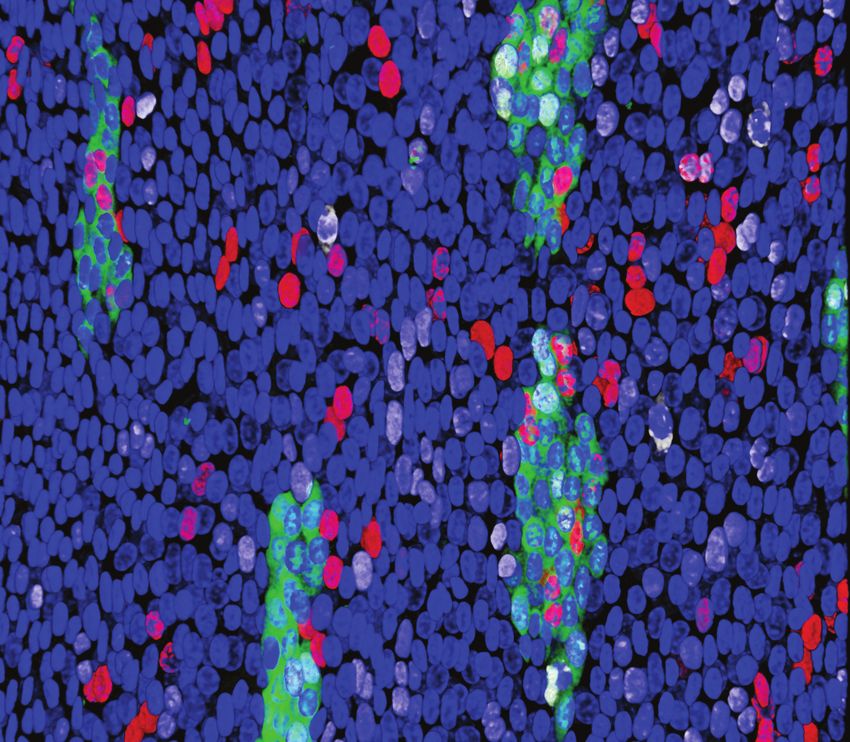
4   About the Institute

         About the Institute
         The Wellcome/ Cancer Research UK Gurdon Institute is a world-leading centre for
         research at the interface between developmental biology and cancer biology.

         Our research is focussed in four overlapping areas:

                                                               our focus

                  Cell division,  Function and regulation              Mechanisms of cell           Cell biological
                proliferation and   of the genome and                  fate determination,       processes underlying
              genome maintenance        epigenome                       multipotency and         organ development
                                                                             plasticity              and function                 and interactions across Cambridge’s   •   A wealth of stimulating           Join us
                                                                                                                                  vibrant research environment,             seminars and masterclasses,       We have a thriving community of
                                                                                                                                  including through department              an annual Institute retreat,      graduate students and postdocs
                                                                                                                                  affiliations and teaching.                and Institute postdoc and PhD     who contribute to and benefit from
                                                                                                                                                                            student groups (pp. 62–65)        our exciting research environment.
         We investigate these areas in both      findings have been successfully         sources including national and           We benefit from:                      •   Award-winning public              We welcome enquiries from those
         normal development and cancer           translated to drug discovery            international governmental and           •   Core facilities with state-of-        engagement between our            interested in joining us, which can
         using multiple model systems, from      through spinout companies.              charitable grants. Scientific progress       the-art equipment and support         scientists and the wider public   be done by writing to the relevant
         yeast to human organoids (pp.                                                   and future plans are assessed at             including super-resolution            (pp. 16–17).                      group leader.
         20–50).                                 The Gurdon Institute’s principal        regular intervals by our International       microscopy, next-generation       •   An on-site canteen, social
                                                 sponsors are Wellcome and Cancer        Scientific Advisory Board (p. 77) .          sequencing and bioinformatics         events and sports groups, which   Find out about the latest
         Since our formation in 1991, our        Research UK, who support our                                                         (pp. 56–57)                           enhance our welcoming and         opportunities on our website.
         research has led to major insights      excellent infrastructure through core   The Institute is embedded within         •   Central services providing            inclusive environment.
         into the molecular and cellular         grants, and our research through        the University of Cambridge,                 administration, computing and     •   An Athena SWAN Bronze Award
         defects that give rise to cancer        direct grants to group leaders. Our     providing unparalleled                       IT, stores, media preparation         for promoting equality and
         and other diseases of ageing, and       research is also funded by other        opportunities for collaborations             and glass-washing.                    diversity across our workforce.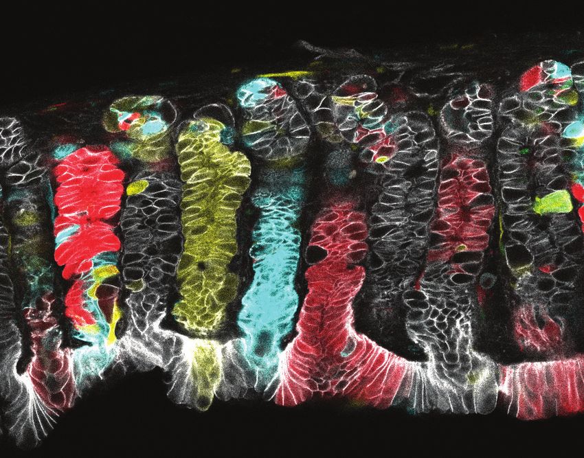
6 COVID stories The Gurdon Institute 7
COVID stories
quickly increased, with home tests “Then, of course, there was the
arriving from all over the country, wider shortage across the country,
we were pushed to the limit and at so Charles and I looked at what we
one point caused a bottleneck of could do.”
the whole pipeline. The solidarity
Once our building opened again
and sense of responsibility of
on 15th June under University
What did we do during the coronavirus pandemic in 2020? everyone in the team meant that we
guidance, the two set up a no-touch
would ensure all the samples were
log-in log-out system allowing the
Along with institutions and Some of our scientists paediatrician at Addenbrooke’s The roles taken by Weronika Fic, processed each day, and we know
Institute to track numbers of people
businesses across England, the re-focussed their research Hospital from March to September. Dmitry Nashchekin, Helen Zenner we played an important part in
on site in real time, ensuring we
Institute shut its doors at 5pm on Omer Ziv with Miska lab colleagues It was actually quite nice to have and Mihoko Tame (St Johnston lab) helping contain the first COVID-19
stuck to the strict rules on numbers
20th March for Lockdown 1.0. Only and collaborators at Justus Liebig something to do during that first and Paolo Amaral (Kouzarides lab) wave.”
in different lab spaces.
minimal maintenance and technical University worked to produce a lockdown, although being coughed were in sample preparation, RT-PCR
staff remained, regularly checking map and database of the short on by feverish children all day wasn’t tests and data analysis, the full team
the building and the fly stocks, and long-range interactions of ideal!” eventually processing over 8000
while researchers could no longer the SARS-CoV-2 RNA genome nasopharyngeal swab samples daily.
access their benches and had to (details in ‘Research highlights
call a sudden halt to hundreds of 2020’). Ben Simons was involved in
experiments. Administrative staff epidemiological modelling.
took computers and files home.
And that’s how it remained until
15th June when we re-opened
at one quarter occupancy, slowly
moving up to 50% occupancy by
September. Many different staff,
and especially researchers, were
delighted to return once more
to the building. Lockdown 2.0 in Core staff made Personal
November sent some more staff Protective Equipment: Alex
back home again. Sossick and Charles Bradshaw of
the Imaging and Bioinformatics Family members sewed face
The core staff have done an teams each took home a 3D printer. coverings: The mother of a staff
incredible job to keep as much of The machines were set up to run member sent about 600 of her
the Institute open and functioning 24-hours a day, making in total hand-sewn, eco-friendly and
as possible (and legal) at all times, 500 visor headbands, which were washable face coverings all the way
Five researchers worked shifts
in terms of maintenance and safety distributed locally to key workers from her village in Italy, enough for
as volunteers at the Cambridge
in the building, supplying media, in GP surgeries, hospitals and care everyone to have two each.
Others intermitted from research Testing Centre that ran seven days
and enabling computing services for homes. “The Institute had given a
and returned to the clinic: Ben a week from 6am to midnight to
remote working. Meanwhile, other Paolo recalls: “Volunteering at the lot of our own PPE, especially visors,
Fisher (Miska lab) says “I returned support the national effort to boost
Institute members have contributed centre felt like a call of duty. As the to Addenbrookes,” says Alex.
to full-time clinical work as a COVID-19 testing capacity.
directly to fighting the pandemic: number of samples to be processed
8   About the Institute             The Gurdon Institute   9

                          Highlights in
                           2019/2020
10 Awards                                                                                                                                                                                                                                     The Gurdon Institute 11

       Awards
       Feb '19: Steve Jackson receives          Three of the Institute’s labs are           November and Steve Jackson is
       cancer research prize                    among more than a dozen across              among them, awarded funding
       The 47th ARC Foundation                  the UK working together to                  for a project on the DNA damage
       Léopold Griffuel Award in                generate data, develop new tools            response in collaboration with
       Translational and Clinical Research      and build a ‘family tree’ of cell           partners in Switzerland and Austria.
       was presented to Steve Jackson at a      divisions during development,               This was the first of the new
       ceremony in Paris on 10th April. The     starting at fertilisation. Azim Surani is   Horizon 2020 grants to come to
       award was given "for his work on         a co-lead for ‘cell lineage in human        Cambridge.
       DNA damage repair and his role in        epiblast specification and early
       the development of medicines such        differentiation’; Emma Rawlins is a         Dec '19: Song for the Unsung
       as PARP1 and 2 inhibitors used [for]     co-lead for ‘human lineage analysis         Heroine
       cancer treatment."                       in a 3D spatial context: cardio-            The Institute's Office Manager,
                                                pulmonary system development’;              Lynda Lockey, won the Unsung
       May '19: Pisa Honorary Doctorate         and Ben Simons is a lead for one            Heroine Award in the University
       for John Gurdon                          of three cross-cutting technology           of Cambridge Professional
                                                platforms – computational biology           Services Recognition Scheme.
       John Gurdon received a Doctorate
                                                and data analysis.                          Institute Director Julie Ahringer said
       Honoris Causa in Translational
       Medicine from the Scuola Superiore                                                   "Lynda is a very deserving recipient
                                                Sep '19: Meri Huch wins BINDER              of the award...Her dedicated and
       Sant’Anna of Pisa. He gave a lecture
                                                Innovation Prize                            understated work makes things run
       there and at Bologna University.
                                                The 2019 BINDER Innovation                  smoothly, and positively impacts         Aug ‘20: Award for research           Prize 2020 by the Leverhulme              The awardees join a four-year
       While in Bologna he was interviewed
                                                Prize was awarded to Group                  everyone".                               contributing to national prosperity   Trust. The prizes of £100,000             programme that provides financial
       by national newspaper 'Il Resto del
                                                Leader Meri Huch for her research                                                    The Royal Society Mullard Award       "recognise the achievement of             support, training and networking
       Carlino'.
                                                on liver organoids for the study            Jan ‘20: Ahringer honoured by            2020 was awarded to Steve Jackson     outstanding researchers whose work        opportunities.
       Jul '19: Gurdon researchers in           of liver biology and disease. The           Genetics Society of America              for his research that led to the      has already attracted international
                                                award is given for "outstanding cell                                                                                       recognition and whose future career       Dec ’20: Aztekin wins ELISIR
       Wellcome’s new £10M project on                                                       Julie Ahringer was honoured with         discovery of the drug olaparib,
                                                biological research with a focus on                                                                                        is exceptionally promising".              scholarship at EPFL
       human development                                                                    the Genetics Society of America's        which has reached blockbuster
                                                cell culture," and awarded by the           George W. Beadle Award                   status for the treatment of ovarian                                             PhD student in the Gurdon lab,
       The Human Developmental                                                                                                                                             Dec ‘20: Ma selected as EMBO
                                                German Society for Cell Biology.            "for outstanding contributions           and breast cancers.                                                             Can Aztekin, moves directly to an
       Biology Initiative aims to provide                                                                                                                                  Young Investigator
                                                                                            to the community of genetics                                                                                             independent principal researcher
       insights into how humans develop –
                                                Nov '19: Steve Jackson awarded              researchers...beyond an exemplary        Oct ‘20: Philip Leverhulme Prize      Hansong Ma was selected as one of         position at the Swiss Federal Institute
       from one cell to billions of different
                                                ERC Synergy Grant                           research career".                        for Ma                                30 new EMBO Young Investigators,          of Technology in Lausanne (EPFL), as
       cells that make up our tissues and
                                                Recipients of the ERC Synergy                                                        Hansong Ma was awarded a              judged to be "among the next              an EPFL Life Sciences Independent
       organs.
                                                Grants were announced in                                                             prestigious Philip Leverhulme         generation of leading life scientists".   Research (ELISIR) scholar.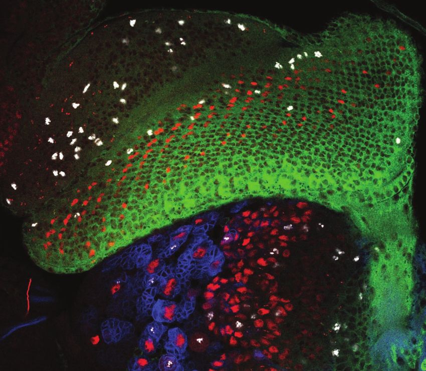
12 Research highlights The Gurdon Institute 13
Research in 2019 liver diseases where regeneration is
impaired.
Aloia L et al. (2019) Nature Cell
Biology 21: 1321–1333.
an 'epitranscriptomic' pathway with
effects on lung cancer cell behaviour
in vitro. They developed a
technique to precisely locate which
guanosine on a micro RNA called
nucleocytoplasmic transport,
uncovering new links between
different forms of dementia.
Paonessa F et al. (2019) Cell Reports
26: P582–593.E5.
let-7 was modified with a methyl
does not depend on the protein generation DNA sequencing group, regulating its processing and
H2AX. They show that MDC1, to over 4500 yeast strains in the downstream action to suppress cell
Jelly secretion
which was previously believed to Gene Knockout Collection. The migration. RNA uptake to
the feeding glands
with RNA
work only when interacting with resulting comprehensive resource Pandolfini L, Barbieri I et al. (2019) Jelly
Molecular Cell 74: P1278–1290.E9.
with
RNA
phosphorylated H2AX, in fact identifies new genes responsible
retains its capacity to recruit repair for maintaining the stability of DNA
factors to the site of DNA damage in cells, and whose absence or
even when H2AX is absent. This mutation leads to a variety of effects, Brain location determines stem
may enable DNA repair in areas of from changes in short sequence cell activation speed
the genome known to be depleted repeats to the loss of whole Otsuki and Brand revealed that
of H2AX. chromosomes. These 'mutational stem cells activate rapidly or slowly
Salguero I et al. (2019) Nature signatures' can now also be studied depending on where they reside systemic
Communications 10: 5191.
RNA spread
in human cells. in the brain. G2 quiescent stem RNA ingestion
Fly gene provides clue to Puddu F et al. (2019) Nature 573:
New cell type in tail cells, which activate first and have
reversing mitochondrial disease 416–420. RNA uptake to the hemolymph
regeneration high regenerative potential, reside
Don't get your DNA in twist primarily in fruit fly ventral brain Transmissible RNA pathway in
The Ma lab have identified a Researchers in the Gurdon and
The Zegerman lab’s new publication regions. G0 quiescent stem cells are honey bees
protein in fruit flies (Drosophila) Simons labs working under
shows that limiting the rate of DNA more numerous in the dorsal brain.
that can be targeted to reverse Jerome Jullien identified a new The Miska lab's Eyal Maori along
duplication - by limiting the number This is an important consideration in
the effects of disease-causing cell type involved in regeneration with colleagues in UK, Israel and
of DNA replication initiation events designing regenerative therapies.
mutations in mitochondrial genes. of tadpole tails. They've named the USA have discovered a pathway
- is important to prevent intertwining Otsuki L & Brand AH (2019)
The discovery could provide clues the Regeneration-Organizing Cells
between the newly replicated Developmental Cell 49: 1–8. by which honey bees share RNA
about how to counteract human for their role in promoting and through secretion and ingestion of
mitochondrial diseases, for which chromosomes. This work may be coordinating new tissue growth after worker and royal jellies, offering a
there is currently no cure. relevant for the treatment of cancer How to boost adult liver amputation, and hope to find clues in
Chiang A C-Y et al. (2019) Current cells, which are characterised by Nuclear membrane dysfunction promising route for administering
regeneration these cells to inform new approaches
underlying dementia bee 'vaccines'. In addition, the
Biology 29: 1–7. high rates of DNA duplication. to regeneration in mammals. researchers identified a specific
Morafraile EC et al. (2019) Genes & A paper from the Huch lab - with
Aztekin C et al. (2019) Science 364: The Livesey and Jackson groups protein in royal jelly that binds and
Development 33: 21–22. collaborators in the Gurdon Institute,
653–658. pooled expertise, studying patient- protects the RNA in granules while
Alternative DNA repair pathway UK and Germany - describes the
derived neurons in the lab to outside the body.
for MDC1 molecular mechanism triggered by
investigate how mutations in the Maori E et al. (2019) Molecular Cell
New DNA stability genes TET1 that allows damaged adult
RNA modification pathway tau gene cause frontotemporal 74: P598–608.E6.
Salguero and colleagues in uncovered in systematic study liver cells to regenerate. This paves
the Jackson lab have found an the way for design of drugs to affects cancer cell migration dementia (FTD). They found that, in
of Yeast Knockout Collection FTD neurons, microtubules deform
alternative pathway to elicit the boost regeneration in conditions Researchers in the Kouzarides lab
The Jackson lab applied next- the nuclear membrane and perturb
DNA-damage response that such as cirrhosis or other chronic and colleagues have characterised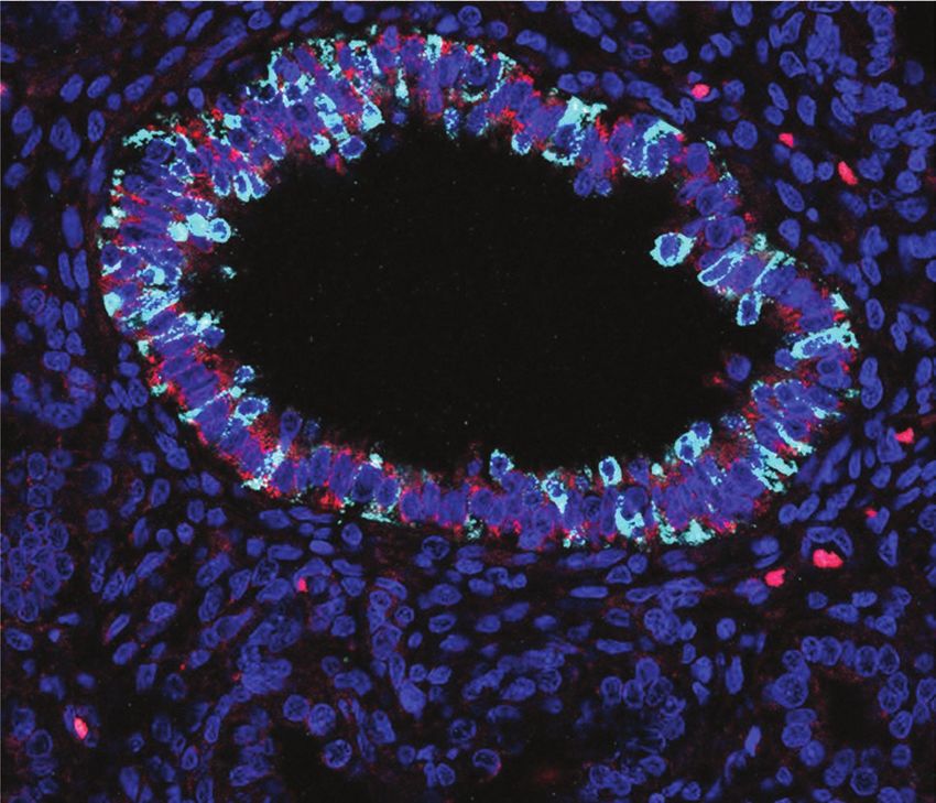
14 Research highlights The Gurdon Institute 15
Research in 2020
collaborative studies by the Simons
lab have shown - at single-cell
resolution - how stem cells react
to regenerate tissue and restore
homeostasis. Stretching induces
skin expansion by creating a
transient bias in the renewal activity
of epidermal stem cells, while a Long-range interactions in SARS-
transcription factor Ascl1, which is and Simons labs. These organoids
a determinant for nerve, resides grown in chemically defined culture second subpopulation of basal Sperm populations show CoV-2 RNA
progenitors remains committed to
on chromatin to direct gene medium provide an important homogeneous epigenetic marks Omer Ziv from the Miska lab, in
expression. While previous studies model for research into the healthy differentiation.
collaboration with Justus-Liebig
suggest that residency times are and diseased pancreas, including Aragona M et al. (2020) Nature 584: Gurdon lab and colleagues, led by
University colleagues, has revealed
only seconds or minutes, this conditions such as cystic fibrosis, 268–273. Jerome Jullien, examined histones
precise details of the base-pairing
experiment showed a long-term pancreatitis, cancer and diabetes. in sperm to uncover a conserved
patterns formed by the long RNA
association of hours or days, which Georgakopoulos N et al. (2020) mechanism for transmission of
genome of the SARS-CoV-2 virus,
could explain the stability of cell BMC Developmental Biology 20: 4. epigenetic information to the
responsible for the COVID-19
fate commitment. embryo. As sperm develop they
pandemic. Ziv devised the method
Gurdon JB et al. (2020) Proc Natl lose a large proportion of the
that takes a snapshot of both short-
histones found in somatic cells,
Acad Sci 117 (26): 15075-15084. Tailless/TLX directs cell fate and long-range interactions in the
but the remainder are retained
change in tumourigenesis in the same position across the
RNA, which are essential for viral
function and therefore present
Hakes and Brand uncover the cell sperm population, indicating the
How inflammation affects potential to prime transcription for
potential therapeutic targets.
fate changes that occur during Ziv O et al. (2020) Molecular Cell 80
regeneration embryonic development.
brain tumour initiation. They (6): 1067-1077.E5
Why can regeneration-incompetent show that high levels of Tailless/ Oikawa M et al. (2020) Nat Comms
Cancer drug hope for genetic 11: 349.
tadpoles not regenerate their TLX, known to be associated
disease
tails? The Gurdon lab show that with aggressive glioblastomas,
Gene regulatory architectures in
immune cells behave differently revert intermediate progenitors to Berquez, Gadsby, Festa and Gallop
Embryo polarisation link to cell germline and somatic tissues
for regeneration-competent and neural stem cells as a first step to lab colleagues discovered that
-incompetent tadpoles. Successful tumourigenesis. Their findings also adjusting membrane composition cycle Jacques Serizay and Ahringer lab
suppression of inflammation is support enforced differentiation as with the PI3K inhibitor alpelisib colleagues profiled and compared
The Zegerman lab, with Gurdon
required for the multiple cellular an effective treatment for Tailless/ rebalances actin cytoskeletal transcriptional and regulatory
Institute colleagues, provided the
mechanisms necessary for new tail TLX-induced brain tumours. organisation in cell culture and element activities across five tissues
first direct molecular mechanism
growth. Hakes AE & Brand AH. (2020) Elife alleviates absorption defects in of the adult nematode worm, C.
through which polarisation of the
Aztekin C et al. (2020) Development 9: e53377. an in vivo mouse model of Lowe elegans. The results demonstrate
embryo is coordinated with DNA
147: dev.185496. Pancreas organoids to model syndrome/Dent disease. Their
replication initiation factors, linking
fundamental differences in
disease findings provide proof-of-concept
developmental cues with changes
regulatory architectures of germline
How does stretching skin make for the first disease-modifying
in the cell cycle, in the nematode C.
and somatic tissue-specific genes,
Pancreas organoids can be
Is TF residency time the key to it grow? treatment.
elegans.
and provide a tissue-specific
successfully generated from single Berquez M et al. (2020) Kidney
cell fate commitment? Gaggioli V et al. (2020) PLoS
resource for future studies.
cells, or fresh and frozen tissue, By tracing the dynamics of cells International 98 (4): 883-896. Serizay J et al. (2020) Genome Res
The Gurdon lab used a competition then expanded and maintained during stretch-mediated expansion Genetics 16 (12): e1008948.
30: 1752–1765.
assay to test how long the long-term in culture, say the Huch of the mouse skin epidermis,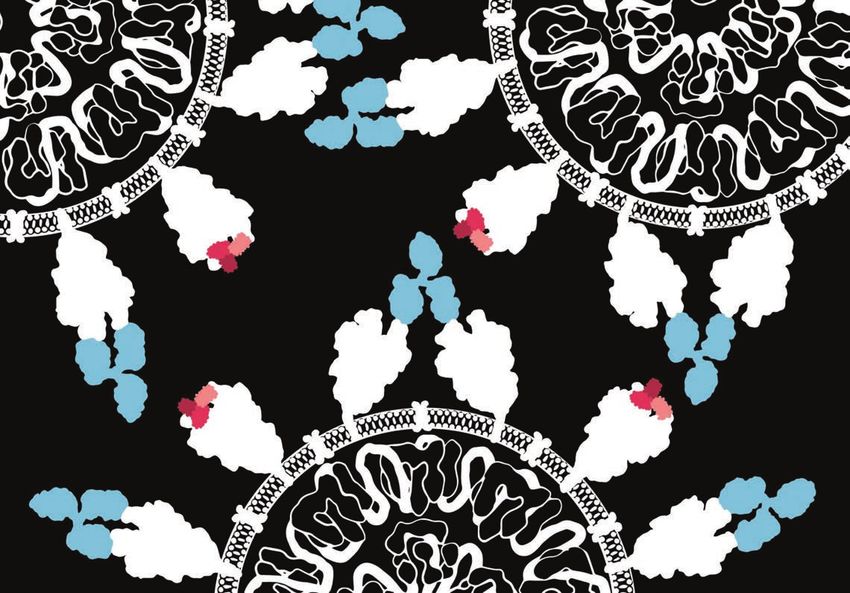
16 Public engagement

        Public engagement
        Mission: to make our fundamental        classrooms with support for teachers
        biological research accessible          and students to deepen their
        and responsive to the public for        knowledge of fundamental biology
        the mutual benefits of inspiration,     and current research. Teachers
        knowledge-exchange and trust.           and our scientists co-created four
                                                innovative teaching 'toolkits', free
                                                to use in classrooms across the
                                                        UK: The Cell Explorer (online      temporary tattoos. Visitors to        Institute provided accommodation,      Sixth-form workshops
                                                             interactive 3D cell model),   festivals and events could choose     travel expenses and food to all        Our Sixth-form workshops aim to
                                                                 Explore Epigenetics       a tattoo from our collection of       participants. Students told us "It     inspire A-level biology students.
                                                                    (an online             designs and then have a chance        gave me an experience hard to find     State schools from across the
                                                                      game about           to discuss the research with the      elsewhere" and "I learned that I can   country can bring groups of Year 12
                                                                        epigenetics),      scientist applying their tattoo.      become a scientist."
      Generate increased trust in fundamental                                                                                                                           biology students to the Institute to
                                                                         a kit for Fruit   Afterwards, they could show off
     research and ensure our research remains                                                                                                                           learn about our research. The visit
                relevant to society                                       Fly Larvae       their new tattoo to friends and       Stitching Science                      includes a tour of our facilities, a
                                                  Empower                  Dissection      share their new science knowledge.    The Public Engagement Seed             seminar about the history of science
                                                 and inspire               (teaching       Thank you to the Wellcome Centre
                             Public
                            Public                 the next                                                                      Fund Project for 2019 was devised      and the future of cancer research, a
                                                                           about the       for Cell Biology in Edinburgh for
                         Engagement
                         engagement              generation                                                                      and led by Stephanie Norwood,          hands-on workshop where students
                           strategy                                        size and        sharing their idea!
                           strategy                                                                                              a former PhD student. The              can test their skills at identifying
                                                                          scale of
                                                                                                                                 project aims to create an informal     cancerous tissue with microscopes,
                                                                         cells, tissues    Aspiring Scientists Training          environment for scientists to          and a Q&A with our PhD students.
                                                                        and organs)        Programme                             interact with local communities
                                                                       and Unlocking
                 Embed public                                                              Providing an inspiring, immersive     and learn a new craft, disseminate
                                                                     Genetic Editing (a
                 engagement in                                                             experience to encourage groups        information about research
                 research culture
                                                                 hands-on problem-
                                                                                           that are underrepresented in          projects, and increase public trust                       Silver Engage
                                                              solving game). The
                                                                                           science, we welcomed 11 Sixth-        in fundamental research. The                              Watermark
                                                         project was funded by
                                                                                           form students for a week at the       project engaged crafters through a                          The Gurdon Institute
                                                 Wellcome and evaluated by the
                                                                                           Institute. Students attended          series of knitting workshops, craft                         was awarded a Silver
        Our projects in 2019 to support         University of Cambridge Faculty of
                                                                                           morning workshops about scientific    fairs and other events. Participants                        Engage Watermark in
        the public engagement strategy          Education. SCoPE website:
                                                                                           topics or presentation skills. Then   create a detailed crochet cell           December 2020, the first such award
        included:                               https://scopegurdoninstitute.co.uk
                                                                                           they spent the rest of the day in a   containing various organelles            at the University of Cambridge. The
                                                                                           lab to talk with lab members about    (mitochondria, cytoplasm and             Silver Engage Watermark, awarded
        Scientists' Collaborative Project       Tattoo My Science
                                                                                           their research and science careers.   membrane) and discuss the                by the National Co-ordinating Centre
        with Educators (SCoPE)                  Our scientists created designs                                                                                            for Public Engagement, recognises
                                                                                           The project was funded by the         different parts of the cell with
        We aim to bring contemporary            based on their biology research,           University of Cambridge Widening      scientists as they knit. Website:        the Institute's “robust and committed
        research into GCSE and A-level          and we turned these into fun,              Participation Project. The Gurdon     https://bitly.com/StitchingSci           approach to public engagement”.
18 Activities and impacts in 2019 The Gurdon Institute 19
Focus on
research20 Group leaders The Gurdon Institute 21
JULIE AHRINGER
Developmental regulation of chromatin structure and function H3K9me2
2-cell 6-cell 32-cell 200-cell
How is chromatin regulated to direct correct gene expression
programmes? Animal development is a remarkable process during
which a single-celled totipotent zygote produces a myriad of different
cell types. A driving force is the differential control of chromatin activity,
which establishes gene expression programmes that drive cellular
identity. Deciphering this control is necessary for understanding how
the genome directs development and the diseases that result from
chromatin dysregulation.
We study how cell-type specific gene expression and chromatin
organisation are achieved using the simple C. elegans model, focusing
on controls and interactions at regulatory elements, the formation and
function of euchromatin and heterochromatin, and the regulation of 3D
nuclear organization. Taking advantage of the experimental amenability
and defined lineage of C. elegans, we apply high-throughput genomics, DAPI
super-resolution microscopy, single-cell analyses, and computational
approaches to understand core mechanisms of gene expression
regulation in development.
Selected publications: Heterochromatin in early
Serizay J et al. (2020) Tissue-specific profiling reveals distinctive regulatory development in C. elegans
architectures at ubiquitious, germline and somatic genes. BioRxiv DOI: Embryonic nuclei imaged
10.1101/2020.02.20.958579v1. using STED super-resolution
Janes J et al. (2018) Chromatin accessibility dynamics across C. elegans microscopy reveals that
development and ageing. Elife 7:e37344. H3K9me2 is found in
McMurchy AN et al. (2017) A team of heterochromatin factors collaborates distinct foci.
Co-workers: Alex Appert, Francesco with small RNA pathways to combat repetitive elements and germline
Carelli, Marie de la Burgade, Yan stress. Elife 6:e21666.
Dong, Martin Fabry, Andrea Frapporti,
Rhys McDonough, Arianna Pezzuolo,
Evans KJ et al. (2016) Stable C. elegans chromatin domains separate
Roopali Pradhan, Anna Townley, broadly expressed and developmentally regulated genes. Proc Natl Acad
Ser van der Burght, Connie Xiao Sci USA 113(45): E7020–7029.22 Group leaders The Gurdon Institute 23
ANDREA BRAND
Time to wake up: regulation of stem cell quiescence and proliferation
Stem cell populations in tissues as varied as blood, gut and brain
spend much of their time in a mitotically dormant, quiescent, state.
A key point of regulation is the decision between quiescence and
proliferation. The ability to reactivate neural stem cells in situ raises the
prospect of potential future therapies for brain repair after damage or
neurodegenerative disease. Understanding the molecular basis for stem
cell reactivation is an essential first step in this quest.
In Drosophila, quiescent neural stem cells are easily identifiable and
amenable to genetic manipulation, making them a powerful model with
which to study the transition between quiescence and proliferation.
These stem cells exit quiescence in response to a nutrition-dependent
signal from the fat body, a tissue that plays a key role in the regulation
of metabolism and growth. My lab combines cutting-edge genetic and
molecular approaches with advanced imaging techniques to study the
reactivation of Drosophila neural stem cells in vivo. This enables us to
deduce the sequence of events from the level of the organism, to the
tissue, the cell, and finally the genome.
Selected publications:
Hakes AE & Brand AH (2020) Tailless/TLX reverts intermediate neural
progenitors to stem cells driving tumourigenesis via repression of asense/
ASCL1. Elife 9:e53377.
Otsuki L& Brand AH (2019) Dorsal-ventral differences in neural stem cell
quiescence are induced by p57KIP2/Dacapo. Dev Cell 49(2): 293-300.e3.
Otsuki L & Brand AH (2018) Cell cycle heterogeneity directs the timing of
Co-workers: Neha Agrawal, Diana Arman, neural stem cell activation from quiescence. Science 360: 99–102.
Maire Brace, Catherine Davidson,
Bernardo Delarue Bizzini, Alex Donovan, Amy Cheetham SW & Brand AH (2018) RNA-DamID reveals cell-type-specific
Foreman, Thomas Genais, Leia Judge, Oriol binding of roX RNAs at chromatin-entry sites. Nat Struct Mol Biol 25:109–114.
Llorà Batlle, Anna Malkowska, Tara Srinivas, The developing visual system
Jocelyn Tang, Christine Turner, Marloes van
Wezel, Rebecca Yakob, Nemira Zilinskaite The Drosophila central brain (red and blue) and eye imaginal disc (green and red) with dividing cells labelled
in white (van den Ameele and Brand).24 Group leaders The Gurdon Institute 25
DAVID FERNANDEZ-ANTORAN
Radiation biology and cell competition
How does ionising radiation affect tissue homeostasis? Healthy
adult epithelial tissues progressively accumulate clones of cells carrying
mutations implicated in cancer. Expansion of the clones follows
Darwinian evolution rules, where some mutations can increase cell
fitness and promote the growth of clones at the expense of the non-
mutated normal adjacent cells, in a process of clonal competition.
Ionising radiation has long been studied as one of the most common
environmental mutagenic agents that promotes tumour formation by
damaging DNA and creating new oncogenic mutations. However, little
is known about its effects on clonal evolution and tissue dynamics. We
have recently shown that radiation can act as an environmental selective
pressure, affecting cell competition mechanisms and promoting
expansion of pre-existing oncogenic mutations, which might increase
the risk of cancer development.
We use long-term human and mouse 3D primary epithelial cultures, in vivo
cell lineage tracing, mathematical modelling, next generation sequencing
methods and state-of-the-art confocal microscopy techniques to unravel
the molecular responses and the cellular interactions that control normal
and mutant cell behaviours after exposure to ionising radiation.
Our final aim is to set the basis for designing external interventions that
can modulate cell competition outcomes during radiation exposure,
eliminate oncogenic mutations and reduce the risk of cancer initiation
and progression.
Co-workers: Inês Ferreira, Jose Selected publications:
Antonio Valverde-Lopez
Fowler JC et al. (2020) Selection of oncogenic mutant clones in normal human
skin varies with body site. Cancer Discov DOI: 10.1158/2159-8290.CD-20-1092.
Piedrafita G et al. (2020) A single-progenitor model as the unifying paradigm of The fight for space during ionising radiation exposure
epidermal and esophageal epithelial maintenance in mice. Nat Comms 11: 1429. This rendered image shows an irradiated mouse oesophageal epithelium populated by fitter oncogenic
Fernandez-Antoran D et al. (2019) Outcompeting p53-mutant cells in the mutant clones (green) that are expanding at the expense of non-mutated normal adjacent cells. Proliferation
normal esophagus by redox manipulation. Cell Stem Cell, 25: 329–341.e6. markers are shown in white and red; cell nuclei in blue.26 Group leaders
JENNY GALLOP
Signalling to the actin cytoskeleton
How do cells control their movement? Cells move during embryonic
development and throughout the life of an organism. When they move,
cells reorganise a system of filaments - the actin cytoskeleton - that gives
them their shape and exerts force on the surrounding tissues. When
regulation of the actin cytoskeleton is disrupted it can lead to cancer
metastasis, intellectual disability, kidney dysfunction and other problems.
We study how the actin cytoskeleton is assembled in different ways. The
cell membrane is an important site of control of actin rearrangements
because it is the boundary between the outside and inside of the cell
and is responsible for initiating communication between and within
cells, which is called signalling.
We have developed cell-free systems using phospholipid bilayers and
frog egg extracts that allow us to find out how signalling lipids in the
cell membrane precisely control the molecular events of actin assembly.
We use combine these cell-free systems with the use of fruit flies, frog
embryos and cultured human cells to test and generate hypotheses
about the molecular events underlying actin regulation during
development and disease.
Selected publications:
Berquez M et al. (2020) The phosphoinositide 3-kinase inhibitor alpelisib
restores actin organization and improves proximal tubule dysfunction in
vitro and in a mouse model of Lowe syndrome and Dent disease. Kidney
Int. 98: 883–896. Watching filopodia grow
Co-workers: Thomas Blake, Jonathan Jarsch IK et al. (2020) A role for SNX9 in the biogenesis of filopodia. J Cell Three-dimensional reconstruction of filopodia-like structures growing from a supported lipid bilayer. The
Gadsby, Pantelis Savvas Ioannou, Biol 219(4): e201909178. structures were segmented based on fluorescent actin intensity in a stack of microscopy images of size 76.13 x
Julia Mason, Kathy Oswald, Kazimir Richier B et al. (2018) Integrin signaling downregulates filopodia during 76.13 x 30 microns.
Uzwyshyn-Jones, Pankti Vaishnav
muscle-tendon attachment. J Cell Sci 131: jcs21733. Colours were randomly assigned as a guide for the eye. The segmentation was performed using a custom
Daste F et al. (2017) Control of actin polymerization via the coincidence of image-analysis pipeline.
phosphoinositides and high membrane curvature. J Cell Biol 216: 3745–3765.28 Group leaders The Gurdon Institute 29
JOHN GURDON Somatic cell nuclear transfer in Amphibia
Nucleus of differentiated cell
Nuclear reprogramming by oocytes and eggs
Transplanted nucleus
2 days 1 year
and egg cytoplasm
Can we make cell reprogramming more efficient? Our group focuses generate wide range of
on somatic cell nuclear transfer to amphibian eggs and oocytes from different cell types
two complementary points of view. One aims to identify the molecules unfertilised and tadpole frog
enucleated egg
and mechanisms by which the cytoplasm of an egg or oocyte can
reprogramme the nucleus of a differentiating somatic cell to behave
like that of an embryo. From this state, many different kinds of cells for
replacement can be generated. Nuclei of differentiated cells
The other aim is to identify the molecules and mechanisms that stabilise Transplanted nuclei have
the differentiated state of somatic cells, as a result of which they resist 1 day 1 day changed donor gene
reprogramming procedures. For these purposes we use single nuclear expression
transfer to unfertilised eggs or multiple nuclear transfer to ovarian
oocyte and its injected nuclei
oocytes.
nucleus enlarge
We make use of the special properties of an amphibian oocyte to inject
messenger RNA that codes for a transcription factor protein. When this Design of competition experiments to analyse transcription factor action using Xenopus oocytes
has been synthesised, it concentrates in the oocyte nucleus. The next
day we inject plasmid DNA directly into the oocyte nucleus, where the
factor causes transcription, and later expression, of a reporter gene in
the plasmid. 24 hrs 24 hrs 1-24 hrs
Freeze and assay for
reporter expression
Selected publications:
Gurdon JB et al. (2020) Long-term association of a transcription factor mRNA Plasmid DNA with Plasmid DNA as compe-
with its chromatin binding site can stabilize gene expression and cell fate for Ascl1 an Ascl binding site titor with an Ascl binding
commitment. Proc Natl Acad Sci USA 117: 15075–15084. and a Firefly site and a Renilla
reporter reporter
Aztekin C et al. (2019) Identification of a regeneration-organizing cell in the
Xenopus tail. Science 364: 653–658.
Co-workers: Hector Barbosa Triana,
Dilly Bradford, Frances Connor, Nigel Hörmanseder E et al. (2017) H3K4 methylation-dependent memory of
Garrett, Khayam Javed, Toshiaki somatic cell identify inhibits reprogramming and development of nuclear Molecules and mechanisms
Shigeoka, Ming-Hsuan Wen transfer embryos. Cell Stem Cell 21: 135–143.e6.
Top: Two types of nuclear transfer experiments, with eggs or oocytes.
Bottom: The Xenopus oocyte can be used to provide a functional test for the binding of a cell-fate-
determining transcription factor, such as Ascl1 for nerve. Once expression of the first plasmid DNA is
established, a second plasmid is not able to compete because of the stable binding of the factor.30 Group leaders The Gurdon Institute 31
STEVE JACKSON
Maintenance of genome stability MDC1 has H2AX-independent roles in the DNA damage response
100
MDC1
DNA is constantly damaged by environmental and endogenously
Cell survival (%)
recruitment of
arising agents. Cell survival and genome integrity are promoted repair factors 10
by the DNA-damage response (DDR), which detects, signals the H2AX
presence of and repairs DNA damage. DDR defects are associated
ATM 1 WT
with developmental disorders, immunodeficiencies, infertility, P H2AX KO
premature ageing and cancer. Our research aims to characterise the cell IR MDC1 KO
H2AX/MDC1 KO
biology and mechanisms of established and new DDR pathways and 0.1
components, and to apply this knowledge to better understand and 0 1 2 3 4 5
treat human diseases. H2AX IR dose (Gy)
We have taken the global approach of cataloguing the >4,500 knockout After DNA damage, phosphorylated histone H2AX recruits MDC1, Unexpectedly, eliminating MDC1
genes of the diploid yeast knockout collection, using next-generation allowing the accumulation of additional repair factors results in more DNA damage
sequencing to identify those genes that have an impact on genome sensitivity than eliminating H2AX
stability. Analysing this dataset revealed genes affecting repetitive
element maintenance, and nuclear and mitochondrial genome stability, Repair factors, like 53BP1, accumulate at DNA damage sites even in the absence of H2AX, but this
and showed how strains adapt to loss of non-essential genes. At the requires the PTS-repeat region of MDC1
other end of the scale spectrum, we determined what structural features
of the DDR factors PALB2 and MDC1 associate with chromatin in ways MDC1 − wt MDC1 − ∆PST
that are crucial for effective DDR in the absence of BRCA1 and H2AX,
H2AX-free genomic regions
respectively.
ATM
Selected publications: H2AX+/+
Belotserkovskaya R et al. (2020) PALB2 chromatin recruitment restores
Co-workers: Samah Awad Diab, Rimma
Belotserkovskaya, Ramsay Bowden, homologous recombination in BRCA1-deficient cells depleted of 53BP1.
Chris Carnie, Julia Coates, Sabrina Nat Commun 11: 819.
PST recruitment of
Collier, Giuseppina D'Alessandro, Salguero I et al. (2019) MDC1 PST-repeat region promotes histone H2AX-
Muku Demir, Kate Dry, Yaron Galanty, H2AX repair factors
independent chromatin association and DNA damage tolerance. Nat
-/-
Maryam Ghaderi Najafabadi, Nadia
Gueorguieva, Vipul Gupta, Soren Commun 10: 5191. P
MDC1
Hough, Rebecca Lloyd, Donna Lowe, Puddu F et al., (2019) Genome architecture and stability in the 53BP1
David Morales, Francisco Muñoz Saccharomyces cerevisiae knockout collection Nature 537:416-420.
Martinez, Domenic Pilger, Fabio Puddu,
Helen Reed, Matylda Sczaniecka-Clift,
Almudena Serrano Benitez, George
Spooner, John Thomas, Andrea Voigt,
Mike Woods, Guido Zagnoli-Vieira32 Group leaders The Gurdon Institute 33
TONY KOUZARIDES
Epigenetic modifications and cancer Epigenetic targets for cancer therapy
Do enzymes that modify chromatin and RNA offer therapeutic
targets? DNA exists in the cell nucleus wrapped around histone
proteins to form chromatin. The DNA and histones are decorated
with many types of covalent chemical modifications, which can affect RNA
transcription and other cellular processes. In addition, non-coding RNAs
that regulate chromatin function can be similarly chemically modified. histone-modifying RNA-modifying
Our lab is involved in characterising the pathways that mediate and enzyme enzyme
control DNA, RNA and histone modifications. We try to understand the
cellular processes they regulate, their mechanism of action and their
involvement in cancer.
Our focus at the moment is modifications of messenger RNA (mRNA) Post-transcriptional
and non-coding RNA. There are very few modifications identified on control
these low-abundance RNAs, unlike on transfer RNA and ribosomal RNA,
where there are many. We have been developing sensitive technologies Transcriptional
to detect modifications, such as specific antibodies, chemical reactivity control
assays and mass spectrometry. Using these, we have been able to detect
a number of novel modifications on mRNA and microRNA (short-length
non-coding RNAs) and have shown that these function to regulate DNA
mRNA translation and microRNA processing. Furthermore, we have
shown that the enzymes that mediate these modifications are implicated modification
in cancer. We are developing small-molecule inhibitors against some of
these enzymes in collaboration with STORM Therapeutics.
Co-transcriptional
Selected publications: control
Barbieri I & Kouzarides T (2020) Role of RNA modifications in cancer. Nat
Co-workers: Minaam Abbas, Andrej Rev Cancer 20, 303–322.
Alendar, Andrew Bannister, Francisco Pandofini L et al. (2019) METTL1 Promotes let-7 MicroRNA Processing via
José Campos Laborie, Alistair Cook, m7G Methylation. Mol Cell Bio 74(6): 1278-1290. The crucial role of modifications
Elena Everatt, Marie Klimontova, Sri
Lestari, Nikki Mann, Carlos Melo, Barbieri I et al. (2017) Promoter-bound METTL3 maintains myeloid Gene expression can be regulated by chemical modifications before and during transcription, including of
Helena Santos Rosa, Konstantinos leukaemia by m6A-dependent translation control. Nature 552: 126–131. non-coding RNA.
Tzelepis, Daniel Wing34 Group leaders The Gurdon Institute 35
HANSONG MA
Mitochondrial DNA transmission and maintenance wild type
mtDNA mutant mtDNA
maternal
How mitochondrial genomes are transmitted and maintained. In inheritance
addition to the nuclear genome, all animals have another genome
packed inside the mitochondrion called mtDNA. This maternally
inherited genome encodes important proteins for energy production.
During development and ageing, as mtDNA continues to replicate and
turnover, mutations can occur to some of the copies. The subsequent
prevalence of these mutants, which determines the progression and
inheritance of the clinical abnormalities of mitochondrial disorders,
depends on how they compete with the co-existing wild-type genomes
for transmission. To date, over 50 mtDNA-linked disorders have been mtDNA
described in humans. repair
We have developed genetic systems in Drosophila to study the rules
governing the transmission of mtDNA mutations. By creating fruit flies
carrying both functional and pathogenic mitochondrial genomes, we heteroplasmic
reveal nuclear factors and mtDNA sequence polymorphisms that bias transmission
the transmission of one genome over the other. We also study repair
mechanisms that safeguard the integrity of mtDNA during development
and ageing.
The maternally transmitted
Selected publications: mitochondrial DNA (mtDNA) is a
multi-copy genome that shows complex
Chiang A et al. (2019) A Genome-wide screen reveals that reducing transmission patterns during developing
mitochondrial DNA polymerase can promote elimination of deleterious and aging, and between generations
mitochondrial mutations. Curr Biol 29: 4330-36. due to relaxed replication, random
Klucnika A & Ma H (2019) A battle for transmission: the selfish and segregation, and selection that favours
corporative animal mitochondrial genomes. Open Biol 9: 180267. the transmission of one genome over
Co-workers: Ason Chiang, Ivy Ma H & O’Farrell PH (2016) Selfish drive can trump function when animal another in the pool. In addition to
Di, Beitong Gao, Jan Jezek, mitochondrial genomes compete. Nat Genet 48: 798–802. mitochondrial diseases caused by
Anna Klucnika, Andy Li, Eleanor accumulation of a particular mutant,
McCartney, Matthew McCormack,
random mtDNA mutations have been
Kathy Oswald, Sumaera Rathore,
Ziming Wang shown to increase with age, contributing
to mitochondrial dysfunction and various
age-related conditions.36 Group leaders The Gurdon Institute 37
ERIC MISKA
Non-coding RNA and genome dynamics
What does non-coding RNA do in development and disease?
Most of the RNA transcribed from the DNA in our genome is not 5′CS
translated into protein but instead has direct functions in regulating
biological processes. This paradigm shift in nucleic acid biology has 5′
been supported by technical advances in high-throughput sequencing, miR-21
molecular genetics and computational biology, which can be combined
with more traditional biochemical analyses.
Many species and roles of non-coding RNA have been identified. Our
goal is to understand how non-coding RNAs regulate development,
physiology and disease. We are exploring microRNA in the pathology
of cancer and other diseases, RNA interference in viral immunity, Piwi-
interacting RNA in germline development and genome integrity, and
endogenous small interfering RNA in epigenetic inheritance – where 5′
we predict a big impact in understanding human health. Our model 5′CS
V
organisms are the nematode worm, the cichlid fishes of the Rift Lakes N
3′CS
of East Africa, mouse, and human cell culture. More recently we have M
L
developed a technology to assess RNA structure and RNA–RNA
interactions in living systems. We used this to uncover unexpected 3′
biology for the Zika virus, and key regulatory mechanisms for SARS-
CoV-2. 5′CS
5′
Selected publications:
Ziv O et al. (2020) The Short- and Long-Range RNA–RNA Interactome of
Co-workers: Harry Baird, Sarah Buddle, SARS-CoV-2. Mol Cell 80: 1067–1077.e5. The Zika virus genomic structure
Nicholas Burton, Alexandra Dallaire,
Benjamin Fisher, Giulia Furlan, David
Maori E et al. (2019) A Secreted RNA Binding Protein Forms RNA-Stabilizing inside human cells
Jordan, Tsveta Kamenova, Joanna Granules in the Honeybee Royal Jelly. Mol Cell 74: 598-608.e6.
The Zika virus genome adopts
Kosalka, Lisa Lampersberger, Miranda Ziv O et al. (2018) COMRADES determines in vivo RNA structures and alternating structures as the 5'
Landgraf, Bethan Manley, Ragini Medhi, interactions. Nat Methods 15: 785–788. cyclization sequence (CS) participates
Narendra Meena, Harris Papadopoulou,
Jon Price, Audrey Putman, Fu Xiang in interaction with host microRNA
Quah, Navin Brian Ramakrishna, Cristian miR-21 (top), capsid translation
Riccio, Marc Ridyard, Miguel Vasconcelos (right), and genome cyclization (left).
Almeida, Gregoire Vernaz, Archana Yerra,
Chengwei (Ulrika) Yuan, Omer Ziv38 Group leaders
EMMA RAWLINS
Stem and progenitor cells in the mammalian lung
How do stem cells build and maintain the lung? The complicated
three-dimensional structure of our lungs is essential for respiration and
host defence. Building this structure relies on the correct sequence
of division and differentiation events by lung progenitor cells, which
also maintain the slowly turning-over airway epithelium in the adult.
How is the production of different cell types controlled in embryonic
development and adult maintenance? We apply mouse genetics, live
imaging, single-cell molecular analysis and mathematical modelling
to understand lung stem cells, with a longer-term aim of directing
endogenous lung cells to repair, or regenerate, diseased tissue.
In the adult lung we focus on the cellular mechanisms that maintain stem
cell quiescence at steady-state, but allow a rapid repair response when
needed. In the embryonic lung we study a population of multipotent
progenitors that undergo steroid-induced changes in competence
during development. In the embryo, we have recently switched our focus
to normal human lung development, primarily using an organoid system
that we developed. We combine the analysis of fresh human embryonic
tissue with gene-targeting in the organoids, to determine the molecular
and cellular mechanisms of normal human lung development. This will
provide insights into conditions related to premature birth and into the
possibility of therapeutic lung regeneration.
Selected publications:
Nikolic´ M et al. (2018) Human lung development: recent progress and new
challenges. Development 145: dev163485.
Co-workers: Ana Lucia Cabriales Nikolic´ M et al. (2017) Human embryonic lung epithelial tips are multipotent
Torrijos, Ziqi Dong, Tessa Hughes, progenitors that can be expanded in vitro as long-term self-renewing
Quitz Jeng, Florence Leroy, Kyungtae
organoids. Elife 6: e26575.
Lim, Shuyu Liu, Vishal Menon, Ziming
Shao, Vanesa Sokleva, Dawei Sun Balasooriya GI et al. (2016) An FGFR1-SPRY2 Signaling Axis Limits Basal Cell How does the lung build itself?
Proliferation in the Steady-State Airway Epithelium. Dev Cell 37: 85–97. In this 17-weeks-gestation human embryonic lung, the differentiating airway epithelium has been stained to
visualise mRNA. Differentiating secretory cells (cyan) and ciliated cells (red) are visible. Cell nuclei in blue. (Credit:
Dr Kyungtae Lim.)40 Group leaders
DANIEL ST JOHNSTON domain
apical
Polarising epithelial cells and body axes secretion
apical
How do cells know ‘up’ from ‘down’? Most cells in the body perform
different functions at opposite sides of the cell. This cell polarity is lateral
essential in development, for example: in determining the head-to-tail
axis of many animals, for cell migration and for asymmetric stem-cell
divisions. Furthermore, loss of polarity is a hallmark of tumour cells and
is thought to contribute to tissue invasion and metastasis. Our work forming sheets basal
focuses on epithelia, the sheets of polarised cells that form barriers that create a
between compartments and make up most of our organs and tissues. barrier
We study the factors that mark different sides of epithelial cells and
how these organise the internal cell architecture, using the Drosophila loss of polarity
in cancer
intestine and the follicle cell epithelium as models.
We have recently discovered that the gut epithelium polarises by a
fundamentally different mechanism from other fly epithelia, and is
forming tubes
much more similar to mammalian epithelia. We are now identifying new
polarity factors in the fly gut and are testing whether these play similar
roles in mouse intestinal organoids. We are also using live microscopy
to visualise polarised secretion in epithelial cells, and quantitative super-
resolution microscopy to examine the clustering and co-localisation of
polarity proteins.
Selected publications:
Lovegrove H et al. (2019) The role of integrins in Drosophila egg chamber
morphogenesis. Development 146: dev182774
Fic W et al. (2019) Drosophila IMP regulates Kuzbanian to control the timing of
Notch signalling in the follicle cells. Development 146: dev168963. The special properties of epithelia
Co-workers: Edward Allgeyer, Jia Chen, Chen J et al. (2018) An alternative mode of epithelial polarity in the Drosophila
Jin Mei Cheng, Helene Doerflinger, midgut. PLoS Biol 16: e3000041. A drawing showing how epithelial cells stick together to form epithelial sheets, with their free apical surfaces
Weronika Fic, Xiao Li He, Nathan facing towards the outside or the lumen of an epithelial tube or gland. The lateral junctions (yellow) create a
Hervieux, Florence Leroy, Dmitry barrier between cells so that fluids, solutes and pathogens cannot leak across the epithelium. Most cancers
Nashchekin, John Overton, Amandine
arise from epithelial tissues and are characterised by a loss of apical–basal polarity (red cells).
Palandri, Jenny Richens, George
Sirinakis, Mihoko Tame, Joseph Jose
Thottachery, Helen Zenner, Xixi Zhu42 Group leaders The Gurdon Institute 43
BEN SIMONS
Mechanisms of stem cell fate in tissue development, maintenance and disease
How do stem and progenitor cells regulate their fate behaviour to
specify and maintain tissues? During development, cell proliferation
and differentiation must be coordinated with collective cell movements
to specify organs of the correct size, pattern and composition. In the
adult, stem cells must regulate a precise balance between proliferation
and differentiation to maintain tissue homeostasis.
To address the mechanisms that regulate stem and progenitor cell
fate, we combine cell lineage-tracing approaches and single-cell
gene expression profiling with concepts and methods from statistical
physics and mathematics. Applied to epithelial tissues, we have shown
how common principles of self-organisation and emergence provide
predictive insights into the cellular mechanisms that regulate tissue
development, maintenance and repair. As well as questioning the
nature of stem cell identity and function, these studies emphasize
the role of cell fate stochasticity and state flexibility, and establish
a quantitative platform to investigate pathways leading to cancer
initiation and progression.
Selected publications:
Han S et al. (2019) Defining the identity and dynamics of adult gastric isthmus
stem cells. Cell Stem Cell 25: 342–356.
Kitadate Y et al. (2019) Competition for mitogens regulates spermatogenic
stem cell homeostasis in an open niche. Cell Stem Cell 24: 79–92.
Hannezo E et al. (2017) A unifying theory of branching morphogenesis. Cell
171: 242–255.
Co-workers: Ignacio Bordeu, Lemonia Cell lineage tracing in the stomach corpus
Chatzeli, Catherine Dabrowska, Frances
England, Adrien Hallou, Seungmin Genetic lineage tracing using a multicolour confetti reporter system reveals the compartmentalisation of the
Han, Daniel Kunz, Jamie McGinn, Kathy mouse stomach corpus gland. (Credit: Juergen Fink and Seungmin Han.)
Oswald, Bart Theeuwes, Yanbo Yin,
Min Kyu YumYou can also read