Radiomics and Radiogenomics in Evaluation of Colorectal Cancer Liver Metastasis
←
→
Page content transcription
If your browser does not render page correctly, please read the page content below
REVIEW
                                                                                                                                      published: 07 January 2022
                                                                                                                                 doi: 10.3389/fonc.2021.689509

                                              Radiomics and Radiogenomics
                                              in Evaluation of Colorectal
                                              Cancer Liver Metastasis
                                              Yun Wang , Lu-Yao Ma , Xiao-Ping Yin * and Bu-Lang Gao

                                              CT-MRI Room, Affiliated Hospital of Hebei University, Baoding, China

                                              Colorectal cancer is one common digestive malignancy, and the most common approach
                                              of blood metastasis of colorectal cancer is through the portal vein system to the liver. Early
                                              detection and treatment of liver metastasis is the key to improving the prognosis of the
                                              patients. Radiomics and radiogenomics use non-invasive methods to evaluate the
                                              biological properties of tumors by deeply mining the texture features of images and
                                              quantifying the heterogeneity of metastatic tumors. Radiomics and radiogenomics have
                                              been applied widely in the detection, treatment, and prognostic evaluation of colorectal
                                              cancer liver metastases. Based on the imaging features of the liver, this paper reviews the
                                              current application of radiomics and radiogenomics in the diagnosis, treatment, monitor of
                                              disease progression, and prognosis of patients with colorectal cancer liver metastases.
                          Edited by:
                          Oliver Diaz,        Keywords: colorectal cancer, liver metastasis, radiomics, gene, treatment, prognosis
       University of Barcelona, Spain

                      Reviewed by:
                    Zhongxiang Ding,
            Zhejiang University, China
                                              1 INTRODUCTION
                              Fan Xia,
              Fudan University, China
                                              Colorectal cancer (CRC) is the third most prevalent malignancy and the second commonest cause of
                                              cancer-related deaths throughout the world (1), with the incidence and mortality still on the rise in
                  *Correspondence:
                        Xiao-Ping Yin
                                              recent years (2). Because of the hepatic unique blood circulation characteristics, the liver has become
             yinxiaoping78@sina.com           the most common organ for blood metastasis of cancers, accounting for 25% of all cancer metastasis
                                              (3) and approximately 35%–55% of CRC (4, 5). The liver has uniquely favorable conditions for
                   Specialty section:         stagnation and growth of cancerous cells, with double blood supply from the visceral and portal
         This article was submitted to        vascular systems and natural spaces among adjacent endothelial sinusoidal cells that are deficient of
                   Cancer Imaging and         a typical basement membrane for covering (3, 6, 7). Hepatic metastasis is a critical indicator of
        Image-directed Interventions,         prognosis for patients with primary cancers, and the life expectancy of patients with hepatic
               a section of the journal       metastases from gastrointestinal cancers is only 6 months without appropriate treatment (8).
                 Frontiers in Oncology
                                              Accurate prediction and differentiation of liver metastases from CRC is critical to making an
           Received: 01 April 2021            appropriate therapeutic plan and improving the prognosis of the patients. Ultrasound, computed
      Accepted: 03 December 2021              tomography (CT), magnetic resonance imaging (MRI), and positron emission tomography (PET)
       Published: 07 January 2022
                                              have been routinely applied to detect and assess liver lesions, including metastases of cancer (9, 10).
                            Citation:         Some liver metastatic lesions from primary cancers of different systems may have common
       Wang Y, Ma L-Y, Yin X-P and
                                              characteristics, including hyperechoic lesions surrounded by a hypoechoic halo (targeted ring
      Gao B-L (2022) Radiomics and
      Radiogenomics in Evaluation of
                                              sign) in primary gastrointestinal and vascular carcinomas on ultrasound imaging and presence of
  Colorectal Cancer Liver Metastasis.         calcification in CRC or ovarian carcinomas (7, 11, 12). Metastatic lesions with typical imaging
            Front. Oncol. 11:689509.          features may be easily identified from specific primary carcinomas; however, this kind of lesion
     doi: 10.3389/fonc.2021.689509            accounts for only a small proportion of metastatic lesions, with most of the metastatic lesions being

Frontiers in Oncology | www.frontiersin.org                                         1                                January 2022 | Volume 11 | Article 689509
Wang et al. Radiomics of Colorectal Liver Metastasis
atypical on imaging, whose specific origin cannot be identified combination of genomic information and radiomic features to
easily. Thus, thorough laboratory and physical examinations, enable decision support rather than whole-genome analysis to
molecular genetic test, and tissue biopsy have been applied to determine the genetic causes of radiosensitive variations in the
assess the primary origin of liver metastases even though these scope of radiation oncology. Radiogenomics is important
tests and examinations are costly, invasive, or time-consuming because not all patients have had their cancerous diseases
(13, 14). genomically profiled even though they may undergo imaging
With the development of great-volume computing capability, examinations during the course of disease. Radiogenomic data
it is currently feasible to quickly extract countless quantitative can provide gene expression or mutation information to increase
characteristics from three-dimensional imaging data of MRI, CT, diagnostic, predictive, and prognostic capability and to enable
ultrasound, and PET for evaluation of the nature of different precision therapy because these radiomics data are originated
lesions, because digital medical images contain considerable from the complete tumor lesion rather than a small sample
information that reflects potential pathophysiology. This of tissue.
technology of transforming digital medical imaging data into In patients with CRC, one factor significantly affecting the
high-dimensional data for assessment and decision support is prognosis is the proper management of colorectal cancer liver
referred to as radiomics (15). The framework of radiomics metastases (CRLM), and surgical treatment stands for the only
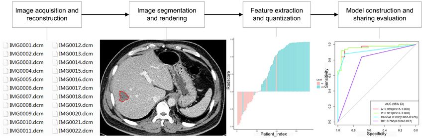
application is shown in Figure 1. The radiomics technology opportunity of long-term survival. The 5-year survival rate of
has been motivated by the notion that biomedical images CRC patients with complete resection of liver metastases has
comprise information that mirrors and can be used to reveal been reported to be approximately 30% higher than that without
basic pathophysiology through quantitative analysis. It has been appropriate treatment of the liver metastases (16). Therefore, one
applied in many conditions, but the most developed field of of the keys to improving the prognosis of CRC patients is to
application is in oncology. Quantitative features of imaging are detect liver metastases for initiating appropriate treatment as
based on imaging shape, intensity, volume, size, and texture, soon as possible. Currently, few studies have been performed on
which provide detailed information on tumor microenvironment radiomics or radiogenomics of CRLM, and this review focused
and phenotype distinct from that offered by laboratory results, on the radiomics and radiogenomic features of CRLM, trying to
clinical reports, and genomic or proteomic analyses. Combined facilitate early detection and appropriate treatment of CRLM
with other clinical information, these features can be used for besides evaluation of its genetic factors and response to
correlation analysis with clinical results and decision-making, treatment for improving the prognosis. The flow chart of the
and radiomics can thus provide countless imaging biomarkers to content of this paper is shown in Figure 2.
potentially help cancer diagnosis, detection, prognosis
evaluation, prediction of treatment response, and monitoring
of disease progression. Radiomics is a young field of study and
will undergo a slow progress because of technical complexity, 2 RADIOMICS PROGRESS IN THE
datum overfitting, deficiency of standards for outcome DIAGNOSIS AND TREATMENT OF CRLM
validation, incomplete presentation of outcomes, and
unrecognizable confounding factors in the databases. In recent years, the field of medical image analysis has developed
Radiogenomics refers to the exploring of radiomics data to rapidly, and the development of pattern recognition tools has
find correlations with genomic modes and has aroused promoted fast progress of quantitative feature extraction. By
considerable interest in the research field of oncology (15). extracting a great deal of quantitative features from medical
Here, in this paper, radiogenomics only indicates the imaging data, radiomics can be used to analyze image
FIGURE 1 | Framework of radiomics application.
Frontiers in Oncology | www.frontiersin.org 2 January 2022 | Volume 11 | Article 689509Wang et al. Radiomics of Colorectal Liver Metastasis
FIGURE 2 | Flow chart of this paper.
information in detail. Compared with traditional approaches of metastasis (n = 10), heterochronous hepatic metastasis within
imaging diagnosis, it can significantly improve tumor diagnosis, 18 months after initial staging (n = 4), or no hepatic metastasis
grading and staging, evaluation of responses to chemotherapy, (n = 15), Rao et al. (19) found that the mean entropy of the whole
and prognosis prediction (15, 17, 18), providing professional liver was significantly (p < 0.05) higher in patients with
guidance for treatment planning. synchronous metastases than those without hepatic metastases,
whereas the mean uniformity of the whole liver was significantly
2.1 Radiomics in the Diagnosis of CRLM (p < 0.05) lower in patients with synchronous metastases than
With the progress of imaging technology, conventional imaging those without liver metastases. This study indicated that texture
approaches can effectively detect large and typical CRLM. evaluation of seemingly disease-free liver is promising to
However, due to the complexity of hepatic hemodynamics and distinguish CRC patients with or without hepatic metastases.
differences of liver parenchymal background on imaging among After analyzing the texture in non-enhanced CT imaging in
patients, different imaging modalities perform differently in seemingly non-diseased regions of liver for impact of hepatic
diagnosis of atypical or tiny liver metastases. It is hard to texture by presence of malignant tumors in patients with CRC,
detect tiny or occult metastases by using the existing imaging Ganeshan et al. (20) found that the fine to medium texture ratio
approaches; however, identification of these lesions is crucial to after imaging filtration was significantly (p < 0.05) different in
early management and improved prognoses. Radiomics features, seemingly non-diseased hepatic areas in patients with hepatic
including entropy, texture and texture ratio, uniformity, and metastasis compared with those without liver metastasis
convolutional neural networks (CNNs), have been effectively (entropy, p = 0.0257) or those with extra-liver disease
applied for diagnosis of CRLM. In assessing the capability of (uniformity, p = 0.0143). Imaging textures of entropy and
whole-liver CT imaging texture analyses of hepatic parenchyma uniformity have been found to be more advantageous to other
in distinguishing CRC patients with simultaneous hepatic features in the diagnosis of CRLM.
Frontiers in Oncology | www.frontiersin.org 3 January 2022 | Volume 11 | Article 689509Wang et al. Radiomics of Colorectal Liver Metastasis
CNNs are able to generate useful characteristics from imaging discrimination demonstrated in the clinical-radiomics
data and have been proven to have high values in predicting combined model (C-indices of 0.941 and 0.833, respectively, in
oncological outcomes (5, 21–23). Lee et al. used CNNs to the training and validating set). Han et al. (25) investigated MRI
generate imaging features from the liver parenchyma in 2019 data of 182 resected CRLM lesions in chemotherapy-free patients
patients with stage I–III CRC for predicting metachronous liver including desmoplastic HGPs in 59 patients and replacement
metastasis based on preoperative abdominal CT imaging (5). HGPs in 123, with the decision tree algorithm being used for
They found that the radiomics model combining clinical radiomics modeling, fused radiomics model being reconstructed
variables with the top principal components of imaging had from combination of radiomics signatures of all sequences, and
the greatest performance (mean AUC = 0.747) to predict 5-year clinical and combined models being constructed via multivariate
metachronous liver metastasis compared with the model using logistic regression analysis. They found that the fused radiomics
clinical features only. Even though no hepatic metastasis was model of tumor zone and the radiomics model of tumor–hepatic
found during the initial colectomy, the radiomics features using interface zone exhibited superior performance to any single
the CNNs could be used to predict possible metachronous sequence or the clinical model and that the radiomics model of
liver metastasis. tumor–liver interface zone was better than that of the tumor
zone (AUC of 0.912 vs. 0.879). The combined model had good
2.2 Differentiation of Histopathological discriminating capability, with the AUC of nomogram being
Growth Patterns of CRLM 0.971, 0.909, and 0.905, respectively, in the training, internal
The heterogeneities of genetic, phenotypic, epigenetic, and validating, and external validating set. Their study (25) revealed
morphological features inside and outside the CRLM lesion that MRI-based radiomics is capable of predicting the
result in different responses to systemic treatment (24, 25). The predominant CRLM HGPs as a potential biomarker for
histopathological growth pattern (HGP) is one such therapeutic strategy. Through analysis of the above studies, it
heterogeneity with corresponding microvasculatures. Based on was found that the combination model of radiomics and clinical
the interface of cancerous cells with adjacent hepatic texture, information can show better discrimination ability than the
CRLM has two primary kinds of HGPs: replacement and single radiomics model.
desmoplastic, with other uncommon kinds of mixed and
pushing HGPs (26). The desmoplastic HGP is characterized by 2.3 Evaluation of HGPs for Treatment
separation of the cancerous cells from the hepatic texture by a Effect on CRLM
fibrous band with lymphocytic infiltration and sprouting Metastases are the major death cause in most patients with solid
angiogenesis in the microvasculature. In this pattern, the malignancies, and hepatic metastasis is the critical factor for
cancerous cells initiate a reaction similar to the healing of survival of patients with advanced malignant tumors (27, 28).
wounds: scar tissues are created with presence of inflammation Histological presentations of liver metastases are heterogeneous
and new blood vessels. In the replacement HGP, the cancerous and reflected by different HGPs that affect clinical outcomes. The
cells constitute cellular plates that are in continuity with the desmoplastic HGP is a positive prognostic biomarker while the
hepatocytic plate, allowing the cancerous cells to displace replacement HGP is a negative one (27). A retrospective study
hepatocytes and co-opt the sinusoidal blood vessels at the enrolling 732 patients found that the exclusive desmoplastic
cancer–liver interface, without disturbing the hepatic stromal growth serves as a positive prognostic marker for patients with
architecture or inducing sprouting angiogenesis (25, 27, 28). CRLM, which is not matched by any other factors evaluated (31).
Desmoplastic metastases are frequently well or moderately In this study, 19% of patients without chemotherapy (n = 367)
differentiated, whereas replacement liver metastases are of poor had desmoplastic growth in the whole tumor–hepatic interface
differentiation, lacking immune reaction and secondary and were independently associated with 50% 5-year survival rate
glandular structures (27, 29). The pushing HGP is less without progression (hazard ratio or HR: 0.54, p = 0.001) and
common with the hepatocyte plate being compressed and 78% 5-year overall survival (HR: 0.39, p < 0.001). CRLM lesions
pushed away by the metastatic cancer cells, with no with this kind of HGP are more suitable for regional metastases-
desmoplastic rim around the cancerous cells or direct contact directed treatment. On the contrary, replacement HGP is linked
of the cancerous cells with the hepatocytes. to poor pathological responses, with the presence of a large
The HGPs of CRLM can be effectively differentiated using proportion of cancerous cells after chemotherapy, and bad
multidetector CT-based radiomics and MRI-based radiomics ima ging r eaction on C T in p atients with p rima ry
(multi-habitat and multi-sequence) (25, 30). After studying 126 chemotherapy and anti-angiogenesis therapy before surgery for
patients with CRLM lesions who had undergone abdominal CRLM (32). This type of HGP occurs more often in new hepatic
contract-enhanced CT imaging followed by partial metastatic lesions that grow even during systemic therapy. The
hepatectomy with histopathologically confirmed HGPs replacement HGP indicates not only worse overall and
including desmoplastic HGP in 68 patients and replacement progression-free survival (31, 33, 34), but also resistance to
HGP in 58, Cheng et al. (30) found that the fused radiomics systemic therapy in patients with CRLM (32). A possible
signature had the best predictive performance in differentiating reason for the resistance to systemic therapy of the
replacement from desmoplastic HGPs (AUC of 0.926 and 0.939, replacement type of HGP is vessel co-option, which serves as
respectively, in the training and validating set), with good an approach of continuous blood supply when the vascular
Frontiers in Oncology | www.frontiersin.org 4 January 2022 | Volume 11 | Article 689509Wang et al. Radiomics of Colorectal Liver Metastasis
endothelial growth factor is inhibited by treatment (35). response (cutoff ≥ 0.42; OR = 20, 95%CI = 1.85–217.4) (46).
Moreover, different HGPs have varied immune phenotypes Two measures for homogeneity of lower angular second moment
that contribute to varied responses to immune therapy. and a higher variance had been demonstrated to associate with
Evidence has indicated that tumors with limited numbers of good responding CRLM lesions rather than non-responding
infiltrated T cells are in essence frequently resistant to immune lesions on T2 MRI imaging, with the variance of 446.07 ±
therapy (36). Vascular co-opting hepatic lesions of metastasis 329.60 in patients with good responses vs. 210.23 ± 183.39 in
usually have low infiltration of immune cells or inflammatory non-responding patients (p < 0.001) and the angular second
cells as demonstrated in lesions with the replacement type of moment of 0.96 ± 0.02 vs. 0.98 ± 0.01, respectively (p < 0.001).
HGP in contrast to those with desmoplastic HGP which are After investigating therapeutic radiomics features for predicting
frequently surrounded by a lot of lymphocytes in the dense rime tumor sensitivity in 667 patients with CRLM to 5-fluorouracil,
(29, 37). Thus, the types of HGPs differentiated using the irinotecan, and folinic acid alone or combined with cetuximab,
multidetector CT-based radiomics and MRI-based radiomics Dercle et al. (42) found that the radiomics response signature
(25, 30) may indicate the prognosis of patients with relevant outperformed known biomarkers of the KRAS mutation status
types of HGP in CRLM lesions. In studying the HGP types of and tumor contraction rate in the early prediction of therapeutic
CRLM using MRI-based radiomics in comparison with the sensitivity and for guiding decisions of cetuximab therapy. In
histopathological types, Han et al. (25) found that more tissue evaluating the significance of pre-treatment CT texture analyses
types were presented in the desmoplastic HGP lesion of CRLM, for predicting treatment responses in 82 patients with CRLM after
including inflammatory, fibrosis, tumor, and hepatic cells, combined targeting chemotherapy, Zhang et al. (49) found
indicating greater heterogeneity than lesions of replacement significant (p < 0.05) differences in Entropy, Energy, Variance,
HGP. Replacement and desmoplastic HGPs may be able to Standard deviation, Quantile 95, and sumEntropy between the
predict responses to bevacizumab and long-term prognosis. response and non-response groups in pre-treatment lesions.
Galjart et al. have convincingly demonstrated that patients Lesions with higher Entropy, lower Energy, higher Variance,
with CRLM and an exclusive desmoplastic HGP (100% of the higher Standard Deviation, and higher sumEntropy seemed to
tumor–hepatic interface) undergoing partial hepatectomy have indicate a better therapeutic response. Good diagnostic efficiency
outstandingly good outcomes (31). was obtained when sumEntropy > 0.867, with a sensitivity of 60.5%
and a specificity of 79.5%. Radiomics texture indexes originating
2.4 Evaluation of Response to from basic CT imaging data of CRLM lesions had the potential
Chemotherapy of CRLM capability of imaging biomarkers for predicting cancer response to
In CRLM patients, less than 30% were initially resectable (38). In targeted chemotherapy. By comparing the image features before
some patients, the metastatic foci, which could not be removed, and after diagnosis and treatment, we found statistically significant
might disappear on imaging after appropriate therapy, but some radiomics features, such as Entropy, Energy, Variance, Standard
metastases could still be detected during radical surgery. Because deviation, and Quantile, which can all be used to evaluate the
radiomics can explore subtle changes of tumor and liver texture remission effect of drugs on CRLM lesions. In the future, these
before and after treatment, it can be used to evaluate the response radiomics features can be used clinically as a relatively cheap and
of CRLM lesions to chemotherapy (39–48). The CRLM lesion noninvasive monitoring means for patients with CRLM or
uniformity, entropy, homogeneity (variance and angular second other malignancies.
moment), gray-tone difference, matrix contrast and shape, Most of the reported studies on radiomics are based on CT
skewness, narrowed standard deviation, mean attenuation, images, and radiomics features from MRI images can also be
density of major hepatic lesion, and histogram parameters for used to predict the treatment effect on liver metastases. In order
apparent diffusion coefficient maps have all be used to predict to determine the predictive value of pre-treated MR texture
responses to chemotherapy. Good responses have been features of CRLM lesions for therapeutic response to
associated with decreased entropy, increased uniformity, higher chemotherapy, Zhang et al. (48) extracted five histogram
variance, lower angular second moment, lower baseline skewness features (variance, mean, kurtosis, skewness, and entropy) and
value, narrowed standard deviation, high mean attenuation, five co-occurrence matrix features of gray level (GLCM;
mean values of histogram parameters for apparent diffusion entropy), angular second moment, correlation, inverse
coefficient maps, and high baseline density of dominant difference moment, and contrast) from whole liver MRI T2WI
hepatic lesions. data of 26 patients with CRLM before chemotherapy. After
The entropy of CRLM lesions had been reported to decrease careful evaluation, a higher variance, contrast, entropy,
in patients with good responses while the uniformity increased entropy, a lower angular second moment, correlation, and
after chemotherapy (entropy: −5.13 in good responding patients inverse difference moment were revealed to significantly (p <
and +1.27 in non-responding patients, OR = 1.34; uniformity: 0.05) independently associate with good responses to
+30.84 vs. −0.44, respectively, OR = 0.95) (45). However, a chemotherapy (AUCs 0.602–0.784). Multivariable logistic
higher entropy had also been associated slightly with regression demonstrated that variance (p < 0.001) and angular
therapeutic success (6.65 ± 0.26 in patients with good second moment (p = 0.001) remained predictive parameters to
responses vs. 6.51 ± 0.34 in non-responding patients, P = 0.08) distinguish responding from non-responding tumors, with the
(41), and a low baseline uniformity was related to a good highest AUC of 0.814 (48).
Frontiers in Oncology | www.frontiersin.org 5 January 2022 | Volume 11 | Article 689509Wang et al. Radiomics of Colorectal Liver Metastasis
2.5 Prognosis Prediction of CRLM evaluation criteria for solid tumors. The radiomic signature
After active surgical resection, radiofrequency, and with the combination of decreases in sum of target liver
chemotherapeutic targeted therapy, some patients with CRLM lesions, density, and texture analyses of dominant liver lesion
can achieve a high-quality survival of up to 10 years, whereas at baseline and 2-month CT imaging data could predict the
others only obtain a short tumor-free survival. Individual overall survival and detect tumors with good responses better
differences make the application of personalized treatment than the RECIST1.1 criteria for CRLM treated by bevacizumab
strategy particularly important, and identifying risk factors and FOLFIRI as first-line medicines.
allows clinicians to develop surveillance strategies for patients Other radiomics features have also been related to the
who are at a higher risk of recurrence. Researchers all over the survival. The combination of CRLM correlation and contrast
world have proposed many scoring systems for grading and into a single texture parameter had been reported to associate
predicting prognosis of CRLM patients with different tumor with overall survival (HR 2.35) (53). One texture analysis score
loads (17–19), but the ultimate effects on prognosis may be quite combining three features of high baseline density of dominant
different. The radiomics features of heterogeneity, homogeneity, hepatic lesion, reduction in kurtosis, and decrease in the sum of
uniformity, Graytone difference matrix contrast, spatial target hepatic lesions assessed 2 months after chemotherapy had
heterogeneity, entropy, texture, and gray level size zone matrix been demonstrated to strongly associate with overall survival
have been used to evaluate the prognoses of patients with CRLM. (SPECTRA score >0.02 vs. ≤0.02, with the HR of 2.82 in the
Radiomics features have been used to predict the survival of training set and 2.07 in the validating set) (43). Radiomic
patients with CRLM who have undergone chemotherapy or evaluation score 2 months later had the same prediction value
hepatic surgery because radiomics can assess subtle liver of prognosis as the RECIST criteria following chemotherapy for
texture differences on different images (40–43, 46, 47, 50–53). 6 months. In the gray level size zone matrix, the small area
An association had been revealed between CRLM heterogeneity/ emphasis (positive parameter of prognosis, HR 0.62) and the
homogeneity and survival. Patients with a greater uniformity of minimal pixel value (negative parameter, HR 1.66) had been
CRLM on CT imaging (cutoff value ≥ 0.42 with a relative risk of revealed to be related to progression-free survival (52).
6.94 for overall survival and a relative risk of 5.05 for In addition to the above mentioned radiomics features,
progression-free survival) had been reported to have poor CRLM density on CT imaging (46), ShapeSI4 (in a radiomic
overall survival and progression-free survival (46). A shorter signature) (42), standard deviation (40), future hepatic residual
overall survival had also been demonstrated to associate with energy and entropy combined as a single linear predictor (53),
metastatic homogeneity on CT imaging (HR: 1.5 × 10²⁰–1.3 × and AUC of volume histograms at PET-CT (47) have also been
1049) (40). After comparing with before chemotherapy, a reported to associate with the overall survival.
radiomic signature based on two heterogeneity features,
Graytone Difference Matrix contrast and spatial heterogeneity,
had been related to overall survival (HR = 44.3 for patients with 3 RADIOGENOMICS IN DIAGNOSIS AND
superior image quality; HR = 6.5 for patients with conventional TREATMENT OF CRLM
image quality) (42), with the radiomic signature having a better
value in predicting survival than the 8-week tumor shrinkage or Radiogenomics can be used to discover the radiomics features
KRAS-mutational status assessed in accordance with the RECIST that reflect gene expression or polymorphism for further
criteria (AUC 0.80 vs. 0.67 for KRAS and 0.75 for RECIST, p < understanding the occurrence and development of diseases
0.001) in the validation setting. The CRLM heterogeneity at 18F- (54). Radiogenomics promises to understand gene expression
FDG PET/CT was also confirmed to be a predictor of shorter of diseases through noninvasive and conventional imaging
overall survival (HR 4.29) at multivariant analysis (51), and a methods. With continuous progress of the technology,
model constructed with numbers of metastases, histogram radiogenomics has been widely studied in systemic diseases in
uniformity, and metabolic cancer volume was constructed to recent years. Many scholars have reported a correlation between
predict shorter event-free survival (HR 3.20, p < 0.001) (51). radiomics features and EGFR (epidermal growth factor receptor)
Entropy of the metastatic lesions had been associated with the mutation (55–57) or ALK (anaplastic lymphoma kinase)
prognosis of patients with CRLM (40, 41, 50). It had been rearrangement of lung cancer (58, 59). In detection and
reported that the overall survival was in a positive correlation management of breast cancers, many researchers have found
with the entropy of CRLM [HR: 0.16–0.63 (40), and HR = 0.65, that breast cancer is associated with radiomics features at the
95% CI = 0.44–0.95 (50)]. The value of entropy ratio between gene sequence level (60), gene expression level (61), and
CRLM and liver texture had also been demonstrated to relate to molecular subtype level (62). Marigliano et al. (63) analyzed
the prognosis, with a negative correlation between the value and multiphase CT images (arterial phase, portal-venous phase, and
overall survival (HR 1.9) (41). After studying the tumor and liver urinary tract phase) of 20 patients with clear cell renal cancer and
texture on CT portal venous-phase images in 230 patients with found that the radiogenomics data derived from these images
CRLM (120 in the training and 110 in the validation group) were well correlated with expressions of some microRNAs (miR-
before and 2 months after chemotherapy, Dohan et al. (43) 185-5p, miR-21-5p, miR-210-3p, miR-221-3p, and miR-145-5p),
established a predictive model of efficacy after 6 months of especially between entropy and miR-21-5p. Similarly, progress
chemotherapy, which is as effective as the RECIST1.1 has also been made in radiogenomics for prostate cancer (64).
Frontiers in Oncology | www.frontiersin.org 6 January 2022 | Volume 11 | Article 689509Wang et al. Radiomics of Colorectal Liver Metastasis
Currently, there are only limited studies on radiogenomics of approximately 85% of mutations in the RAS gene in human
tumors involving the liver. Segal et al. were the first in 2007 to malignancies, NRAS accounts for approximately 15%, and
explore the correlation of gene expression pattern of a HRAS accounts for below 1% (72). In CRC, RAS mutations
hepatocellular carcinoma with the imaging features, identifying primarily take place in the KRAS gene, and approximately 45%
32 image characteristics from enhanced CT imaging of three of metastatic CRCs contain activated KRAS mutations (73).
phases to be correlated to the expression degrees of 116 genetic NRAS mutation happens in 2%–7% patients with metastatic
biomarkers among 6,732 genes confirmed by microarray analysis CRC (71). KRAS gene mutations are related to right-sided
(65). However, only three imaging features on average were colonic cancers, but NRAS gene mutation is related to left-
required to catch expression variations of any genetic marker, sided primary malignancies and female gender, indicating
and the use of 28 image features combined could explain distinctive biology for NRAS and KRAS mutant molecule
variations of all 116 genetic markers (65). Moreover, it was subsets of metastatic CRC (74).
found that the genes in some particular molecular profiles had KRAS gene is related to the pathogenesis and progression of
common physiological function, including cellular proliferation CRC, and mutation of this gene may cause resistance to EGFR
and hepatic enzyme syntheses, which could correlate to specific inhibitors and poor tumor response to molecular targeted drugs
imaging characteristics. Thus, two image features, presence of (75, 76). De Macedo et al. (77) studied the DNA of primary
arteries and absence of low-density halos, were found to correlate tumor and metastatic tissue in 102 cases of CRLM and found that
with “venous invasion signatures”, which are image patterns to the KRAS gene was highly homogeneous across the primary
predict microscopic venous invasion and OS (65). Kuo et al. (66) CRC cancer areas and consistent in the original cancer lesion
also conducted radiogenomic analysis to identify imaging traits with the metastatic tissues in the same person. KRAS mutation is
in hepatocellular carcinomas, which were related to a genetic an independent risk factor for the prognosis of patients with
expression profile of 61 genes to detect tumor responses to CRC (78). Therefore, understanding the KRAS mutation rate in
doxorubicin. The enhanced CT imaging data of 30 patients with CRC will help treatment planning and
hepatocellular carcinomas had been studied for six image prognosis evaluation.
features, which were found to correlate with the microarray of NRAS defines a group of molecules with different clinical
18,000 genes. features from KRAS-mutant and wild-type metastatic CRC (71).
CRC is a heterogeneous tumor, and its occurrence and NRAS gene mutation can cause disordered malignant proliferation
development are affected by a variety of factors. Lifestyle habits and promote metastasis (71), thus associating with worse survival
such as high-fat diet are important risk factors to increase the and outcomes than KRAS-mutant or wild-type metastatic CRC.
incidence of CRC (67). Besides external factors, intrinsic genetic Activating mutations in NRAS take place in 30% of cases with skin
factors also affect the occurrence and development of CRC (68). melanoma, and BRAF mutation happens at a high incidence in
Knowing the status of gene mutation in CRC can effectively these malignancies (74). BRAF or NRAS gene mutation is related to
provide guidelines for clinical treatment and prognosis poor survival of metastatic melanoma patients. However, BRAF
evaluation, thus formulating a recurrence surveillance strategy mutation is reciprocally exclusive with melanoma NRAS mutation
for patients (69). and with CRC KRAS mutation.
BRAF mutations take place in 7% of cancers, and
3.1 Radiogenomics of KRAS/NRAS/BRAF approximately 8%–12% of metastatic CRC cases contain BRAF
Mutations in CRLM mutations (79). BRAF gene mutation can cause poor drug effect
3.1.1 Clinical Significance of KRAS/NRAS/BRAF and worse prognosis, and reduce the effect of cancer cell
Mutation in CRLM apoptosis, thus aggravating the condition of patients with
The RAS/RAF/MEK/extracellular signal-regulated kinase cancers. Some studies (80) found that the mutation rate of the
signaling cascade is referred to as the pathway of mitogen- BRAF gene is higher in patients with lower tumor differentiation.
activated protein kinase (MAPK), which controls cellular
differentiation, proliferation, angiogenesis, migration, and 3.1.2 Radiogenomics of KRAS/NRAS/BRAF
survival. Dysregulation of the pathway constitutes the bases for Mutations in CRLM
tumorigenesis (70). This pathway consists of RAS small Yang et al. (81) studied 346 radiomic features extracted from
guanidine triphosphatases (GTPase) and can activate the portal venous-phase CT imaging data of primary tumors and
family proteins of RAF (ARAF, CRAF, and BRAF). Abnormal KRAS/NRAS/BRAF gene mutation in 117 patients with CRC,
activation or signaling of the MAPK pathway had been including 61 cases in the training and 56 in the verification group
demonstrated in many tumors, including CRC, through some before treatment. The support vector machine methods and
distinctive mechanisms, like mutations in BRAF and RAS (70), RELIEFF were constructed to choose important features and
which most frequently occur in human neoplasms. establish the radiomic features. It was found that the radiomic
KRAS, NRAS, and HRAS are the RAS oncogenes to encode a signature was significantly associated with the KRAS/NRAS/
family of GTP-adjusted switches and can repeatedly mutate in BRAF mutation (p < 0.001), with the AUC, sensitivity, and
human cancers (71). Once activated, these genes will cause specificity for predicting KRAS/NRAS/BRAF mutation as 0.869,
pleiotropic effects in cells, leading to cellular differentiation, 0.757, and 0.833 in the primary group, and 0.829, 0.686, and
proliferation, and survival. KRAS mutations take up 0.857 in the validation group, respectively.
Frontiers in Oncology | www.frontiersin.org 7 January 2022 | Volume 11 | Article 689509Wang et al. Radiomics of Colorectal Liver Metastasis
Lubner et al. (50) investigated tumor texture analysis on single applied. It is therefore necessary to develop non-invasive and
CRLM lesion on contrast-enhanced CT imaging in 77 patients cost-effective methods to predict the MSI status and guide
before treatment. It was found that entropy (spatial scaling factor further therapeutic strategies. By extracting 254 radiomics
or SSF 4, p = 0.007), mean positive pixels (SSF 3, p = 0.002), and features of intensity from CT imaging of the CRC cancer
standard deviation (SSF 3, p = 0.004) of medium filtration were region in combination with clinical features in 198 patients
significantly associated with the tumor stage. Skewness was found including 134 patients with microsatellite stable tumors and
to negatively associate with KRAS mutations (p = 0.02), whereas 64with MSI tumors, Golia Pernicka et al. (89) were able to
the coarse filtration entropy was significantly (p = 0.03) associated develop three prediction models with clinical features only,
with survival (HR for death 0.65). Therefore, radiogenomics is radiomic features only, and combination of radiomic and
expected to understand the gene expression profile of the disease clinical features. The combined radiomics model outperformed
through noninvasive and routine imaging examination and may the other two models in predicting MSI, with the AUC of 0.80
be a breakthrough in the diagnosis, treatment, disease monitor, and 0.79 for the training and testing set, respectively (specificity
and prognosis evaluation of CRC and CRLM. 96.8% and 92.5%, respectively).
Fan et al. studied 119 patients with stage II CRC confirmed
3.2 Radiogenomics of Microsatellite pathologically, known MSI status, and preoperative enhanced CT
Instability in CRLM images for extracting radiomics features (90). In their study, the
3.2.1 Clinical Significance of Microsatellite radiomics features were obtained from the portal-vein phase CT
Instability of CRLM imaging data of segmented tissues of each complete primary
Some kinds of genomic instability are able to drive the initiation cancer lesion with the Matrix Laboratory software while the
and development of CRC. The most common type is radiomic signatures were constructed using the selection
chromosomal instability, which is found in 85% of CRC, and operator logistic regression and least absolute shrinkage model.
another is microsatellite instability (MSI) which occurs in 15% Six radiomics and 11 clinical features were chosen for predicting
patients with CRC. MSI tumors are a subset of CRC the MSI status. The model combining both radiomic and clinical
characterized by malfunction of mismatch repair genes features achieved the overall best performance in predicting the
(MMR), which can cause failure to repair errors in short MSI status than either the radiomics or clinical feature model
tandem repetitive DNA sequences known as microsatellites alone, yielding the AUC, sensitivity, and specificity of 0.752, 0.663,
(82, 83). In the microsatellite sequences, the DNA replication and 0.841 for the combined model, 0.598, 0.371, and 0.825 for
stability is poor and is prone to mismatches. MSI is caused by clinical model alone, and 0.688, 0.517, and 0.858 for radiomics
lack of DNA mismatch repair (MMR) system, arising from model alone, respectively. Combined analyses of radiomic and
germline mutations in the MMR gene, which is prone to the clinical features improved the predictive efficacy and helped
Lynch syndrome, or from epigenetic inactivation of MLH1 in selecting appropriate patients for personalized therapy.
sporadic malignancies. Approximately 5% metastatic CRCs In exploring the value of radiomics analysis derived from
showed MSI or deficient MMR, and sporadic CRC patients dual-energy CT imaging to preoperatively evaluate the MSI
with MSI were often related to BRAFV600E mutation via its status in CRC, Wu et al. (88) investigated 102 CRC patients
association with CpG methylator phenotype (84). with pathologically confirmed MSI status and selected nine top
High-frequency MSI (MSI-H) refers to the occurrence of MSI features to constitute the radiomic model. They found that
at two or more sites; low-frequency MSI (MSI-L) is MSI radiomic analyses of iodine-based material decomposition
occurring only at one site; microsatellite stability (MSS) imaging data with dual-energy CT has a great capability to
indicates MSI, which does not occur at any site (85, 86). MSI predict the MSI status in patients with CRC, with the AUC,
has a guiding role in predicting the malignant degree and accuracy, sensitivity, and specificity of 0.961, 0.875, 1.000, and
pathogenesis of tumor, and can also provide direction for 0.812 in the training set, and 0.875, 0.788, 0.909, and 0.727 in the
clinical selection of treatment plan and prognosis evaluation. testing set, respectively. Good clinical application and calibration
Studies have shown that MSI-H can be used as a biomarker to were demonstrated with the decision curve and calibration
guide clinical immunotherapy for CRLM patients (83, 84, 87). analyses, respectively.
Through transformation therapy of immune drugs, it is possible Although there is consistency between CRC MSI status and
to remove the metastatic foci so as to further improve survival liver metastasis, there were currently no correlation studies
and quality of life for cancer patients. between MSI status and radiomics of liver metastasis.
3.2.2 Radiomics Combined With MSI in CRLM
Understanding the MSI status is necessary because CRC tissues 4 SUMMARY
with MSI have specific biological behavior and may indicate
better prognoses and benefit from immunotherapy or resistance In the diagnosis, treatment, monitor of disease progression, and
to fluorouracil treatment (88). However, the approaches for prognosis of CRLM, thousands of radiomics features can be
evaluating MSI status using polymerase chain reaction and extracted, such as image intensity features, high-order features,
immunohistochemistry are performed on pathological tissues texture features, and shape features. Due to the lack of unified
from invasive biopsies or surgeries and have not been extensively standards at present, different research teams choose different
Frontiers in Oncology | www.frontiersin.org 8 January 2022 | Volume 11 | Article 689509Wang et al. Radiomics of Colorectal Liver Metastasis
radiomics features in the selection of features. Through review of responses to chemotherapy and survival in addition to accurate
published studies in the literature, it is found that the most and early prediction compared to standard biomarkers.
widely used radiomics features include entropy, uniformity, Continuous surveillance of the radiomics and radiogenomics
variance, and skewness. At present, the unity of the results is biomarkers will provide adequate information to monitor cancer
relatively poor, but all these results show the feasibility and recurrence and individualized treatment to the constantly
significance of the application of radiomics and radiogenomics in changing genome of cancer. The current research results in
the diagnosis, treatment, monitor of disease progression, and radiomics and radiogenomics of CRLM warrant further
prognosis of CRLM. exploration into wider application in other fields.
Radiomics and radiogenomics can be widely used in clinical
medicine research with noninvasiveness and low cost. However,
as a new field, it is still in its infancy, with many limitations. For
example, the research data for radiomics mostly come from small
AUTHOR CONTRIBUTIONS
samples and single centers, whereas some big data from Study design: X-PY and B-LG. Data collection: YW and L-YM.
multicenters are different because of use of different scanning Supervision: L-YM. Writing of the original version of article:
equipment and scanning conditions. Moreover, imaging YW. Revision of the original version: B-LG. All authors
delineation segmentation approaches may also differ from contributed to the article and approved the submitted version.
center to center or from study to study. Future development
and research in radiomics and radiogenomics will have to solve
these issues for better outcomes.
As an innovative arena in medical imaging, radiomics and FUNDING
radiogenomics can be used to identify pathological process,
reveal the underlying pathophysiological mechanisms through This study is funded by Hebei Natural Science Foundation
medical imaging and clinical data, and identify hidden imaging Project (H20212017), Project of Hebei Provincial Department
patterns that can be used to predict tumor biological behavior of finance (361007), and Medical Discipline Cultivation Project
and patients’ prognoses, providing efficient prediction of of Hebei University (2020b05).
12. Virmani J, Kumar V, Kalra N, Khandelwal N. Characterization of Primary
REFERENCES and Secondary Malignant Liver Lesions From B-Mode Ultrasound. J Digit
1. Li M, Li X, Guo Y, Miao Z, Liu X, Guo S, et al. Development and Assessment Imaging (2013) 26:1058–70. doi: 10.1007/s10278-013-9578-7
of an Individualized Nomogram to Predict Colorectal Cancer Liver 13. Hainsworth JD, Rubin MS, Spigel DR, Boccia RV, Raby S, Quinn R, et al.
Metastases. Quant Imaging Med Surg (2020) 10:397–414. doi: 10.21037/ Molecular Gene Expression Profiling to Predict the Tissue of Origin and
qims.2019.12.16 Direct Site-Specific Therapy in Patients With Carcinoma of Unknown
2. Chen W, Zheng R, Baade PD, Zhang S, Zeng H, Bray F, et al. Cancer Statistics Primary Site: A Prospective Trial of the Sarah Cannon Research Institute.
in China, 2015. CA Cancer J Clin (2016) 66:115–32. doi: 10.3322/caac.21338 J Clin Oncol (2013) 31:217–23. doi: 10.1200/JCO.2012.43.3755
3. Qin H, Wu YQ, Lin P, Gao RZ, Li X, Wang XR, et al. Ultrasound Image-Based 14. Weiss LM, Chu P, Schroeder BE, Singh V, Zhang Y, Erlander MG, et al.
Radiomics: An Innovative Method to Identify Primary Tumorous Sources Blinded Comparator Study of Immunohistochemical Analysis Versus a 92-
of Liver Metastases. J Ultrasound Med (2020) 40(6):1229–44. doi: 10.1002/ Gene Cancer Classifier in the Diagnosis of the Primary Site in Metastatic
jum.15506 Tumors. J Mol Diagn (2013) 15:263–9. doi: 10.1016/j.jmoldx.2012.10.001
4. Akgul O, Cetinkaya E, Ersoz S, Tez M. Role of Surgery in Colorectal Cancer 15. Gillies RJ, Kinahan PE, Hricak H. Radiomics: Images Are More Than Pictures,
Liver Metastases. World J Gastroenterol (2014) 20:6113–22. doi: 10.3748/ They Are Data. Radiology (2016) 278:563–77. doi: 10.1148/radiol.2015151169
wjg.v20.i20.6113 16. House MG, Ito H, Gonen M, Fong Y, Allen PJ, DeMatteo RP, et al. Survival
5. Lee S, Choe EK, Kim SY, Kim HS, Park KJ, Kim D. Liver Imaging Features by After Hepatic Resection for Metastatic Colorectal Cancer: Trends in
Convolutional Neural Network to Predict the Metachronous Liver Metastasis Outcomes for 1,600 Patients During Two Decades at a Single Institution.
in Stage I-III Colorectal Cancer Patients Based on Preoperative Abdominal Ct J Am Coll Surg (2010) 210:744–752, 752-745. doi: 10.1016/j.jamcollsurg.
Scan. BMC Bioinformatics (2020) 21:382. doi: 10.1186/s12859-020-03686-0 2009.12.040
6. Harvey CJ, Albrecht T. Ultrasound of Focal Liver Lesions. Eur Radiol (2001) 17. Bibault JE, Xing L, Giraud P, El Ayachy R, Giraud N, Decazes P, et al.
11:1578–93. doi: 10.1007/s003300101002 Radiomics: A Primer for the Radiation Oncologist. Cancer Radiother (2020)
7. Paley MR, Ros PR. Hepatic Metastases. Radiol Clin North Am (1998) 36:349– 24:403–10. doi: 10.1016/j.canrad.2020.01.011
63. doi: 10.1016/S0033-8389(05)70027-0 18. Lambin P, Leijenaar RTH, Deist TM, Peerlings J, de Jong EEC, van Timmeren
8. Brenner H, Kloor M, Pox CP. Colorectal Cancer. Lancet (2014) 383:1490–502. J, et al. Radiomics: The Bridge Between Medical Imaging and Personalized
doi: 10.1016/S0140-6736(13)61649-9 Medicine. Nat Rev Clin Oncol (2017) 14:749–62. doi: 10.1038/nrclinonc.
9. Kirchner J, Sawicki LM, Deuschl C, Gruneisen J, Beiderwellen K, Lauenstein 2017.141
TC, et al. 18 F-Fdg Pet/Mr Imaging in Patients With Suspected Liver Lesions: 19. Rao SX, Lambregts DM, Schnerr RS, van Ommen W, van Nijnatten TJ,
Value of Liver-Specific Contrast Agent Gadobenate Dimeglumine. PloS One Martens MH, et al. Whole-Liver Ct Texture Analysis in Colorectal Cancer:
(2017) 12:e0180349. doi: 10.1371/journal.pone.0180349 Does the Presence of Liver Metastases Affect the Texture of the Remaining
10. Montoya JC, Eckel LJ, DeLone DR, Kotsenas AL, Diehn FE, Yu L, et al. Low- Liver? United Eur Gastroenterol J (2014) 2:530–8. doi: 10.1177/
Dose Ct for Craniosynostosis: Preserving Diagnostic Benefit With Substantial 2050640614552463
Radiation Dose Reduction. AJNR Am J Neuroradiol (2017) 38:672–7. doi: 20. Ganeshan B, Miles KA, Young RC, Chatwin CR. Texture Analysis in Non-
10.3174/ajnr.A5063 Contrast Enhanced Ct: Impact of Malignancy on Texture in Apparently
11. Katragadda CS, Goldstein HM, Green B. Gray Scale Ultrasonography of Calcified Disease-Free Areas of the Liver. Eur J Radiol (2009) 70:101–10. doi: 10.1016/
Liver Metastases. AJR Am J Roentgenol (1977) 129:591–3. doi: 10.2214/ajr.129.4.591 j.ejrad.2007.12.005
Frontiers in Oncology | www.frontiersin.org 9 January 2022 | Volume 11 | Article 689509Wang et al. Radiomics of Colorectal Liver Metastasis
21. Li Q, Kim J, Balagurunathan Y, Liu Y, Latifi K, Stringfield O, et al. Imaging 39. Ahn SJ, Kim JH, Park SJ, Han JK. Prediction of the Therapeutic Response
Features From Pretreatment Ct Scans Are Associated With Clinical Outcomes After Folfox and Folfiri Treatment for Patients With Liver Metastasis From
in Nonsmall-Cell Lung Cancer Patients Treated With Stereotactic Body Colorectal Cancer Using Computerized Ct Texture Analysis. Eur J Radiol
Radiotherapy. Med Phys (2017) 44:4341–9. doi: 10.1002/mp.12309 (2016) 85:1867–74. doi: 10.1016/j.ejrad.2016.08.014
22. Litjens G, Kooi T, Bejnordi BE, Setio AAA, Ciompi F, Ghafoorian M, et al. A 40. Andersen IR, Thorup K, Andersen MB, Olesen R, Mortensen FV, Nielsen DT,
Survey on Deep Learning in Medical Image Analysis. Med Image Anal (2017) et al. Texture in the Monitoring of Regorafenib Therapy in Patients With
42:60–88. doi: 10.1016/j.media.2017.07.005 Colorectal Liver Metastases. Acta Radiol (2019) 60:1084–93. doi: 10.1177/
23. Meng Y, Zhang Y, Dong D, Li C, Liang X, Zhang C, et al. Novel Radiomic 0284185118817940
Signature as a Prognostic Biomarker for Locally Advanced Rectal Cancer. 41. Beckers RCJ, Trebeschi S, Maas M, Schnerr RS, Sijmons JML, Beets GL, et al.
J Magn Reson Imaging (2018) 48(3):605–14. doi: 10.1002/jmri.25968 Ct Texture Analysis in Colorectal Liver Metastases and the Surrounding Liver
24. Goey KKH, Sorbye H, Glimelius B, Adams RA, Andre T, Arnold D, et al. Parenchyma and Its Potential as an Imaging Biomarker of Disease
Consensus Statement on Essential Patient Characteristics in Systemic Aggressiveness, Response and Survival. Eur J Radiol (2018) 102:15–21. doi:
Treatment Trials for Metastatic Colorectal Cancer: Supported by the Arcad 10.1016/j.ejrad.2018.02.031
Group. Eur J Cancer (2018) 100:35–45. doi: 10.1016/j.ejca.2018.05.010 42. Dercle L, Lu L, Schwartz LH, Qian M, Tejpar S, Eggleton P, et al. Radiomics
25. Han Y, Chai F, Wei J, Yue Y, Cheng J, Gu D, et al. Identification of Response Signature for Identification of Metastatic Colorectal Cancer
Predominant Histopathological Growth Patterns of Colorectal Liver Sensitive to Therapies Targeting Egfr Pathway. J Natl Cancer Inst (2020)
Metastasis by Multi-Habitat and Multi-Sequence Based Radiomics Analysis. 112:902–12. doi: 10.1093/jnci/djaa017
Front Oncol (2020) 10:1363. doi: 10.3389/fonc.2020.01363 43. Dohan A, Gallix B, Guiu B, Le Malicot K, Reinhold C, Soyer P, et al. Early
26. Vermeulen PB, Colpaert C, Salgado R, Royers R, Hellemans H, Van Den Evaluation Using a Radiomic Signature of Unresectable Hepatic Metastases to
Heuvel E, et al. Liver Metastases From Colorectal Adenocarcinomas Grow in Predict Outcome in Patients With Colorectal Cancer Treated With Folfiri and
Three Patterns With Different Angiogenesis and Desmoplasia. J Pathol (2001) Bevacizumab. Gut (2020) 69:531–9. doi: 10.1136/gutjnl-2018-316407
195:336–42. doi: 10.1002/path.966 44. Liang HY, Huang YQ, Yang ZX, Ying D, Zeng MS, Rao SX. Potential of Mr
27. Latacz E, van Dam PJ, Vanhove C, Llado L, Descamps B, Ruiz N, et al. Can Histogram Analyses for Prediction of Response to Chemotherapy in Patients
Medical Imaging Identify the Histopathological Growth Patterns of Liver With Colorectal Hepatic Metastases. Eur Radiol (2016) 26:2009–18. doi:
Metastases? Semin Cancer Biol (2021) 71:33–41. doi: 10.1016/j.semcancer. 10.1007/s00330-015-4043-2
2020.07.002 45. Rao SX, Lambregts DM, Schnerr RS, Beckers RC, Maas M, Albarello F, et al.
28. van Dam PJ, van der Stok EP, Teuwen LA, Van den Eynden GG, Illemann M, Ct Texture Analysis in Colorectal Liver Metastases: A Better Way Than Size
Frentzas S, et al. International Consensus Guidelines for Scoring the and Volume Measurements to Assess Response to Chemotherapy? United Eur
Histopathological Growth Patterns of Liver Metastasis. Br J Cancer (2017) Gastroenterol J (2016) 4:257–63. doi: 10.1177/2050640615601603
117:1427–41. doi: 10.1038/bjc.2017.334 46. Ravanelli M, Agazzi GM, Tononcelli E, Roca E, Cabassa P, Baiocchi G, et al.
29. van Dam PJ, Daelemans S, Ross E, Waumans Y, Van Laere S, Latacz E, et al. Texture Features of Colorectal Liver Metastases on Pretreatment Contrast-
Histopathological Growth Patterns as a Candidate Biomarker for Enhanced Ct May Predict Response and Prognosis in Patients Treated With
Immunomodulatory Therapy. Semin Cancer Biol (2018) 52:86–93. doi: Bevacizumab-Containing Chemotherapy: A Pilot Study Including
10.1016/j.semcancer.2018.01.009 Comparison With Standard Chemotherapy. Radiol Med (2019) 124:877–86.
30. Cheng J WJ, Tong T, Sheng W, Zhang Y, Han Y, Gu D, et al. Prediction of doi: 10.1007/s11547-019-01046-4
Histopathologic Growth Patterns of Colorectal Liver Metastases With a 47. van Helden EJ, Vacher YJL, van Wieringen WN, van Velden FHP, Verheul
Noninvasive Imaging Method. Ann Surg Oncol (2019) 26:4587–98. doi: HMW, Hoekstra OS, et al. Radiomics Analysis of Pre-Treatment [(18)F]Fdg
10.1245/s10434-019-07910-x Pet/Ct for Patients With Metastatic Colorectal Cancer Undergoing Palliative
31. Galjart B, Nierop PMH, van der Stok EP, van den Braak R, Hoppener DJ, Systemic Treatment. Eur J Nucl Med Mol Imaging (2018) 45:2307–17. doi:
Daelemans S, et al. Angiogenic Desmoplastic Histopathological Growth 10.1007/s00259-018-4100-6
Pattern as a Prognostic Marker of Good Outcome in Patients With 48. Zhang H, Li W, Hu F, Sun Y, Hu T, Tong T. Mr Texture Analysis: Potential
Colorectal Liver Metastases. Angiogenesis (2019) 22:355–68. doi: 10.1007/ Imaging Biomarker for Predicting the Chemotherapeutic Response of Patients
s10456-019-09661-5 With Colorectal Liver Metastases. Abdom Radiol (NY) (2019) 44:65–71. doi:
32. Frentzas S, Simoneau E, Bridgeman VL, Vermeulen PB, Foo S, Kostaras E, 10.1007/s00261-018-1682-1
et al. Vessel Co-Option Mediates Resistance to Anti-Angiogenic Therapy in 49. Zhang J, Zhou Y, Qiu M, Wu B. Prediction of the Therapeutic Response After
Liver Metastases. Nat Med (2016) 22:1294–302. doi: 10.1038/nm.4197 Target-Combined Chemotherapy Treatment for Patients With Liver
33. Barnhill R, van Dam PJ, Vermeulen P, Champenois G, Nicolas A, Rawson RV, Metastasis From Colorectal Cancer Using Computed Tomography Texture
et al. Replacement and Desmoplastic Histopathological Growth Patterns in Analysis. Sheng Wu Yi Xue Gong Cheng Xue Za Zhi (2018) 35:914–20. doi:
Cutaneous Melanoma Liver Metastases: Frequency, Characteristics, and 10.7507/1001-5515.201801062
Robust Prognostic Value. J Pathol Clin Res (2020) 6:195–206. doi: 10.1002/ 50. Lubner MG, Stabo N, Lubner SJ, del Rio AM, Song C, Halberg RB, et al. Ct
cjp2.161 Textural Analysis of Hepatic Metastatic Colorectal Cancer: Pre-Treatment
34. Barnhill R, Vermeulen P, Daelemans S, van Dam PJ, Roman-Roman S, Servois Tumor Heterogeneity Correlates With Pathology and Clinical Outcomes.
V, et al. Replacement and Desmoplastic Histopathological Growth Patterns: A Abdom Imaging (2015) 40:2331–7. doi: 10.1007/s00261-015-0438-4
Pilot Study of Prediction of Outcome in Patients With Uveal Melanoma Liver 51. Rahmim A, Bak-Fredslund KP, Ashrafinia S, Lu L, Schmidtlein CR,
Metastases. J Pathol Clin Res (2018) 4:227–40. doi: 10.1002/cjp2.105 Subramaniam RM, et al. Prognostic Modeling for Patients With Colorectal
35. Donnem T, Reynolds AR, Kuczynski EA, Gatter K, Vermeulen PB, Kerbel RS, Liver Metastases Incorporating Fdg Pet Radiomic Features. Eur J Radiol
et al. Non-Angiogenic Tumours and Their Influence on Cancer Biology. Nat (2019) 113:101–9. doi: 10.1016/j.ejrad.2019.02.006
Rev Cancer (2018) 18:323–36. doi: 10.1038/nrc.2018.14 52. Shur J, Orton M, Connor A, Fischer S, Moulton CA, Gallinger S, et al. A
36. Chen DS, Mellman I. Elements of Cancer Immunity and the Cancer-Immune Clinical-Radiomic Model for Improved Prognostication of Surgical
Set Point. Nature (2017) 541:321–30. doi: 10.1038/nature21349 Candidates With Colorectal Liver Metastases. J Surg Oncol (2019) 121:357–
37. Hoppener DJ, Nierop PMH, Hof J, Sideras K, Zhou G, Visser L, et al. 364. doi: 10.1002/jso.25783
Enrichment of the Tumour Immune Microenvironment in Patients With 53. Simpson AL, Doussot A, Creasy JM, Adams LB, Allen PJ, DeMatteo RP, et al.
Desmoplastic Colorectal Liver Metastasis. Br J Cancer (2020) 123:196–206. Computed Tomography Image Texture: A Noninvasive Prognostic Marker of
doi: 10.1038/s41416-020-0881-z Hepatic Recurrence After Hepatectomy for Metastatic Colorectal Cancer. Ann
38. Adams RB, Aloia TA, Loyer E, Pawlik TM, Taouli B, Vauthey JN, et al. Surg Oncol (2017) 24:2482–90. doi: 10.1245/s10434-017-5896-1
Selection for Hepatic Resection of Colorectal Liver Metastases: Expert 54. Bodalal Z, Trebeschi S, Nguyen-Kim TDL, Schats W, Beets-Tan R.
Consensus Statement. HPB (Oxford) (2013) 15:91–103. doi: 10.1111/j.1477- Radiogenomics: Bridging Imaging and Genomics. Abdom Radiol (NY)
2574.2012.00557.x (2019) 44:1960–84. doi: 10.1007/s00261-019-02028-w
Frontiers in Oncology | www.frontiersin.org 10 January 2022 | Volume 11 | Article 689509You can also read